Reversibly cross-linked poly(ethylene glycol)–poly(amino acid)s copolymer micelles: a promising approach to overcome the extracellular stability versus intracellular drug release challenge

Yuling Li*a, Sai Wanga, Dandan Zhua, Yuling Shena, Baixiang Dua, Xiaojun Liua and Yuanlin Zhengb
aSchool of Chemistry and Chemical Engineering, Jiangsu Normal University, Xuzhou 221116, P. R. China. E-mail: ylli19722@163.com; Tel: +86 516 83403165
bKey Laboratory of Biotechnology for Medicinal Plant of Jangsu Province, Jiangsu Normal University, Xuzhou 221116, P. R. China

Received 5th November 2014 , Accepted 11th February 2015

First published on 12th February 2015


Abstract

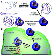
Reversibly shell cross-linked micelles based on a lipoic acid (LA) decorated triblock copolymer poly(ethylene glycol)-b-poly(γ-benzyl-L-glutamate)-b-poly(L-phenylalanine) (PEG–PGlu(EDA–LA)–PPhe) have been developed for active loading and efficient intracellular delivery of DOX. The triblock copolymer was synthesized through consecutive ring-opening polymerization of cyclic monomers γ-benzyl-L-glutamate N-carboxyanhydride (BLG-NCA) and L-phenylalanine N-carboxyanhydride (Phe-NCA) using amino-terminated poly(ethylene glycol) (PEG–NH2) as macroinitiator, followed by conjugation with LA for reversible cross-linking. The amphiphilic polymer was self-assembled to core shell corona micelles, which could be further crosslinked in the presence of a catalytic amount of dithiothreitol (DTT) in phosphate buffer (pH 7.4) to form shell-cross-linking micelles (SCLM). The SCLM showed excellent stability under physiological conditions but rapid dissociation and drug release in reductive environments mimicking those of the cytoplasm and the cell nucleus. Confocal laser scanning microscopy further demonstrated that DOX was delivered and released into the nuclei of HeLa cells following 12 h incubation with DOX-loaded SCLM. MTT assays revealed that DOX-loaded SCLM had similar anti-tumor activity as non-cross-linked micelles (NCLM) for HeLa cells following 48 h incubation. PEG–PGlu(EDA–LA)–PPhe micelles displayed low cytotoxicity up to a concentration of 1.0 mg mL−1. These biodegradable reversibly shell-cross-linked micelles provide a promising platform for intelligent intracellular drug delivery in clinical chemotherapy.


Introduction

Recently, various nanocarriers have been developed to improve efficiency of cancer chemotherapy taking advantages of the capability of target-controlled release of anticancer drugs resulted from enhanced permeability and retention (EPR) effect.1–3 Polymeric micelles, a kind of antitumour drug nanocarriers based on amphiphilic block copolymers, have been widely used for drug delivery. Polymeric micelles have presented many significant characteristics including long circulation time, good drug solubility, and passive targeting ability of tumor sites.4,5 However, a practical challenge associated with polymer micelles self-assembled from the amphiphilic block polymers is their low structural stability and may fall apart upon dilution, resulting in liberation of the payload at unwanted sites. An effective method to resolve this problem is to stabilize micelles by covalent cross-linking. Various cross-linking techniques occurring in either hydrophilic shell or hydrophobic core have been exploited to stabilize micelle structure.6,7 Ideally, the cross-linking of micelles can be cleaved to release entrapped drugs within target cells in order to overcome possible barrier to drug release from covalent cross-linking structures. Reversing cross-linking triggered by an environmental factor is a way to do it, and has become an area of great interest. Some environmental-responsive cross-linked micelles have been reported. These smart nanocarriers can be tailored to be responsive to a change in surrounding environment, such as temperature, pH, enzyme, ionic strength, and light.8–10 For example, using a well-defined double hydrophilic block copolymer, Liu's group reported the first example of cross-linked micelles containing thermoresponsive core for selective ratiometric fluorescent detection of Hg2+ ions.11 Chen's group prepared pH-sensitive micelles based on PEG-block-acetalated-dextran (PEG-b-AC-Dex) for acid-triggered intracellular release of anticancer drug DOX.12 Hu et al. utilized dually responsive shell cross-linked micelles synthesized via two types of amphiphilic diblock copolymers of P(CL-g-CPT)-b-P(OEGMA-co-MAEBA)–CPT and PCL-b-P-(OEGMA-co-MAEBA-co-FA) for photo-triggered release of caged camptothecin prodrugs.13

Among them, cross-linking with disulfide have been intensively investigated because disulfide bonds can reversibly cleavaged into free thiols responding to reductases, such as glutathione (GSH), which are stable under normal physiological conditions. Owing to the obvious difference in GSH concentrations, this distinct concentration gradient naturally occurring in the body has been used as a trigger for release of bioactive agents including drugs, genes, and proteins.14–16 For example, Lam's group prepared thiolated linear-dendritic polymers based disulfide cross-linked micelles with a high loading capacity up to 35.5%, which could be triggered to release PTX at the tumor site or in cancer cells.17 Zhong's group prepared reduction-sensitive reversibly core-cross-linked micelles based on water-soluble poly(ethylene glycol)-b-poly(N-2-hyaroxypropyl methacrylamide)–lipoic acid (PEG-b-PHPMA–LA) conjugates actively delivering doxorubicin (DOX) into the nuclei of cancer cell.18 Wang et al. developed a micellar system from disulfide linked block copolymers of PCL and poly(ethyl ethylene phosphate) aiming at intracellular DOX release triggered by GSH in tumor cells.19 Han et al. developed a redox-responsive core cross-linked (CCL) micelle conjugated with cypate moieties and cisplatin prodrugs within the cores for synergistic photothermal and chemotherapy based on diblock copolymer poly[(2-(2-methoxyethoxy)ethyl methacrylate)-co-(N-methacryloxy succinimide)]-block-poly(N-(2-hydroxypropyl) methacrylamide) synthesized via reversible addition-fragment chain transfer (RAFT) polymerization.20 Stimuli-responsive crosslinked micelles could minimize its premature release in the circulating blood pool and lead to site-specific release of the drug modulated by the specific intracellular microenvironments.21

Herein, a type of novel stabilized reduction-responsive poly(amino acid)s micelles based on LA decorated poly(ethylene glycol)-b-poly(γ-benzyl-L-glutamate)-b-poly(L-phenylalanine) (PEG–PGlu(EDA–LA)–PPhe) triblock copolymer are developed for active loading and efficient intracellular delivery of DOX (Scheme 1). These triblock polymeric micelles containing disulfide linkages were designed on account of the following considerations: (i) in materials, both PEG and poly(amino acid)s are selected as building blocks for our reversibly cross-linked micelles due to their inherently non-cytotoxicity, immunogenicity, and biodegradability have been practically used for efficient intracellular delivery of multifarious antitumor drugs such as DOX, paclitaxel (PTX), and cis-platin.22–24 (ii) in aqueous solution, PEG–PGlu(EDA–LA)–PPhe could be self-assembled into micelles with the PEG corona, the PBLG middle shell, and the PPhe inner core. The middle shell could be cross-linked using a catalytic amount of dithiothreitol (DTT) to enhance structural stability. Meanwhile, the PEG outer corona could protect the micelles forming into intermicellar bridges that lead micelles to be instable during the shell cross-linking process. And the PPhe core is offered as a reservoir of hydrophobic anticancer drugs.25 (iii) We use lipoic acid to achieve reduction sensitivity, which is produced naturally in the human body and can be easily cross-linked in the presence of a catalytic amount of DTT.26,27 (iv) Due to the large concentration gradient of GSH between the intracellular and the extracellular environment, the disulfide-cross-linked delivery vehicles modified with LA are prone to rapidly de-cross-link and release DOX intracellularly at target tissues.


image file: c4ra12255k-s1.tif
Scheme 1 Schematic illustration of the preparation of DOX-loaded PEG–PGlu(EDA–LA)–PPhe crosslinked micelles for reduction responsive intracellular drug release.

Experimental section

Materials

α-Methoxy-ω-amine-poly(ethylene glycol) (mPEG–NH2, Mn = 5000 g mol−1, Suzhou PegBio Co., Ltd.), γ-benzyl-L-glutamate (BLG) and L-phenylalanine (Phe) (GL Biochem Ltd.), lipoic acid (LA, 98%, Acros), 1,4-dithio-D,L-threitol (DTT, 99%, Merck), 1,3-dicyclohexyl carbodiimide (DCC, 99%, Alfa Aesar), N-hydroxysuccinimide (NHS, 98%, Sigma-Aldrich). Doxorubicin hydrochloride (DOX·HCl, 99%, Beijing ZhongShuo Pharmaceutical Technology Development Co., Ltd.) were used as received. Triphosgene (BTC, Shanxi Jiaocheng Jingxin Chemical Factory) was re-crystallized with chloroform prior to use. Tetrahydrofuran (THF) was dried by refluxing over sodium wire and distilled prior to use. N,N-Dimethyl formamide (DMF), ethylenediamine, triethylamine were refluxed by CaH2 and distilled prior to use.

Roswell Park Memorial Institute medium (RPMI-1640, Thermo Fisher Scientific), fetal bovine serum (FBS, Gibco), 24-well and 96-well plates were obtained from Corning Costar (Shanghai, China). 3-(4,5-Dimethyl-2-thiazolyl)-2,5-diphenyl-2H-tetrazolium bromide (MTT, Biosharp), 4′,6-diamidino-2-phenylindole (DAPI, Roche). All other chemicals were used as received.

γ-Benzyl-L-glutamate N-carboxy-anhydride (BLG-NCA) and L-phenylalanine N-carboxy-anhydride (Phe-NCA) were synthesized according to the Fuchs–Farthing method using triphosgene.28

Characterization

The 1H NMR spectra were recorded on a Bruker 400 operating at 400 MHz using deuterated dimethyl sulfoxide (DMSO-d6). The molecular weight and polydispersity of copolymers were determined by a PL GPC 50 instrument equipped with Jordi GPC columns (10E4, 2 M) following a differential refractive-index detector (PL-RI). The measurements were performed using DMF as the eluent at a flow rate of 1 mL min−1 at 50 °C and a series of narrow polystyrene standards for the calibration of the columns. The size and zeta-potential of micelles was determined by a Zetasizer Nano-ZS from Malvern Instruments. Transmission electron microscopy (TEM) measurement was performed on a FEI Tecnai G2 T12 operated with an accelerating voltage of 200 kV. Confocal laser scanning microscopy (CLSM) images were acquired using an FV1000 microscope. The absorbance of each well in MTT assays was measured using a Synergy H4 Hybrid Multi-Mode Microplate Reader. The fluorescence measurements of doxorubicin were performed using F-4500 FL Spectrophotometer at 298 K.

Synthesis of PEG–PBLG–PPhe triblock copolymers

The PEG–PBLG–PPhe triblock copolymers were synthesized using the similar method as reported.29 In a typical synthesis, BLG-NCA (189.4 mg, 0.72 mmol) was added to a stirred solution of mPEG–NH2 (0.3 g, 0.06 mmol) in anhydrous DMF (3 mL) at 35 °C under a N2 atmosphere. After 24 h, Phe-NCA (252.1 mg, 1.32 mmol) in anhydrous DMF (6 mL) were added to the reaction mixture, and the reaction was maintained for another 24 h. The resulting PEG–PBLG–PPhe copolymer was isolated by repeated precipitation in cold diethyl ether and dried overnight under vacuum. Yield: 84%.

Synthesis of PEG–PGlu(EDA)–PPhe (PEG–poly[(2-aminoethyl)glutamamide]–PPhe) triblock copolymers

PEG–PGlu(EDA)–PPhe copolymer was obtained through aminolysis with excess ethylenediamine.30 In brief, PEG–PBLG–PPhe (0.2 g, 0.018 mmol) was dissolved in DMF (4 mL) at 40 °C. Subsequently ethylenediamine (0.656 g, 10.92 mmol) was added, and then the mixture was stirred overnight at 40 °C. The resulting solution was added dropwise into 10% aqueous acetic acid solution (12 mL). The neutralized solution was dialyzed against 0.01 M hydrochloric acid solution (×3) and distilled water (×3). The white powder, PEG–PGlu(EDA)–PPhe was obtained as hydrochloric acid salt after lyophilization. Yield: 92%.

Synthesis of PEG–PGlu(EDA–LA)–PPhe (PEG–poly[(N′-lipoyl-2-aminoethyl) glutamamide]–PPhe) triblock copolymers

In a typical example, PEG–PGlu(EDA)–PPhe (0.08 g, 0.0073 mmol) was dried by azeotropic distillation with toluene and dissolved in anhydrous DMF (4 mL). To this solution was added α-lipoic acid (21.4 mg, 0.10 mmol), N,N′-dicyclohexylcarbodiimide (DCC) (22.3 mg, 0.11 mmol), hydroxysuccinimide (NHS) (12.0 mg, 0.10 mmol) and triethylamine (14.2 mg, 0.14 mmol). The mixture was stirred at room temperature for 48 h, and then filtrated to remove dicyclohexylurea. The PEG–PGlu(EDA–LA)–PPhe polymers were purified by precipitation in diethyl ether for three times and dried under vacuum at 25 °C for 24 h. Yield: 87%.

Preparation and characterisation of micelles

Micelles were self-assembled via slowly addition of distilled water to a homogeneous DMSO solution of PEG–PGlu(EDA–LA)–PPhe with vigorous stirring at room temperature. The resulting solution was transferred into a dialysis bag (MWCO 3500 Da) and dialyzed against deionized water for 24 h.

The CMC was measured by fluorescence spectroscopy using pyrene as probe. The concentration of polymer was varied from 1.0 × 10⁻⁴ to 0.05 g L−1 and the concentration of pyrene in each flask was fixed at 0.6 μM. The fluorescence spectra were recorded using F-4500 FL Spectrophotometer with the excitation wavelength of 330 nm. The emission fluorescence from 340 nm to 500 nm was monitored. The CMC value was obtained as the cross-point when extrapolating the intensity ratio I372/I383 at low and high concentration regions.

TEM observations were performed on a Tecnai G2 T12 at 200 kV. The sample were prepared by dropping 10 μL of 0.1 mg mL−1 micelle solution on a 300-mesh copper grid followed by staining with phosphotungstic acid (1 wt%).

Shell cross-linking micelles (SCLM) and de-cross-linking of micelles

The crosslinking mechanism is based on the thiol-disulfide exchange reaction. Briefly, the pH of prepared micelles dispersion was adjusted to 8.5 with borate buffer (pH = 9.0) and purged with N2 for 10 min. The cross-linking was initiated by adding 10 mol% DTT relative to the amount of lipoyl units under N2 at room temperature. After stirring for 24 h, micelles solution was dialyzed against distilled water for another 24 h. The colloidal stability of the non-cross-linked micelles (NCLM) and shell cross-linked micelles against large volume dilution (100 times dilution) and physiological salt concentration (0.15 M NaCl) was investigated by DLS.

The de-cross-linking of cross-linked micelles in response to a reductive environment was studied by DLS measurement. Briefly, under a N2 atmosphere, predetermined amount of DTT was added to yield the final DTT concentration of 10 mM. The vial was sealed and placed in a shaking bed at 200 rpm and 37 °C. The size of the micelles was determined by DLS at different time intervals.

Loading of DOX and drug loading efficiency (DLE)

DOX was used as a model drug for drug loading. Before loading DOX to the PEG–PGlu(EDA–LA)–PPhe micelles, DOX·HCl (2 mg, 0.0034 mmol) was stirred with TEA (0.99 mL, 6.8 mmol) in DMSO (0.4 mL) overnight in the dark. DOX was loaded into micelles by slowly addition of distilled water to a homogeneous DMSO solution of PEG–PGlu(EDA–LA)–PPhe and DOX (theoretical drug loading content = 15, 20 wt%) with vigorous stirring at room temperature. The unloaded DOX and the organic solvent were removed by dialysis against PB (10 mM, pH 7.4) for 24 h at room temperature. The dialysis medium was changed 5 times. The whole procedure was performed in the dark. The DOX-loaded micelles were cross-linked as described above with a catalytic amount of DTT.

The amount of DOX was determined using fluorescence measurement (excitation at 480 nm). For determination of drug loading content, DOX-loaded non-cross-linked micelles were lyophilized and dissolved in DMSO and analyzed with fluorescence spectroscopy, wherein calibration curve was obtained with DOX/DMSO solutions with different DOX concentrations.

Drug loading content (DLC) and drug loading efficiency (DLE) were calculated according to the following formula:

DLC (wt%) = (weight of loaded drug/total weight of loaded drug and polymer) × 100

DLE (%) = (weight of loaded drug/weight of drug in feed) × 100

In vitro release of DOX

The release profiles of DOX from cross-linked micelles as well as non-cross-linked micelles were studied using a dialysis bag (MWCO 12[thin space (1/6-em)]000 Da) under shaking (200 rpm) at 37 °C in two different media, i.e. PB (10 mM, pH 7.4) with 10 mM DTT or PB (10 mM, pH 7.4). The release studies were performed at a low polymer concentration of 10 mg L−1. Typically, 0.45 mL DOX-loaded micelles solution, either cross-linked or non-cross-linked, were dialysed against 50 mL of release medium. At desired time intervals, 9 mL release media was taken out and replenished with an equal volume of fresh media. The amount of DOX released was determined by using fluorescence measurement (excitation at 480 nm). The release experiments were conducted in triplicate and the results presented were the average data.

Cell viability assay

The cytotoxicity of DOX-loaded PEG–PGlu(EDA–LA)–PPhe micelles was evaluated by MTT assays using HeLa cells. The cells were seeded in a 96-well culture plates at a density of 1 × 10⁴ cells per well in 100 μL RPMI-1640 medium containing 10% fetal bovine serum (FBS), supplemented with 1% L-glutamine, antibiotics penicillin (100 IU mL−1), and streptomycin (100 μg mL−1), and incubated at 37 °C and 5% CO2 for 24 h. The medium was removed and replaced by 80 μL of fresh medium containing 10% FBS. 20 μL of DOX-loaded cross-linked or non-cross-linked micelles in PB buffer (10 mM, pH 7.4) was added to yield final anticancer drug concentrations of 0.1, 0.5, 1, 5, 10, 20 and 40 μg mL−1. The cells were incubated at 37 °C and 5% CO2 for pre-determined time. Then, the medium was removed and replaced by 100 μL of fresh medium. 10 μL of a stock solution containing 5 mg mL−1 of MTT in PBS was added and incubated for another 4 h. The medium was aspirated, the MTT-formazan generated by live cells was dissolved in 150 μL of DMSO, and the absorbance at a wavelength of 490 nm of each well was measured using a microplate reader. The relative cell viability (%) was determined by comparing the absorbance at 490 nm with control wells containing only cell culture medium. Data are presented as average values with standard deviations. The cytotoxicity of empty SCLM and NCLM was determined in a similar way using HeLa cells and MCF-7 cells.

Confocal laser scanning microscopy (CLSM) observation

The cellular uptake and intracellular release behaviors of DOX-loaded cross-linked micelles were observed by confocal laser scanning microscopy (CLSM) using HeLa cells. The cells were cultured on microscope slides in a 24-well plate at a density of 5 × 10⁴ cells per well using RPMI-1640 medium supplemented with 10% FBS, 1% L-glutamine, antibiotics penicillin (100 IU mL−1), and streptomycin (100 μg mL−1). The cells were incubated with prescribed amounts of DOX-loaded micelles or free DOX at 37 °C and 5% CO2-containing atmosphere. After incubation for 4 or 12 h, the culture medium was removed and the cells on microscope plates were washed three times with PBS. The cells were fixed with 4% (w/v) paraformaldehyde for 30 min at 25 °C and the cell nuclei were stained with 4′,6-diamidino-2-phenylindole (DAPI, blue) for 15 min, and then the cells on microscope plates were washed three times with PBS again. CLSM images of cells were obtained using confocal microscope (FV1000).

Results and discussion

Synthesis of PEG–PGlu(EDA–LA)–PPhe copolymers

PEG–PGlu(EDA–LA)–PPhe conjugates were synthesized through modifying PEG–PBLG–PPhe triblock copolymers with LA, and the PEG–PBLG–PPhe copolymers were synthesized via the one-pot two-step polymerization of BLG-NCA and Phe-NCA in the presence of a mPEG–NH2 macroinitiator (Scheme 2).
image file: c4ra12255k-s2.tif
Scheme 2 Synthetic route for the preparation of PEG–PGlu(EDA–LA)–PPhe copolymers.

1H NMR showed the composition ratio of PEG–PBLG–PPhe was calculated to be 113[thin space (1/6-em)]:[thin space (1/6-em)]5[thin space (1/6-em)]:[thin space (1/6-em)]20 and 113[thin space (1/6-em)]:[thin space (1/6-em)]10[thin space (1/6-em)]:[thin space (1/6-em)]20, respectively, by comparing signal intensities of –OCH2CH2– protons of PEG at 3.64 ppm, PhCH2– of PBLG at 5.02 ppm, and phenyl protons of PPhe at 7.17 ppm (Fig. 1). The molecular weights calculated by 1H NMR were close to the design (Table 1). GPC analyses showed a low polydispersities of 1.07–1.05 (Table 1, Fig. 2). Then base deprotection of PEG–PBLG–PPhe in DMF with ethylenediamine at 40 °C stirred overnight yielded PEG–PGlu(EDA)–PPhe. The PEG–PGlu(EDA)–PPhe copolymer signal at 5.02 ppm completely disappeared, which confirmed complete removal of the benzyl groups. Finally, PEG–PGlu(EDA–LA)–PPhe copolymers were achieved by amidation of PEG–PGlu(EDA)–PPhe with lipoic acid in DMF. Based on the ratio of resonances attributable to lipoyl ester moieties at δ 1.26–1.40 and resonances attributable to CONHCH2CH2NH2 at δ 2.93, the grafting efficiencies of LA in polymer PEG113–PGlu(EDA–LA)5–PPhe20 and PEG113–PGlu(EDA–LA)10–PPhe20 are 60% and 70%, respectively.


image file: c4ra12255k-f1.tif
Fig. 1 1H NMR spectra of (A) PEG–PBLG10–PPhe, (B) PEG–PGlu(EDA)10–PPhe and (C) PEG–PGlu(EDA–LA)10–PPhe in DMSO-d6.
Table 1 Characteristics of PEG–PGlu(EDA–LA)–PPhe triblock copolymers
Sample Mn (kg mol−1) Mw/Mnb Conversion of BLGa Conversion of Phea
Design 1H NMRa
a Calculated by 1H NMR spectra.b Measured by GPC with DMF as eluent and polystyrene as standards.
PEG113–PGlu(EDA–LA)5–PPhe20 5.0–2.2–3.2 5.0–1.4–2.9 1.07 83.3% 90.9%
PEG113–PGlu(EDA–LA)10–PPhe20 5.0–3.9–3.1 5.0–3.0–2.9 1.05 90.9% 95.2%


image file: c4ra12255k-f2.tif
Fig. 2 DMF GPC traces of PEG–NH2 and PEG–PGlu(EDA–LA)10–PPhe copolymer.

1H NMR showed besides signals attributable to PBLG–PPhe (δ 1.75–2.21, 2.86–3.02, 3.67, 4.45–4.64, 4.91–5.13 and 7.05–7.43) and PEG (δ 3.24, 3.51) also resonances attributable to lipoyl ester moieties at δ 1.26–1.40, 1.42–1.71, 1.75–1.93, 1.99–2.10 (Fig. 1C). These results supported successful synthesis of PEG–PGlu(EDA–LA)–PPhe triblock copolymer.

Preparation and reduction-triggered destabilization of cross-linked micelles

In an aqueous solution, PEG–PGlu(EDA–LA)–PPhe triblock copolymers can self-assemble into micelles by solvent exchange method. The obtained aggregates possessed the following micelle like structure: the inner hydrophobic PPhe core, the functional PGlu(EDA–LA) middle shell, and the solvated PEG outer corona. The critical micelle concentration (CMC) determined to be 6.56 and 7.38 mg L−1 for PEG–PGlu(EDA–LA)10–PPhe and PEG–PGlu(EDA–LA)5–PPhe triblock copolymers, respectively, by a fluorescence measurement using pyrene as a probe (Table 2). Subsequently, the cross-linking of middle shells was performed by introducing 10 mol% DTT relative to the lipoyl units in the polymers, similar to our previous reports.18,26,31 Dynamic light scattering (DLS) measurements revealed that size of PEG–PGlu(EDA–LA)10–PPhe micelles had slightly reduced from 145.1 nm to 124.0 nm after cross-linking while maintaining low polydispersities (PDI) of 0.198 and 0.239 (Table 2). Both of them showed modest negative surface charges of −19.0 mV and −26.1 mV via the zeta potential measurements (Table 2). TEM micrograph revealed that PEG–PGlu(EDA–LA)10–PPhe micelles and the cross-linked micelles had spherical morphology and size distribution close to that determined by DLS (Fig. 3A and B).
Table 2 Characteristics of non-crosslinked and cross-linked PEG–PGlu(EDA–LA)–PPhe micelles
Sample Non-crosslinked micelles Crosslinked micelles
Sizea (nm) PDIa Zetaa (mV) CMCb (mg L−1) Sizea (nm) PDIa Zetaa (mV)
a Determined by DLS at a concentration of 0.2 mg mL−1 at 25 °C.b Determined by fluorescence measurement using pyrene as a fluorescence probe (pyrene final concentration is 0.6 μM).
PEG–PGlu(EDA–LA)10–PPhe 145.1 0.198 −19.0 6.56 124.0 0.239 −26.1
PEG–PGlu(EDA–LA)5–PPhe 130.2 0.188 −16.8 7.38 113.0 0.219 −12.2


image file: c4ra12255k-f3.tif
Fig. 3 TEM image of the PEG–PGlu(EDA–LA)10–PPhe micelles: (A) NCLM; (B) SCLM; their particles size distribution curves by DLS at a concentration of 0.1 mg mL−1 at 25 °C correspond to (C) and (D), respectively.

The colloidal stability of cross-linked PEG–PGlu(EDA–LA)–PPhe micelles against extensive dilution and physiological salt condition was investigated with DLS. Especially, cross-linked micelles following 100-fold dilution (C < CMC) revealed only slight swelling which can be seen in hydrodynamic size and remained a narrow size distribution, while the non-cross-linked micelles were completely dissociated into small aggregates under otherwise the same conditions (Fig. 4). The size of cross-linked micelles had little change at 0.15 M NaCl, but non-cross-linked micelles had significant aggregation at the same concentration of NaCl. These findings confirm that cross-linking can greatly enhance the colloidal stability of micelles and cross-linked micelles can remain their structure integrity in above harsh conditions.


image file: c4ra12255k-f4.tif
Fig. 4 Stability of crosslinked PEG–PGlu(EDA–LA)10–PPhe micelles against 100-fold dilution (A) and 0.15 M NaCl (B). The corresponding non-cross-linked micelles were used as a control and all samples were incubated for 2 h before the DLS measurement.

The reduction-sensitivity of cross-linked PEG–PGlu(EDA–LA)10–PPhe micelles in response to 10 mM DTT was monitored in PB buffer (pH 7.4, 20 mM) at 37 °C by DLS as reported before.18,26 The results showed that DTT induced rapid dissociation of reduction-sensitive cross-linked micelles to large aggregates, with a contrast, little change of cross-linked micelles size was observed over 15 h in the absence of DTT under otherwise the same conditions (Fig. 5). These results confirmed that cross-linked PEG–PGlu(EDA–LA)–PPhe micelles are apt to rapid dissociation under a reduction condition imitating that of the cytoplasm and cell nucleus.


image file: c4ra12255k-f5.tif
Fig. 5 Time-dependant change of size of cross-linking micelles from PEG–PGlu(EDA–LA)10–PPhe with 10 mM DTT and without 10 mM DTT.

Loading and in vitro release of DOX

DOX is an effective anticancer drug against a wide range of malignancies in clinics. In the current study, DOX was selected as a model drug to study loading and in vitro release behavior. Initial drug-loading experiments were performed at a polymer concentration of 2.5 mg mL−1 using DOX at a theoretical drug loading content of 15 wt% and 20 wt%, respectively. The results showed that PEG–PGlu(EDA–LA)10–PPhe micelles had in general higher drug loading efficiency (DLE) than PEG–PGlu(EDA–LA)5–PPhe micelles (Table 3), likely due to increasing molecular weights of hydrophobic PGlu(EDA–LA) segment, resulting in increasing extent of cross-linking. Similar to pure PEG–PGlu(EDA–LA)–PPhe micelles, Dox-loaded micelles displayed shrinking of micelles by 5–19 nm after crosslinking.
Table 3 Characteristics of DOX-loaded PEG–PGlu(EDA–LA)–PPhe micelles
Sample DLC (wt%) theory Non-crosslinked micelles Crosslinked micelles DLEb (%) DLCb (wt%)
Sizea (nm) PDIa Sizea (nm) PDIa
a Determined by DLS at a concentration of 0.2 mg mL−1 at 25 °C.b Determined by fluorescence measurements.
PEG–PGlu(EDA–LA)10–PPhe 15.0 147.4 0.270 128.6 0.190 72.0 10.8
20.0 149.2 0.153 138.6 0.164 62.7 12.5
PEG–PGlu(EDA–LA)5–PPhe 15.0 130.1 0.220 125.6 0.204 60.3 9.0
20.0 136.4 0.210 129.1 0.212 51.4 10.3


The in vitro drug release from PEG–PGlu(EDA–LA)–PPhe micelles was investigated at pH 7.4 and 37 °C under different conditions mimicked intravenous administration. The results showed that at physiological conditions only ca. 36.4 and 41.4% DOX was released in 24 h from DOX-loaded shell-cross-linked PEG–PGlu(EDA–LA)10–PPhe and PGlu(EDA–LA)5–PPhe micelles, respectively (Fig. 6), likely because of their particularly sturdy middle shell-cross-linked structure. Under a reducing environment containing 10 mM DTT at pH 7.4, however, the release of DOX was significantly accelerated to 91.0 and 88.0% DOX in the presence of otherwise identical conditions, and gave a release profile similar to that of DOX-loaded non-cross-linked micelles, respectively. These results clearly indicate that drug release is rapid and complete under reductive environment. Therefore, the studies well support our assumption that these disulfide cross-linked micelles can reduce the anticancer drug loss under the physiological environment, and they can release the entrapped drugs once they entered the target cells. These disulfide cross-linked micelles can be used as a promising approach to overcome the extracellular stability versus intracellular drug release challenge.


image file: c4ra12255k-f6.tif
Fig. 6 Cumulative DOX release to 10 mM PB from (A) PEG–PGlu(EDA–LA)10–PPhe and (B) PEG–PGlu(EDA–LA)5–PPhe micelles under in vitro conditions at 37 °C over a period of 24 h. Data are presented as mean ± SD (n = 3).

Intracellular drug release of DOX-loaded shell-cross-linked micelles

To demonstrate the feasibility of the micelles for intracellular drug delivery in malignancy therapy, the cellular uptake and intracellular drug release profiles of DOX-loaded PEG–PGlu(EDA–LA)10–PPhe micelles were monitored with confocal laser scanning microscopy (CLSM). DOX-loaded PEG–PGlu(EDA–LA)10–PPhe micelle were incubated with HeLa cells for 4 h or 12 h at 37 °C, using the cells treated with free DOX as the control. The results are shown in Fig. 7. Remarkably, very strong DOX fluorescence in the cells could be observed after just 4 h incubation with DOX-loaded cross-linked PEG–PGlu(EDA–LA)10–PPhe micelles, indicating fast internalization of micelles and rapid release of DOX inside HeLa cells. This is in accordance with our expectation that disulfide bonds are cleaved in the intracellular compartments such as the cytosol and the cell nucleus due to presence of comparatively high concentrations of reducing glutathione tripeptides (2–10 mM). Notably, we found that DOX fluorescence mainly distributed in the cytosol of cells with DOX-loaded SCLM after 4 h incubation and the fluorescence intensity of DOX inside cells increased when increasing the incubation time to 12 h, part of DOX has been transported into the cell nucleus for SCLM. This phenomenon is similar to other delivery systems.17,32 In comparison, the stronger DOX fluorescence could be observed mainly in cell nucleus following incubation with free DOX for 4 h. This is because free drug could enter cell and accumulate in the nuclei via diffusion. The weaker DOX fluorescence observed for cells treated with DOX-loaded micelles than those with free DOX is probably due to poor cellular uptake of micelles that are stealthed by a dense layer of PEG shells, and also sustained release of DOX by the cleavage of disulfide bonds, which was also indicated in the in vitro release experiments. These results strongly support that DOX has been efficiently released from cross-linked PEG–PGlu(EDA–LA)10–PPhe micelles to cytosol within cells.
image file: c4ra12255k-f7.tif
Fig. 7 Confocal microscopy images of HeLa cells incubated with DOX-loaded shell-cross-linked PEG–PGlu(EDA–LA)10–PPhe micelles (SCLM), and free DOX (25 μg mL−1). For each panel, images from left to right show cell nuclei stained by DAPI (blue), DOX fluorescence in cells (red), overlays of two images, and bright field. (A) SCLM, 4 h incubation; (B) free DOX, 4 h incubation; (C) SCLM, 12 h incubation; (D) free DOX, 12 h incubation.

In vitro cytotoxicity of DOX-loaded shell-cross-linked PEG–PGlu(EDA–LA)10–PPhe micelles

We can see that crosslinked micelles of copolymer PEG–PGlu(EDA–LA)–PPhe exhibit improved stability compared to those non-crosslinked micelles, leading to minimal premature release if used for DDS. However, covalently chemical crosslinking also raises increasing concerns on the potential toxicity of those covalent crosslinking residues or the unexpected side effects with drugs loaded during the chemical crosslinking reaction. The in vitro cytotoxicities of the PEG–PGlu(EDA–LA)10–PPhe block copolymers toward HeLa cells and MCF-7 cells were evaluated by MTT assay (Fig. 8). The results displayed that the copolymer did not exhibit apparent inhibition effects on cell proliferation at all the concentrations up to 1.0 mg mL−1, indicating low cytotoxicity and good biocompatibility of block copolymers. As a matter of fact, poly(amino acid)s are a kind of biocompatible and biodegradable synthetic peptide, while LA is natural antioxidant agent existing in human body. The low cytotoxicity of SCLM is favorable for their practical applications in drug delivery.
image file: c4ra12255k-f8.tif
Fig. 8 Cytotoxicity of different concentration of PEG–PGlu(EDA–LA)10–PPhe micelles, against (A) HeLa cells, (B) MCF-7 cells after 24 h incubation using MTT assay. All data represent mean ± SD (n = 4).

The cytotoxicity of DOX to the tumor cells is highly associated with the release of DOX from polymeric micelles. Thus, the time-dependent cell viability of HeLa cells (1 × 10⁴ cells per well) was evaluated by MTT assay. Fig. 9 showed the viability of HeLa cells after incubation with DOX loaded micelles and free DOX at different DOX dosage from 0.1 to 40 μg mL−1. As seen in Fig. 9, the viability of HeLa cells decreased with an increasing amount of both free and encapsulated DOX, suggesting inhibition of cell proliferation in the presence of DOX. The results also showed that DOX-loaded cross-linked PEG–PGlu(EDA–LA)10–PPhe micelles displayed significant antitumor effect with a cytotoxicity profile close to that for DOX-loaded non-cross-linked counterparts towards HeLa cells following 48 h incubation (Fig. 9). The IC50 (i.e. inhibitory concentration to produce 50% cell death) of DOX-loaded cross-linked PEG–PGlu(EDA–LA)10–PPhe micelles was determined to be 0.85 μg mL−1 for HeLa cells, which were very close to those observed for the non-cross-linked counterparts (0.70 μg mL−1). This could be due to the presence of GSH found in HeLa cancer cells that can trigger the degradation of DOX-loaded cross-linked micelles. These results agree well with the in vitro as well as the intracellular DOX release profiles (Fig. 6 and 7). Notably, the antitumor activity of DOX-loaded cross-linked PEG–PGlu(EDA–LA)10–PPhe micelles was much higher than that reported for other DOX-loaded degradable block copolymer micelles (6.96 μg mL−1,29 6.7 μg mL−1,26 1.36 μg mL−1 (ref. 33)). All these results proved that disulfide cross-linking has no unexpected side effects on the intracellular drug release of DOX-loaded PEG–PGlu(EDA–LA)10–PPhe micelles, most likely due to fast de-cross-linking of micelles triggered by the intracellular level of GSH. DOX-loaded micelles showed a little bit lower cytotoxicity than that of free DOX (IC50 of 0.31 μg mL−1 for HeLa cells; Fig. 9), which can be attributed to the slow release of DOX from micelles and delayed nuclear uptake in HeLa cells, as evidenced by the in vitro DOX release (Fig. 6) and cellular uptake studies (Fig. 7) shown above. The antitumor activity of DOX-loaded micelles might be further improved by introducing targeting ligands such as folic acid, antibodies, peptides, and aptamers that enhance in vivo tumor accumulation and facilitate specific cellular uptake of micelles.34,35 These unique features, in addition to biocompatibility, suggest that the new micelles hold great potential for multifunctional drug delivery applications.


image file: c4ra12255k-f9.tif
Fig. 9 Anti-tumor activity of DOX-loaded shell-cross-linked PEG–PGlu(EDA–LA)10–PPhe micelles in HeLa cells. DOX-loaded non-cross-linked PEG–PGlu(EDA–LA)10–PPhe micelles were used as controls. The cells were treated with DOX-loaded micelles or free DOX for 48 h. Data are presented as mean ± SD (n = 4).

Conclusions

We have developed a novel, biocompatible, and degradable shell-cross-linked micelles based on PEG–PGlu(EDA–LA)–PPhe triblock copolymer that efficiently deliver and release doxorubicin into cancer cells, resulting in superior anti-tumor activity. These intelligent disulfide cross-linked micelles obtaining from lipoic acid conjugates have uniquely met the prerequisites of targeted nanocarriers: (i) excellent stability under physiological conditions (pH 7.4, 37 °C) but rapid micelle dissociation and drug release under reductive environments that mimic those of the cytoplasm and the cell nucleus; (ii) high drug-loading efficiency and preferential maximum drug release in response to cytoplasmic glutathione, that resulting in efficient delivery and rapid release of DOX to the cell nucleus; (iii) low cytotoxicity of the empty micelle carrier and high cytotoxicity of reductive DOX-loaded micelles to HeLa cells. These reduction-responsive biodegradable shell-cross-linked polypeptide micelles have great potential for intelligent intracellular drug delivery in clinical chemotherapy.

Acknowledgements

This work is financially supported by research grants from the National Natural Science Foundation of China (no. 21104064 and 21172188) and PAPD of Jiangsu Province.

Notes and references

  1. Z. Wang, G. Niu and X. Chen, Pharm. Res., 2014, 31, 1358–1376 CrossRef CAS PubMed.
  2. D. Wang, B. Lin and H. Ai, Pharm. Res., 2014, 31, 1390–1406 CrossRef CAS PubMed.
  3. A. K. Iyer, A. Singh, S. Ganta and M. M. Amiji, Adv. Drug Delivery Rev., 2013, 65, 1784–1802 CrossRef CAS PubMed.
  4. H. Ding, X. Wang, S. Zhang and X. Liu, J. Nanopart. Res., 2012, 14, 1254–1267 CrossRef.
  5. C. Deng, Y. Jiang, R. Cheng, F. Meng and Z. Zhong, Nano Today, 2012, 7, 467–480 CrossRef CAS PubMed.
  6. S. Wu, H. Kuang, F. Meng, Y. Wu, X. Li, X. Jing and Y. Huang, J. Mater. Chem., 2012, 22, 15348–15356 RSC.
  7. S. Samarajeewa, R. Shrestha, Y. Li and K. L. Wooley, J. Am. Chem. Soc., 2012, 134, 1235–1242 CrossRef CAS PubMed.
  8. Y. Li, K. Xiao, W. Zhu, W. Deng and K. S. Lam, Adv. Drug Delivery Rev., 2014, 66, 58–73 CrossRef CAS PubMed.
  9. C. S. Kim, B. Duncan, B. Creran and V. M. Rotello, Nano Today, 2013, 8, 439–447 CrossRef CAS PubMed.
  10. X. He, J. Li, S. An and C. Jiang, Ther. Delivery, 2013, 4, 1499–1510 CrossRef CAS PubMed.
  11. X. Wan, T. Liu and S. Liu, Langmuir, 2011, 27, 4082–4090 CrossRef CAS PubMed.
  12. Z. Zhang, X. Chen, L. Chen, S. Yu, Y. Cao, C. He and X. Chen, ACS Appl. Mater. Interfaces, 2013, 5, 10760–10766 CAS.
  13. X. Hu, J. Tian, T. Liu, G. Zhang and S. Liu, Macromolecules, 2013, 46, 6243–6256 CrossRef CAS.
  14. Y. Li, T. Liu, G. Zhang, Z. Ge and S. Liu, Macromol. Rapid Commun., 2014, 35, 466–473 CrossRef CAS PubMed.
  15. J. Zhao, J. Liu, S. Xu, J. Zhou, S. Han, L. Deng, J. Zhang, J. Liu, A. Meng and A. Dong, ACS Appl. Mater. Interfaces, 2013, 5, 13216–13226 CAS.
  16. T. Xing, B. Lai and L. Yan, Macromol. Chem. Phys., 2013, 214, 578–588 CrossRef CAS.
  17. Y. Li, K. Xiao, J. Luo, W. Xiao, J. S. Lee, A. M. Gonik, J. Kato, T. A. Dong and K. S. Lam, Biomaterials, 2011, 32, 6633–6645 CrossRef CAS PubMed.
  18. R. Wei, L. Cheng, M. Zheng, R. Cheng, F. Meng, C. Deng and Z. Zhong, Biomacromolecules, 2012, 13, 2429–2438 CrossRef CAS PubMed.
  19. Y.-C. Wang, Y. Li, T.-M. Sun, M.-H. Xiong, J. Wu, Y.-Y. Yang and J. Wang, Macromol. Rapid Commun., 2010, 31, 1201–1206 CrossRef CAS PubMed.
  20. Y. Han, J. Li, M. Zan, S. Luo, Z. Ge and S. Liu, Polym. Chem., 2014, 5, 3707 RSC.
  21. Q. Zhang, N. R. Ko and J. K. Oh, Chem. Commun., 2012, 48, 7542–7552 RSC.
  22. I. W. Hamley, Biomacromolecules, 2014, 15, 1543–1559 CrossRef CAS PubMed.
  23. A. Lalatsa, A. G. Schaetzlein, M. Mazza, L. Thi Bich Hang and I. F. Uchegbu, J. Controlled Release, 2012, 161, 523–536 CrossRef CAS PubMed.
  24. J. Huang and A. Heise, Chem. Soc. Rev., 2013, 42, 7373–7390 RSC.
  25. A. N. Koo, K. H. Min, H. J. Lee, S.-U. Lee, K. Kim, I. Chan Kwon, S. H. Cho, S. Y. Jeong and S. C. Lee, Biomaterials, 2012, 33, 1489–1499 CrossRef CAS PubMed.
  26. Y. L. Li, L. Zhu, Z. Liu, R. Cheng, F. Meng, J. H. Cui, S. J. Ji and Z. Zhong, Angew. Chem., Int. Ed., 2009, 48, 9914–9918 CrossRef CAS PubMed.
  27. A. Zhang, Z. Zhang, F. Shi, J. Ding, C. Xiao, X. Zhuang, C. He, L. Chen and X. Chen, Soft Matter, 2013, 9, 2224–2233 RSC.
  28. J. Sun, X. S. Chen, C. Deng, H. J. Yu, Z. G. Xie and X. B. Jing, Langmuir, 2007, 23, 8308–8315 CrossRef CAS PubMed.
  29. S. J. Lee, K. H. Min, H. J. Lee, A. N. Koo, H. P. Rim, B. J. Jeon, S. Y. Jeong, J. S. Heo and S. C. Lee, Biomacromolecules, 2011, 12, 1224–1233 CrossRef CAS PubMed.
  30. Y. Lee, S. Fukushima, Y. Bae, S. Hiki, T. Ishii and K. Kataoka, J. Am. Chem. Soc., 2007, 129, 5362–5363 CrossRef CAS PubMed.
  31. L. Wu, Y. Zou, C. Deng, R. Cheng, F. Meng and Z. Zhong, Biomaterials, 2013, 34, 5262–5272 CrossRef CAS PubMed.
  32. Y. Bae, S. Fukushima, A. Harada and K. Kataoka, Angew. Chem., Int. Ed., 2003, 42, 4640–4643 CrossRef CAS PubMed.
  33. W. Chen, P. Zhong, F. Meng, R. Cheng, C. Deng, J. Feijen and Z. Zhong, J. Controlled Release, 2013, 169, 171–179 CrossRef CAS PubMed.
  34. N. Kamaly, Z. Xiao, P. M. Valencia, A. F. Radovic-Moreno and O. C. Farokhzad, Chem. Soc. Rev., 2012, 41, 2971–3010 RSC.
  35. S. Yu, R. Dong, J. Chen, F. Chen, W. Jiang, Y. Zhou, X. Zhu and D. Yan, Biomacromolecules, 2014, 15, 1828–1836 CrossRef CAS PubMed.

This journal is © The Royal Society of Chemistry 2015
Click here to see how this site uses Cookies. View our privacy policy here.