Hybrid quadrupolar resonances stimulated at short wavelengths using coupled plasmonic silver nanoparticle aggregation
Ming-Ming
Jiang
a,
Hong-Yu
Chen
ab,
Bing-Hui
Li
a,
Ke-Wei
Liu
a,
Chong-Xin
Shan
a and
De-Zhen
Shen
*a
aState key Laboratory of Luminescence and Applications, Changchun Institute of Optics, Fine Mechanics and Physics, Chinese Academy of Sciences, Dongnanhu Road 3888, Changchun, 130033, People's Republic of China. E-mail: shendz@ciomp.ac.cn
bUniversity of the Chinese Academy of Sciences, Beijing, 100049, People's Republic of China
First published on 28th October 2013
Abstract
The coupling interaction among metal nanoparticles can cause asymmetric distribution of the surface charges, which can lead to the cleavage of surface plasmon resonance. Higher order resonance modes in plasmonic nanostructures exhibit sharp resonance and dramatic line shape shifts. These unique resonant behaviours existing in nanostructures can be used to improve the performance of wide band gap semiconductor devices, such as increasing the output power of lasers. According to the theoretical calculation and simulation, silver nanoparticles have been prepared experimentally, and the corresponding extinction spectra were also tested to verify the simulation results. It is found that the hybrid quadrupolar resonance can indeed occur in the short wavelength region.
1 Introduction
Higher order resonance modes result from interference of broad and narrow excitation modes, and they are typically more sensitive to the coupling interaction of metal nanostructures as well as changes of the refractive index of the environment,1 such as Fano resonance, which is mainly due to the coupling of dark quadrupolar and higher order modes with bright dipolar modes of nanoparticles. In the past decade, it has been reported that Fano resonances can also be generated in plasmonic nanostructures.2–4 Thus, considerable interest has emerged with many promising applications in physical, chemical, and biological science.5–8 Among the topics related to enhancement of the sensitivity, Fano resonance and higher order resonance modes have become the focus in the implementation of enhancements in recent years.9–14 Coupled plasmonic nanostructures provide good tunability to the generation and resonance intensity of higher-order surface plasmon resonance. Among the practical examples reported, disk ring (DR) plasmonic nanostructures are the most typical coupling plasmonic nanostructures, where the ring provides higher order multipolar resonance modes (subradiant modes or narrow dark mode) which are coupled to the disk dipolar mode (superradiant mode or broad bright mode).13,15 Breaking the symmetry by displacing the disk with respect to the ring provides a crucial mechanism for enhancing the coupling of the plasmon modes. It is reported that quadrupolar resonance modes are excited at the normal incidence while the excitation of the higher order modes (e.g., octupolar and hexadecapolar modes) needs an oblique incidence. By variation of the incident angle, the shape of Fano resonance can be altered from asymmetric to symmetric. Despite the design of plasmonic structures exhibiting Fano resonances at specific wavelengths is a challenging task because of their complex nature. A central issue in this design is the spectral engineering of the resonances via controlled hybridization of the available modes.Many of the original studies on plasmonic Fano resonances were carried out on metallic arrays, dolmen-type slab arrangements and the non-concentric ring/disk cavity.1 To implement the Fano resonance, one of the most important things is to produce higher order resonance modes. According to the published results of Fano resonances, experimental conditions are harsh. In metal nanoscale aggregation, the coupling effect among metal nanoparticles can lead to the energy level splitting of surface plasmon resonances, and a shift of the resonance peaks. However, this is difficult in systems where higher order modes are excited in the short wavelength spectral range of interest.2,16 For example, the random distribution of metal nanostructures, such as metal nanospheres, nanorods, triangle nanoparticles and so on, whether higher order resonance modes can be achieved or not is still a topic worth studying.
In recent years, much attention has been given to wide band gap semiconductors, such as ZnO, and GaN, for their wide applications in blue and ultraviolet light emitters and detectors.17–19 However, there is still a lot of room for improvement of the efficiency of luminescence and lasers.20–27 How to make resonance peaks appear in the ultraviolet regime proves to be the dilemma for improvement of the laser luminous efficiency.28–30
In this article, the extinction spectra of the dimer and the heptamer and their corresponding electromagnetic field distribution were studied by using the Finite Difference Time Domain (FDTD) method. Asymmetric distribution of the surface charges, as well as the cleavage of surface plasmon resonance will be caused by the interparticle coupling interaction in the silver nanoparticle aggregates under certain conditions. Due to the energy level splitting, formation of quadrupolar resonance mode and other higher multipolar resonance modes may occur in the short wavelength region. The calculation of the extinction spectrum of the aggregation and the electromagnetic field distribution provide an intuitive approach to study the random distribution of the silver nanoparticles. Experimentally, as long as the size and spacing distance of silver nanoparticles are within the range of theoretical calculation results, as well as the concentration of trimers and tetramers extracted from random silver nanoparticles, hybrid quadrupolar resonances can be produced by using coupled plasmonic silver nanoparticle aggregation in the short wavelength region.
2 Results and discussion
2.1 Theoretical section
The size, shape, and surrounding medium of metal nanoparticles have a very important influence on the localized surface plasmon resonances. Surface plasmon (SP) coupling among nanoparticles can induce redistribution of collective oscillations of the conduction electrons.15,16 The coupling can lead to plasmon-induced photoluminescence. A nanoparticle is one of the most promising nanostructures to form Fano resonances and higher-order dipole resonance. The spectral overlap and destructive interference of these two modes, dipole and quadrupole modes, leads to the formation of the higher-order dipole resonance. Reported results also show that by modifying the interparticle separation, relative particle size, or by breaking the symmetry of plasmonic nanoparticles, the modulation depth as well as the spectral position of the surface plasmon resonances can be highly tuned.31–36Recently, much progress on the Fano resonance has been made as demonstrated by a variety of experimental and theoretical studies in different physical settings.1,3,4,13,15 Feng Hao et al. (2009) demonstrated the tunability of subradiant dipolar and Fano-type plasmon resonances in metallic ring/disk cavities.15 This nanostructure consisted of a ring and a disk, and sustained both subradiant and superradiant dipolar mode set up via hybridization of individual disk and ring plasmons. The subradiant mode exhibits a substantially narrower line width and higher field enhancement than the parent plasmons. Plasmonic heptamers made of silver and all-dielectric oligomers exhibit well-pronounced Fano resonances with strong suppression of the scattering cross-section.2,16,31 The analysis reveals that this type of Fano resonance originates from the optically induced coupling interaction between dipole and quadrupole modes of individual nanoparticles. Based on the results, this asymmetric type coupling interaction between nanoparticles will lead to plasmon redistribution and shifting of the resonance peak position.
The strength of the interaction between the central and the surrounding plasmons is controlled by the distance between the nanospheres.2 Simultaneously, for the sake of prompting the multipole resonance occurred in the short wavelength region, we should focus on the coupling interaction between the central nanoparticle and the surrounding nanoparticles, and the coupling interaction among the surrounding nanoparticles of aggregation.
In order to understand the interparticle coupling interaction of a random distribution of metal nanoparticles, a simple and ideal finite element model consisting of a central sphere and surrounding spheres was built to calculate the extinction spectra using the FDTD method based on the quasi-particle approximation as shown in Fig. 1. The metallic particles in this study are assumed to be silver spheres, and will be modeled by using experimental dielectric data (JC).37 The refractive index of the environment is defined as 1.33. The aggregate changes from the dimer to the heptamer, the radius of the central Ag nanoparticle r1 = 40 nm, the radius of the surrounding Ag nanoparticles r2 = 30 nm, and the radius of silver nanoparticles shown in Fig. 1(b) is denoted as r1 = 40 nm. The gap between the central and the surrounding nanoparticles of different cross-sections of Ag nanoparticle aggregates g ranged from 5 nm to 30 nm, and the type of aggregation from dimer to heptamer. Fig. 1(a) and (b) demonstrate that when the gap is in a certain range, such as 5 nm, the interaction between the nanoparticles is much strong such that interparticle coupling will lead to shifting of the spectral position of the plasmon resonance compared to the case of an isolated particle. The extra format is the quadrupole resonance at around 350 nm. Fig. 1(c) shows that the interparticle coupling between the central nanoparticle and the surrounding nanoparticles is stronger than the interaction between the surrounding nanoparticles of the tetramer. The quadrupole resonance of the tetramer is also relatively strong compared with the dipole mode. So the resonance peak located in the short-wave region will dominate in the extinction spectrum. Fig. 1(d)–(f) reveal the extinction spectra of the pentamer, hexamer and heptamer respectively. From these extinction spectra, we can get that the emergence of the special line shape rooted in the coupling interaction between the plasmons originated from the interparticle coupling between the central nanoparticle and the surrounding nanoparticles, and the plasmons originated from the interparticle coupling between the surrounding nanoparticles. The plasmon hybridization can be used to figure out the multifeatured plasmon response of more complex metallic nanostructures, such as dimers or other nanoparticle aggregates. An aggregate is composed of a central silver nanoparticle and several surrounding nanoparticles, and the associated interaction between the central nanoparticle and the surrounding nanoparticles, leading to higher order resonance modes.
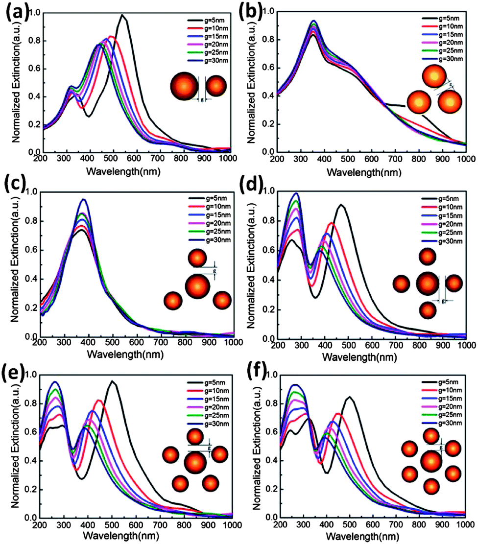 | ||
| Fig. 1 Extinction (scattering + absorption) spectra of silver nanoparticles with a gap between the central and surrounding Ag nanoparticles ranging from 5–30 nm, and the corresponding (a)–(f) are from the dimer to the heptamer respectively. The radius of the central nanoparticle is denoted as r1, the radius of the surrounding nanoparticles is denoted as r2, and the gap distance between the central nanoparticle and the surrounding nanoparticle is denoted as g. | ||
When the gap g is in certain ranges, such as 5 nm, and 10 nm, we can observe the special linear spectral lines. The extinction spectra reveal a special narrow feature in the short wavelength region as shown in Fig. 1(d)–(f). These types of spectral lines are in agreement with the results published in previous studies,10,38 which are called Fano resonance. The quadrupole resonances appear naturally in such systems when the interparticle coupling interaction occurred between the central nanoparticle and the surrounding nanoparticles.3,16 When the two oscillator modes are of similar frequency the interference results in a symmetric antiresonance. If the frequencies are different, the typical asymmetric multiple resonances appear.2 The quadrupole mode of the metal nanoparticle aggregation can overlap with the superradiant bright mode due to the small energy gap, which may help to form Fano resonances.1
The interparticle interaction results in the splitting of the plasmon resonances into two new resonances: the lower energy symmetric and the higher energy antisymmetric plasmons. Their interaction results in other hybridized plasmon resonances as demonstrated in Fig. 1; we have shown that the plasmon response of metal-based nanostructures can be viewed as the collection of plasmons arising from simpler geometries to form an interacting system. The plasmonics of the metallic nanostructures are determined by the electromagnetic interaction between these free plasmons, which leads to mixing (hybridization), splitting, and shifting of the plasmon energies.2,16,31
For further exploration of factors affecting the shift of the peak position, Fig. 2 shows the extinction spectra of the tetramer and heptamer placed on a sapphire substrate surrounded by dielectric embedding media of different permittivities. Fig. 2(a) demonstrates the quadrupole resonance rooted in the interparticle coupling interaction between the central silver nanoparticle and the surrounding nanoparticles. Fig. 2(b) demonstrates the Fano resonance. Both panels demonstrate that the effect of dielectric screening is a strong red-shift of the multiple resonances. Particularly for the tetramer, the LSPR shift for the multiple resonances in Fig. 2(a) is from 250 nm to 350 nm. As the shift of the multipolar format peak position is in the control range of the short wavelength region, the dielectric function of the environment medium should not be too big, for example εd ≤ 2 as shown in Fig. 2.
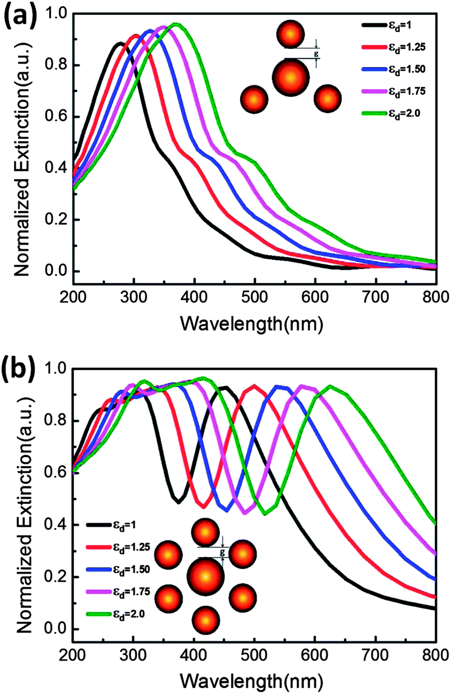 | ||
| Fig. 2 Extinction (scattering + absorption) spectra of silver nanoparticle aggregation: (a) shows the normalized extinction of the tetramer with the radius of the central nanoparticle r1 = 40 nm, the radius of the surrounding nanoparticles r2 = 30 nm, the distance between the central nanoparticle and the surrounding nanoparticles g = 10 nm; (b) shows the normalized extinction of the heptamer with the radius of the central nanoparticle r1 = 40 nm, the radius of the surrounding nanoparticles r2 = 30 nm, and the distance between the central nanoparticle and the surrounding nanoparticles g = 10 nm. | ||
In the following, we used the FDTD method to simulate the distribution of the electromagnetic field for the aggregation through the plasmon hybridization picture. For small particles, the diameter of the particle and the interparticle distance d, g, under the condition d, g ≪ λi. These interparticle interactions among ensembles are essentially of dipolar nature, and the particle ensemble can in the first approximation be treated as an ensemble of interacting dipoles. However, when the size and distribution of nanoparticles in the aggregate appears as asymmetric distribution, along with the size increase of the nanostructure, the electric field of the light can no longer be assumed to be uniform inside the particles, and higher order (quadrupole, octupole, etc.) plasmon modes can directly couple with the electric field of the light simply due to the phase retardation effect. Excitation of multipole plasmon resonances is also caused by the asymmetric distribution of the surface plasmons excited by the electromagnetic interactions between the localized modes. When the gap distance among nanoparticles is large enough, every metal nanoparticle exists as an individual, so the interaction between nanoparticles is very weak. When the gap is small in certain ranges the aggregation almost exists as a whole. So in both cases it is impossible to cause the energy splitting and the resonance peak position shifting. Therefore, the corresponding resonance peak was located in the short wavelength region as shown in Fig. 1. We have simulated the aggregation from dimers to heptamers at the incident wavelength λi = 350 nm as shown in Fig. 3. The radius of the central nanoparticle is 40 nm, and the radius of the surrounding nanoparticles is 30 nm. Close inspection of ring structures of aggregates shows superposed bright features identified as the electromagnetic interaction formed by the quadrupole plasmons and dipole plasmons that is thought to be the hybridization coupling interaction between the dipole modes and the quadrupole modes. Fig. 3(a)–(c) demonstrate that the interparticle coupling among the surrounding nanoparticles is very weak, the main interaction existed between the central nanoparticle and its surrounding nanoparticles. Fig. 3(d)–(f) demonstrate that the interparticle coupling interaction exists between the central nanoparticle and its surrounding nanoparticles, and the interparticle interaction exists between the surrounding nanoparticles. We may safely draw the conclusion that higher order modes, such as quadrupolar resonance, octupolar resonance, hexadecapolar resonance and so on, are derived from the coupling interaction between the central nanoparticle and its surrounding nanoparticles.
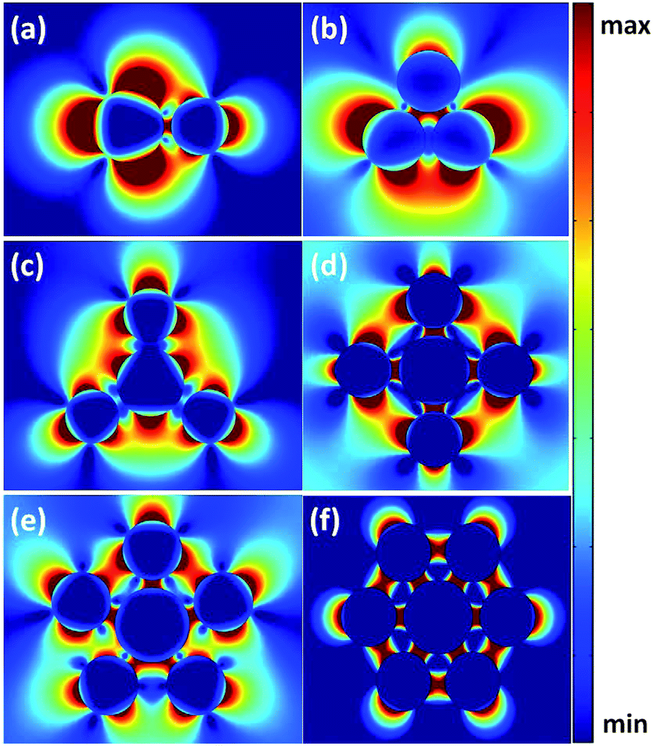 | ||
| Fig. 3 The spatial distribution of the electric field intensity in aggregates with the gap g = 10 nm between the central nanoparticle and the surrounding nanoparticles; the incident wavelength is λi = 350 nm; the light is vertically incident on the cross-section of the aggregate. | ||
Take the heptamer for example, the nanoparticles of the heptamer are silver nanospheres with the diameter d = 50 nm. When the gap distance between nanoparticles is in a certain range, such as 10–30 nm, electromagnetic field distribution was simulated, as shown in Fig. 4. At the incident wavelength λi = 500 nm, with gap g = 20 nm and g = 30 nm, the distribution of the electromagnetic field line is completely symmetrical; the corresponding resonant peak line is also completely symmetrical as shown in Fig. 1. At the incident wavelength λi = 350 nm, with gap g = 20 nm and g = 30 nm, the asymmetric distribution of the electromagnetic field line appears accordingly. This situation will directly lead to the splitting of energy levels. Compared with Fig. 4(a) and (b) and Fig. 4(c) and (d), there is a superimposed region resulting from the interaction shown in Fig. 1(f). The plasmon hybridization picture can be used to describe the sensitive structural tunability of the plasmon resonance as the interaction between plasmons is supported by a nanoscale central nanoparticle and the surrounding nanoparticles. This simple and intuitive picture can also be used to understand the plasmon resonance behavior of composite metallic nanostructures of greater geometrical complexity. The plasmon hybridization picture is important because it provides the nanoscientist with a powerful and general design principle that can be applied both qualitatively and quantitatively, and guides the design of metallic nanostructures and predict their resonant properties.
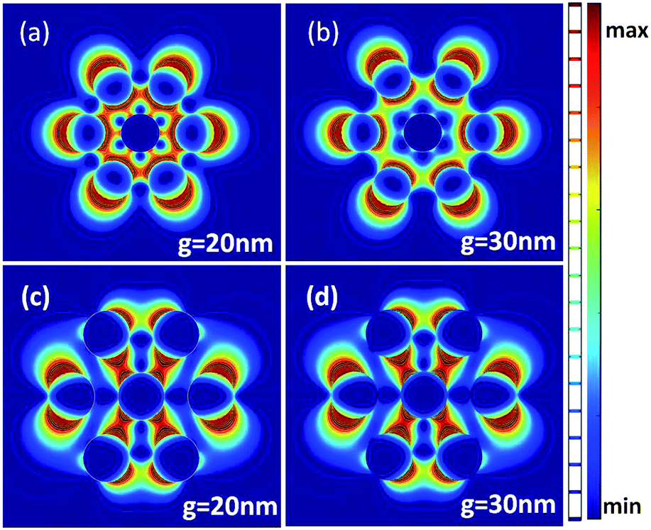 | ||
| Fig. 4 The spatial distribution of the electric field intensity in the heptamer with the diameter of the nanoparticle being 50 nm: (a) and (b) show that at the incident wavelength λi = 500 nm, with different gaps among the nanoparticles 20 nm, 30 nm, compared with the incident wavelength λi = 350 nm (c) and (d). | ||
From the results above, the hybrid quadrupolar resonance stimulated in the short wavelength region should meet some requirements: the interparticle coupling interaction among the surrounding nanoparticles should be weaker than the interparticle coupling interaction between the central nanoparticle and the surrounding nanoparticles. As the distance between the central nanoparticle and the surrounding nanoparticles is smaller than the distance between the surrounding nanoparticles, the number of the surrounding nanoparticles should not be too many, the best is two or three. The effect of a surrounding dielectric medium on the extinction spectrum of the silver nanosphere aggregation also demonstrated that the refractive index of the environment medium should not be too high, otherwise the peak positions of the spectrum will be red shifted. Compared with silver, we also studied gold nanoparticles and found that gold nanoparticle aggregates can exhibit the multipole resonance phenomenon, but not in the short wavelength region. So gold is not suitable for study in the short wavelength band.36,37
2.2 Experimental section
In this article, Ag (99.99%) was deposited on the c-face sapphire by a radio-frequency magnetron sputtering technique at room temperature with a pressure of 5 × 10 Pa, and subsequently annealed in a N2 atmosphere at 350 °C to form Ag nanoparticles. The morphology of the samples was characterized using a scanning electron microscope (SEM), as shown in Fig. 5 with different sputtering times from 1 min to 12 min. The sputtering time will directly determine the spacing distance among the silver nanoparticles and the size of the nanoparticles. Although, silver nanoparticles are randomly distributed during the preparation process, the central silver nanoparticle and the surrounding silver nanoparticle ring-like aggregate can be extracted from the random distribution. With the sputtering time of 3 minutes, silver nanoparticles existed are mainly dimers. With the extension of the sputtering time, the aggregate of silver nanoparticles extracted from dimer to trimer to tetramer can appear even as a heptamer. Detailed SEM images are shown in Fig. 5. Fig. 5(a) demonstrates that the dimer and trimer of silver nanoparticles can easily be extracted as an aggregate, the diameter ranged from 50 nm to 90 nm, and the corresponding gap distance ranged from 10 nm to 40 nm. Naturally, the hybrid quadrupolar resonance can be generated in the short wavelength region according to our calculated results. Just because the sputtering time is relatively short, it leads to the relatively large spacing distance among silver nanoparticles. When we extend the time of sputtering of silver nanoparticles, the concentration of trimers and tetramers collected from random distribution of silver nanoparticles will continue to improve, the corresponding hybrid quadrupolar resonance will also continue to increase. The best experimental results as shown in Fig. 5 involve a sputtering time of 9 minutes. Hence, the diameter change of the silver nanoparticles is not too much, ranged from 50 nm to 100 nm, and the main changes are in the spacing distance among silver nanoparticles and the concentration of trimers and tetramers collected from random distribution of silver nanoparticles, as shown in Fig. 5. So the spacing distance among the silver nanoparticles and the concentration of trimers and tetramers are the most directly influencing factors of the hybrid quadrupolar resonance intensity. Thus two types of dipolar resonances are formed in this structure: hybridized bonding and antibonding linear combinations of the central localized and the surrounding localized modes.2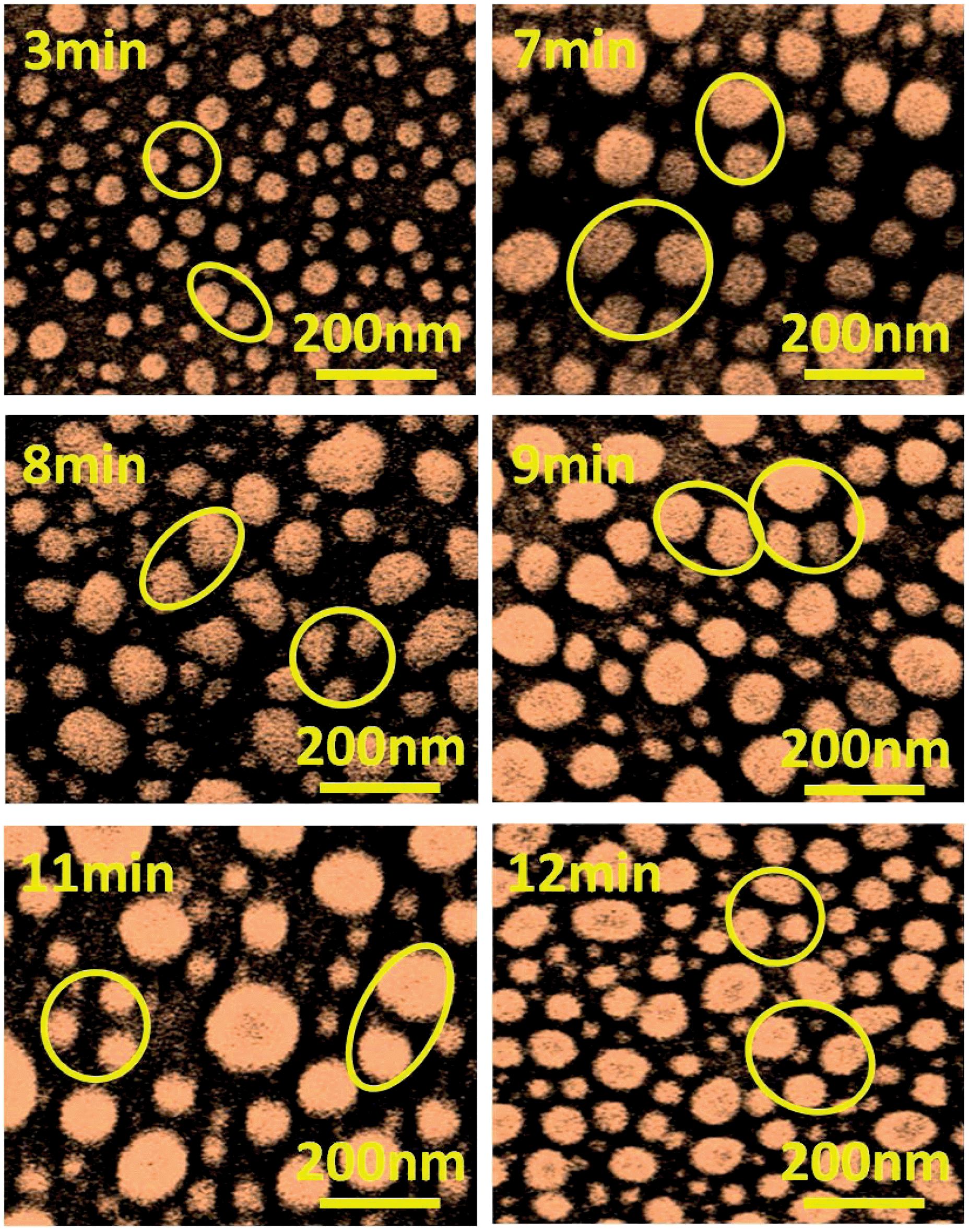 | ||
| Fig. 5 SEM images of nanoparticle aggregates with different sputtering times 3 min, 7 min, 8 min, 9 min, 11 min, and 12 min respectively. | ||
The extinction spectra of random silver nanoparticles were measured using a Shimadzu UV-3101PC scanning spectrophotometer. Fig. 6 demonstrates the extinction spectra of Ag nanoparticles sputtered on the c-plane sapphire with different sputtering times from 1 to 12 min. There are two resonances at around 350 and 500 nm, which are the quadrupole and dipole modes, respectively. From the extinction spectrum graphics, one can know that when the sputtering time is short, such as 1–3 min, the asymmetric profile curve is not very obvious. So there is only one of the main resonance peak positions in the extinction spectra, which is confirmed by the theoretical calculation and simulation results. Silver nanoparticles mainly exist in the form of single nanoparticles and dimer aggregates, but with a large gap distance. As the sputtering time is increased, the multipolar resonance profile curve becomes more and more obvious. So in the short wavelength region, another resonance peak appears, which is caused by the energy level splitting.3,4 And the corresponding surface plasmon resonance is identified as the hybrid quadrupolar resonance. From the extinction spectral curve, the concentration of trimers and tetramers collected from random distribution of silver nanoparticles as well as the gap distance among silver nanoparticles are the crucial factors for the generation and intensity of the hybrid quadrupolar resonance. Therefore, under certain experimental conditions, the hybrid quadrupolar resonance can be produced in the short wavelength region using the random distribution of silver nanoparticles.
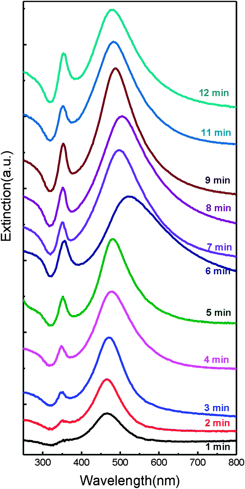 | ||
| Fig. 6 Extinction spectra of silver nanoparticles sputtered on the c-plane sapphire with different sputtering times from 1 min to 12 min. | ||
Compared with silver, gold nanoparticles are more suitable for study in the 500 nm to 600 nm wavelength region.39 Gold nanoparticles also were prepared under the same experimental conditions and environment with different sputtering times of 1 min, 3 min, 6 min, and 9 min respectively.37,40,41 The corresponding extinction spectra are shown in Fig. 7. The obvious quadrupolar resonance peak cannot be observed. There is only one resonance at around 550 nm when sputtering for 1 min, this is dipole resonance mode. When sputtering for 3 min, enhancement of dipole resonance can be derived from strong interparticle interactions among gold nanoparticles. The increase of sputtering time will lead to the decrease of the distance between the nanoparticles. Compared with silver aggregation, the controllability of gold nanoparticles can only enhance the dipole resonance without the cleavage of surface plasmon resonance. With the further extension of the sputtering time of 9 min, the decrease in the spacing distance between nanoparticles will make gold nanoparticles lose individual existence. Therefore, when sputtering for 9 min, obvious dipole resonance cannot be observed. Therefore, hybrid quadrupolar resonance cannot be produced in the short wavelength region using coupled plasmonic gold nanoparticle aggregation.
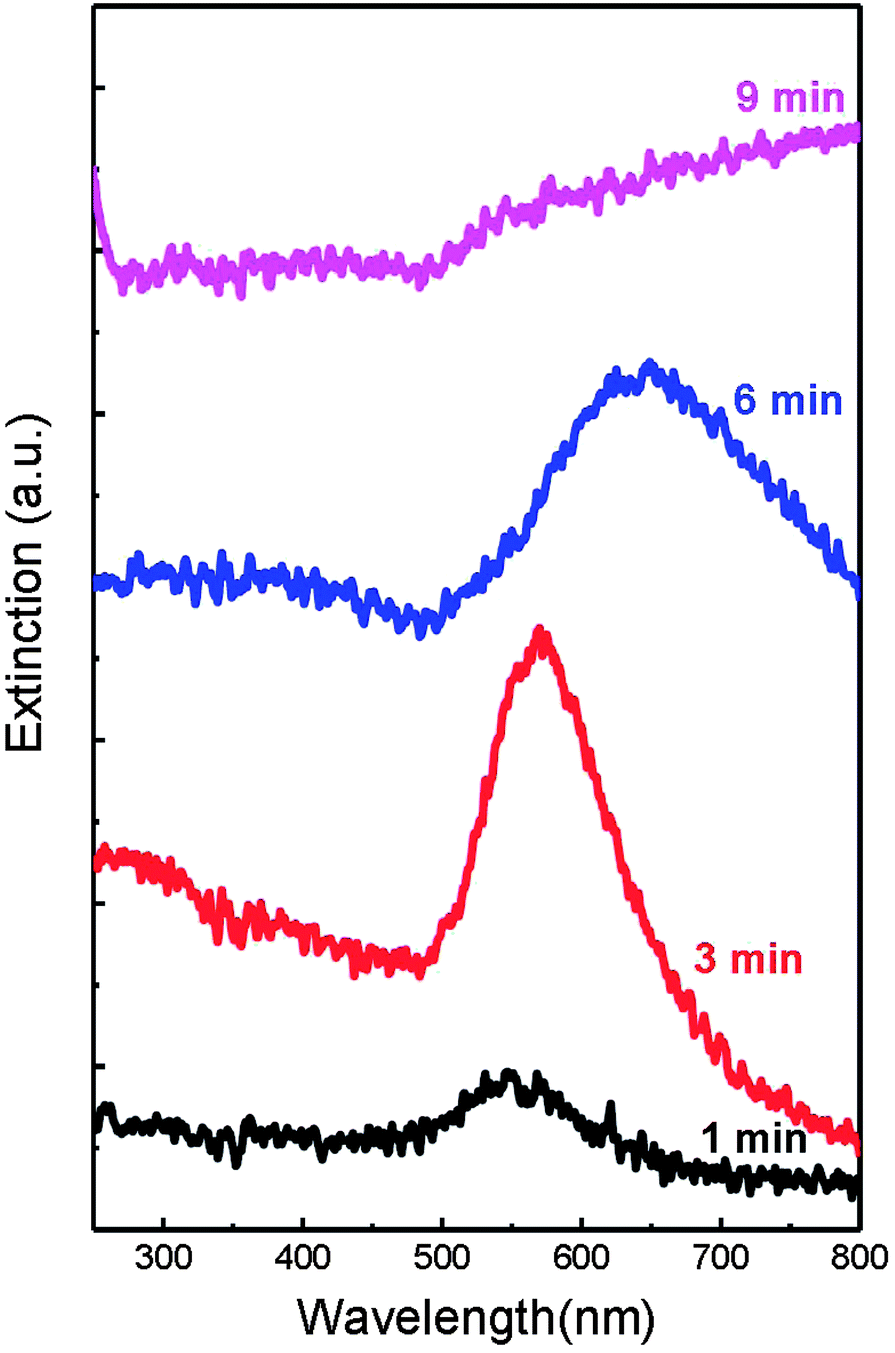 | ||
| Fig. 7 Extinction spectra of gold nanoparticles sputtered on the c-plane sapphire with different sputtering times of 1 min, 3 min, 6 min, and 9 min respectively. | ||
The hybrid quadrupolar resonances appear naturally in such systems when the surrounding localized plasmon modes are coupled to and spectrally overlapped with the central localized plasmon modes. The effective interaction between such two modes is dispersive and can result in a strong interference in the oscillator amplitudes which in turn influences the radiation emitted from the system. When the two oscillator modes have similar frequency, the interference results in a symmetric antiresonance. If the frequencies are different, the typical hybrid quadrupolar resonance appears.38
The peak wavelength, the peak width, and the effect of secondary resonances yield a unique spectral fingerprint for a plasmonic nanoparticle with a specific size and shape. Additionally, UV-Visible spectroscopy provides a mechanism to monitor the changes in nanoparticles over time. When silver nanoparticles aggregate, the metal nanoparticles become electronically coupled and this coupled system has a different SPR from the individual nanoparticles.27,42 In the case of a multi-nanoparticle aggregate, the plasmon resonance will be blue-shifted to a shorter wavelength than the resonance of an individual nanoparticle, and aggregation is observable as an intensity increase in the blue/violet region of the spectrum. This effect can be observed in Fig. 1–4, which displays the optical response of a silver nanoparticle solution destabilized by the addition of saline. Carefully monitoring the UV-Visible spectrum of the silver nanoparticles with the sputtering time is a sensitive technique used for determining if any nanoparticle aggregation has occurred.43
3 Conclusions
In summary, we have shown that the interparticle coupling interaction among the random distribution of silver nanoparticles can lead to mixing (hybridization), splitting, and shifting of the plasmon energies, as well as the hybrid quadrupolar resonances formed by the interaction between the plasmons of the exocyclic metal nanoparticles and the central metal nanoparticle. This interaction also results in the splitting of the plasmon resonances into two new resonances: the lower energy symmetric plasmon and the higher energy antisymmetric plasmon. Particularly the higher energy antisymmetric plasmon embodies the hybrid quadrupolar resonances and can be produced in the short-wavelength region. The physical properties can be applied in wide band gap semiconductor based lasers and detectors. Our work affords a theoretical and experimental method, and provides evidence that silver nanoparticle aggregation produces hybrid quadrupolar resonances in the short-wavelength region.Acknowledgements
This work was supported by the National Basic Research Program of China (973 Program) (no. 2011CB302006, 2011CB302004), the National Natural Science Foundation of China (no. 10974197, 11174273, 11104265, 21101146), and the 100 Talents Program of the Chinese Academy of Sciences.References
- B. Luk'yanchuk, N. I. Zheludev, S. A. Maier, N. J. Halas, P. Nordlander, H. Giessen and C. T. Chong, Nat. Mater., 2010, 9, 707–715 CrossRef CAS PubMed.
- N. A. Mirin, K. Bao and P. Nordlander, J. Phys. Chem. A, 2009, 113, 4028–4034 CrossRef CAS PubMed.
- A. E. Miroshnichenko and Y. S. Kivshar, Nano Lett., 2012, 12, 6459–6463 CrossRef CAS PubMed.
- K. Thyagarajan, J. Butet and O. J. Martin, Nano Lett., 2013, 13, 1847–1851 CAS.
- S. Lal, S. Link and N. J. Halas, Nat. Photonics, 2007, 1, 641–648 CrossRef CAS.
- J. N. Anker, W. P. Hall, O. Lyandres, N. C. Shah, J. Zhao and R. P. Van Duyne, Nat. Mater., 2008, 7, 442–453 CrossRef CAS PubMed.
- Y.-Y. Lai, Y.-P. Lan and T.-C. Lu, Light: Sci. Appl., 2013, 2, e76 CrossRef.
- E. Matioli, S. Brinkley, K. M. Kelchner, Y.-L. Hu, S. Nakamura, S. DenBaars, J. Speck and C. Weisbuch, Light: Sci. Appl., 2012, 1, e22 CrossRef.
- A. N. Poddubny, M. V. Rybin, M. F. Limonov and Y. S. Kivshar, Nat. Commun., 2012, 3, 914 CrossRef PubMed.
- A. E. Miroshnichenko, S. Flach and Y. S. Kivshar, Rev. Mod. Phys., 2010, 82, 2257–2298 CrossRef CAS.
- G. Konstantatos and E. H. Sargent, Nat. Nanotechnol., 2010, 5, 391–400 CrossRef CAS PubMed.
- V. A. Tamma, Y. Cui, J. Zhou and W. Park, Nanoscale, 2013, 5, 1592–1602 RSC.
- Y. H. Fu, J. B. Zhang, Y. F. Yu and B. Luk'yanchuk, ACS Nano, 2012, 6, 5130–5137 CrossRef CAS PubMed.
- D. Dregely, M. Hentschel and H. Giessen, ACS Nano, 2011, 5, 8202–8211 CrossRef CAS PubMed.
- F. Hao, P. Nordlander, Y. Sonnefraud, P. V. Dorpe and S. A. Maier, ACS Nano, 2009, 3, 643–652 CrossRef CAS PubMed.
- E. Prodan, C. Radloff, N. Halas and P. Nordlander, Science, 2003, 302, 419–422 CrossRef CAS PubMed.
- F. Robert, Science, 1997, 276, 895–896 CrossRef.
- D. Li, X. Sun, H. Song, Z. Li, Y. Chen, H. Jiang and G. Miao, Adv. Mater., 2012, 24, 845–849 CrossRef CAS PubMed.
- K. Chung, C.-H. Lee and G.-C. Yi, Science, 2010, 330, 655–657 CrossRef CAS PubMed.
- S. Jiao, Z. Zhang, Y. Lu, D. Shen, B. Yao, J. Zhang, B. Li, D. Zhao, X. Fan and Z. Tang, Appl. Phys. Lett., 2006, 88, 031911 CrossRef.
- K. Ding and C. Ning, Light: Sci. Appl., 2012, 1, e20 CrossRef.
- D. Lepage, A. Jiménez, J. Beauvais and J. J. Dubowski, Light: Sci. Appl., 2013, 2, e62 CrossRef CAS.
- P. Berini and I. De Leon, Nat. Photonics, 2011, 6, 16–24 CrossRef.
- R.-M. Ma, R. F. Oulton, V. J. Sorger, G. Bartal and X. Zhang, Nat. Mater., 2010, 10, 110–113 CrossRef PubMed.
- M. Noginov, G. Zhu, A. Belgrave, R. Bakker, V. Shalaev, E. Narimanov, S. Stout, E. Herz, T. Suteewong and U. Wiesner, Nature, 2009, 460, 1110–1112 CrossRef CAS PubMed.
- R. F. Oulton, V. J. Sorger, T. Zentgraf, R.-M. Ma, C. Gladden, L. Dai, G. Bartal and X. Zhang, Nature, 2009, 461, 629–632 CrossRef CAS PubMed.
- C. Ciraci, R. Hill, J. Mock, Y. Urzhumov, A. Fernandez-Dominguez, S. Maier, J. Pendry, A. Chilkoti and D. Smith, Science, 2012, 337, 1072–1074 CrossRef CAS PubMed.
- S. W. Hwang, D. H. Shin, C. O. Kim, S. H. Hong, M. C. Kim, J. Kim, K. Y. Lim, S. Kim, S.-H. Choi, K. J. Ahn, G. Kim, S. H. Sim and B. H. Hong, Phys. Rev. Lett., 2010, 105, 127403 CrossRef.
- R. Liu, X. Fu, J. Meng, Y.-Q. Bie, D. Yu and Z.-M. Liao, Nanoscale, 2013, 5, 5294–5298 RSC.
- P. Cheng, D. Li, Z. Yuan, P. Chen and D. Yang, Appl. Phys. Lett., 2008, 92, 041119 CrossRef.
- J. A. Fan, C. Wu, K. Bao, J. Bao, R. Bardhan, N. J. Halas, V. N. Manoharan, P. Nordlander, G. Shvets and F. Capasso, Science, 2010, 328, 1135–1138 CrossRef CAS PubMed.
- S. A. Maier, Plasmonics: fundamentals and applications, Springer, 2007 Search PubMed.
- Y.-H. Su, Y.-F. Ke, S.-L. Cai and Q.-Y. Yao, Light: Sci. Appl., 2012, 1, e14 CrossRef.
- M. M. Miller and A. A. Lazarides, J. Opt. A: Pure Appl. Opt., 2006, 8, S239 CrossRef.
- C. Noguez, J. Phys. Chem. C, 2007, 111, 3806–3819 CAS.
- P. R. West, S. Ishii, G. V. Naik, N. K. Emani, V. M. Shalaev and A. Boltasseva, Laser Photonics Rev., 2010, 4, 795–808 CrossRef CAS.
- P. B. Johnson and R.-W. Christy, Phys. Rev. B: Condens. Matter Mater. Phys., 1972, 6, 4370 CrossRef CAS.
- M. Rahmani, B. Luk'yanchuk and M. Hong, Laser Photonics Rev., 2013, 7, 329–349 CrossRef CAS.
- K. Okamoto, I. Niki, A. Shvartser, Y. Narukawa, T. Mukai and A. Scherer, Nat. Mater., 2004, 3, 601–605 CrossRef CAS PubMed.
- E. Hutter and J. H. Fendler, Adv. Mater., 2004, 16, 1685–1706 CrossRef CAS.
- T. Klar, M. Perner, S. Grosse, G. Von Plessen, W. Spirkl and J. Feldmann, Phys. Rev. Lett., 1998, 80, 4249 CrossRef CAS.
- Y.-J. Lu, J. Kim, H.-Y. Chen, C. Wu, N. Dabidian, C. E. Sanders, C.-Y. Wang, M.-Y. Lu, B.-H. Li and X. Qiu, et al. , Science, 2012, 337, 450–453 CrossRef CAS PubMed.
- C. Cheng, E. Sie, B. Liu, C. Huan, T. Sum, H. Sun and H. Fan, Appl. Phys. Lett., 2010, 96, 071107 CrossRef.
| This journal is © The Royal Society of Chemistry 2014 |