Spontaneous and field-induced mesomorphism of a silyl-terminated bent-core liquid crystal as determined from second-harmonic generation and resonant X-ray scattering
C. L.
Folcia
*a,
J.
Ortega
b,
J.
Etxebarria
a,
S.
Rodríguez-Conde
a,
G.
Sanz-Enguita
b,
K.
Geese
c,
C.
Tschierske
c,
V.
Ponsinet
d,
P.
Barois
d,
R.
Pindak
e,
LiDong
Pan
fi,
Z. Q.
Liu
g,
B. K.
McCoy
h and
C. C.
Huang
i
aDepartment of Condensed Matter Physics, University of the Basque Country, UPV/EHU, 48080 Bilbao, Spain. E-mail: cesar.folcia@ehu.es
bDepartment of Applied Physics II, University of the Basque Country, UPV/EHU, 48080 Bilbao, Spain
cInstitute of Chemistry, Organic Chemistry Martin-Luther-University, Halle-Wittenberg Kurt Mothes Str. 2, D-06120 Halle/Saale, Germany
dUniversity of Bordeaux, CNRS, CRPP, UPR8641, F-33600, Pessac, France
ePhoton Sciences Directorate, Brookhaven National Laboratory, Upton, New York 11973, USA
fDepartment of Physics and Astronomy, Johns Hopkins University, Baltimore, Maryland 21218, USA
gDepartment of Chemistry and Physics, St. Cloud State University, St. Cloud, Minnesota 56301, USA
hDepartment of Mathematics and Physics, Azusa Pacific University, Azusa, California 91702, USA
iSchool of Physics and Astronomy, University of Minnesota, Minneapolis, Minnesota 55455, USA
First published on 30th October 2013
Abstract
The polarity and structure of the phases of a liquid crystal constituted by thiophene-based bent-core molecules is investigated by means of optical second-harmonic generation (SHG), and resonant and conventional X-ray diffraction. The material studied is representative of a wide family of mesogens that contain silyl groups at the ends of the chains. These bulky terminal groups have been reported to give rise to smectic phases showing ferroelectric switching. However, the analysis of the SHG signal before and after application of electric fields has allowed us to establish unambiguously that the reported ferroelectricity is not intrinsic to the material but stabilized by the cell substrates once an electric field has been applied. In addition, the results obtained from resonant X-ray diffraction indicate that virgin samples have antiferroelectric undulated synclinic smectic structures.
1. Introduction
Bent core molecules can give rise to liquid crystalline phases that are not found in classical calamitic compounds.1 Remarkably, bent-core liquid crystals can be polar and chiral even though their constituent molecules are achiral.2 Recent studies have demonstrated unusual properties in these materials, which could be used for applications in various fields, such as displays,3 nonlinear optics4 or flexoelectric materials.5 Several papers provide comprehensive reviews of the properties6 and potential applications7 of bent-core liquid crystals.Usually, bent-core molecules assemble in layers, leading to smectic phases. Within each layer the molecular bent shape restricts the rotation of the molecules about their long axes and, therefore, gives rise to strongly polar layers. The polarization is in the layer plane and along the direction of the molecular transverse dipole moment. However, in most of the studied materials, the correlation between the dipoles of neighboring layers is anti-parallel, promoting antiferroelectric (AF) phases (see Fig. 1a). In comparison, ferroelectric (FE) structures are much less common. The AF organization is more stable than the FE one because there are entropic and electrostatic contributions to the free energy which are favorable to the AF structure. In addition to the antiparallel arrangement, more contribution to the entropy arises because molecular positions can fluctuate easily from layer to layer since the molecular tails are synclinic at the interlayer. However such fluctuations are hindered for FE organizations, since in those cases the tails are anticlinic and interdigitation of tails is more difficult at the interlayer interfaces.
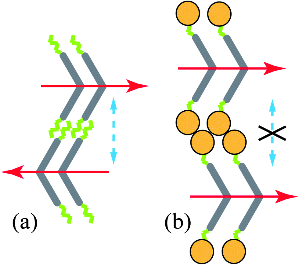 | ||
| Fig. 1 Effect of steric interactions between adjacent layers. The red arrows indicate the polarization of each layer. (a) A synclinic organization of the molecular tails in adjacent layers promotes AF ordering. (b) Bulky groups (orange circles) at the molecular ends decouple adjacent layers suppressing interlayer fluctuations (dashed blue arrows). Thus it will not promote the AF arrangement. | ||
Several approaches have been designed to suppress the entropic penalty for FE phases, for example the introduction of branches in the alkyl chains8 or the incorporation of bulky groups to the chain ends (e.g. oligosiloxanes)9–12 (see Fig. 1b). It is supposed that branched chains or, especially, bulky terminal groups decouple the adjacent layers and suppress the fluctuations, thus reducing the entropic penalty of the FE ordering. However, it is not clear to what extent the rest of the contributions to the free energy actually favor a FE structure (indeed, for example, the electrostatic interaction between adjacent polarized layers remains very important and would still promote AF organizations). In this respect, it has been suggested13 that some of the presumed FE phases have virgin ground states that are really AF, but FE states can be easily achieved by surface stabilization once an electric field has been applied. Since most of the techniques for investigating the type of dipole ordering (parallel or anti-parallel) require the use of electric fields, the material would always behave as FE in the experiments.
With the purpose of clarifying the above problem, we present in this work a study of a thiophene based bent-core mesogen with silyl groups at both ends that can be considered as representative of such family of compounds. The chemical structure is shown in Fig. 2. This material, called T-Bip-Si, contains thiophene units and was recently investigated by polarizing optical microscopy, X-ray diffraction and switching current response.14 The authors found FE behavior between 64 °C and 87 °C, which was attributed to the presence of the large trisiloxane groups at the chain ends. The proposed phase sequence is indicated in Fig. 2.
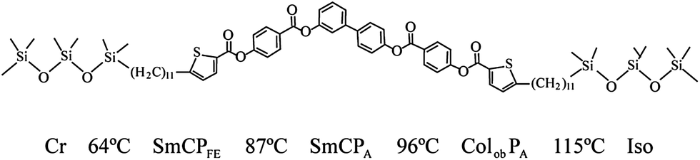 | ||
| Fig. 2 Chemical structure of the studied compound and phase sequence (on heating) according to ref. 14. Abbreviations: Cr = solid crystal, SmCPFE = ferroelectric tilted polar smectic phase, SmCPA = antiferroelectric tilted polar smectic phase, ColobPA = columnar polar oblique antiferroelectric phase, and Iso = isotropic liquid. | ||
In the present work we use optical second harmonic generation (SHG) and SHG interferometry to probe the FE character of the material. These techniques are complementary to those of ref. 14 and allow testing of the existence of macroscopic polarization in the virgin material. Furthermore, the stabilization of a FE state after field application can be easily detected. In addition, we present X-ray structural studies using a goniometer with high angular resolution. Taking advantage of the presence of sulfur atoms in the molecule, we also carried out resonant X-ray scattering. This technique can provide structural information not accessible in ordinary X-ray experiments.15
2. Texture analysis and electro-optical behaviour
According to Geese et al.,14 the transitions between the three mesophases of T-Bip-Si are difficult to detect by means of DSC, X-ray diffraction, or even observation of textures. The optical textures of the virgin samples only show minor changes in the whole temperature range, and just a slight change of color can be noticed. Interestingly, the textures are more typical of columnar or modulated phases than of smectic phases, presenting some features, such as banana-leaf domains, which are characteristic of the former. The phase transitions, however, become evident when an electric field is applied. The transition signatures are based on the electro-optical behavior and measurements of the polarization switching current. The spontaneous polarization values measured at 105 °C, 92 °C, and 71 °C show clear changes: 180, 430 and 620 nC cm−2 respectively.14 In addition, in the temperature range between 115 and 87 °C two polarization current peaks were observed, with different dynamics above and below 96 °C, and only one peak was detected below 87 °C. All these facts suggest two AF phases above 87 °C and a FE phase below that temperature. However, as stated above, it is not clear whether the FE phase is spontaneous or is surface stabilized after field application.13 Then, in order to obtain more information about the structural and polar order within the mesophases, SHG experiments were performed and optical textures were analyzed without and with applied field.To carry out the experiments Linkam commercial cells of 5 μm thickness were used. The electric field was applied perpendicularly to the substrate. The electro-optical behavior under field was studied by means of polarizing optical microscopy. SHG measurements were performed with an experimental set-up described in detail elsewhere.16 The fundamental light was generated by a Q-switched Nd-YAG laser (wavelength 1064 nm) with a pulse width of 6 ns and a frequency of 5 Hz.
When cooling the sample from the isotropic phase under a DC field of 10 V μm−1 synclinic purple domains, characteristic of a columnar phase, are observed. These are also the textures in the whole temperature range if the sample is grown in the absence of field (left-hand side pictures in Fig. 3). In contrast, the field promotes the appearance of low birefringence anticlinic domains (grey) admixed with synclinic domains (purple) in the lower temperature mesophases. As the temperature decreases the former grow at the expense of the latter (right-hand side of Fig. 3). The fact that domains with different clinicity coexist implies that both arrangements have similar free energy probably as a consequence of the layers decoupling due to the siloxane units.9–11,13,17,18 The temperature range of the coexistence is 99–85 °C (these two temperatures should correspond to 96 °C and 87 °C respectively in Fig. 2 and ref. 14). Finally, below 85 °C and under electric field, the sample is full of anticlinic focal conic domains, which are typical of smectic phases. A summary of the above conclusions is depicted in Table 1.
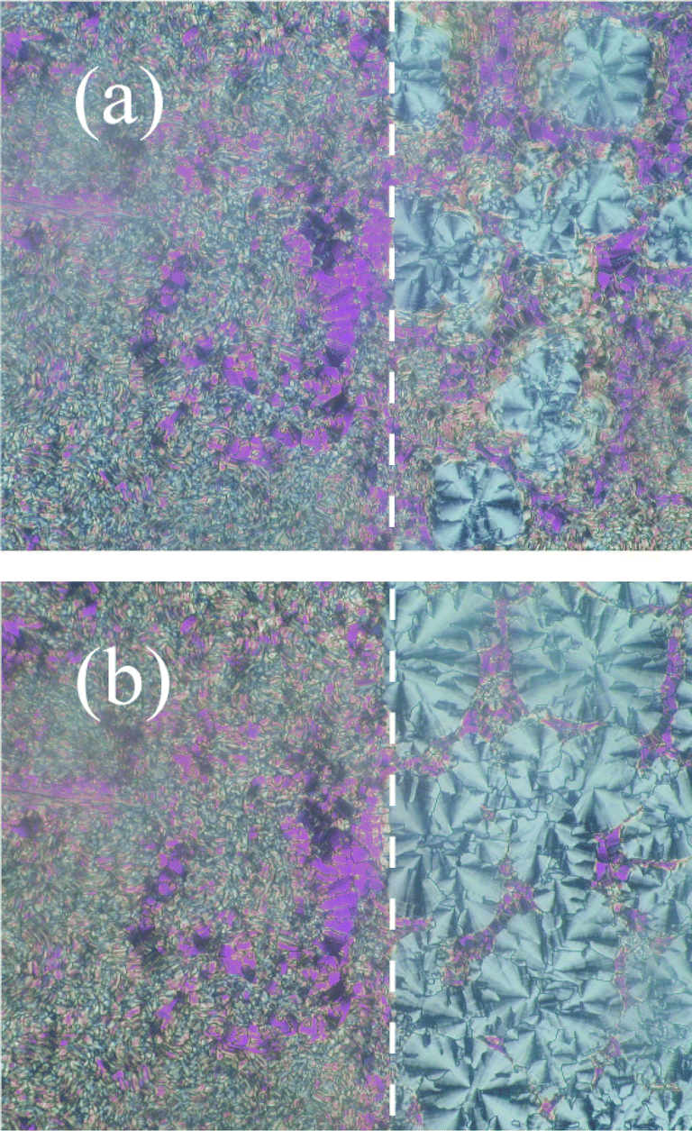 | ||
| Fig. 3 Textures of the studied compound under a DC electric field of 10 V μm−1 at 92 °C [(a), right-hand side] and at 86 °C [(b), right-hand side]. The dashed line indicates the limit of the ITO electrode. The left-hand part corresponds to the area without electrode. The textures at high temperature (118–99 °C) under DC fields cannot be distinguished from those of the left-hand sides of both photographs. The orientations of the polarizer and analyzer are horizontal and vertical respectively. | ||
| ColobPA | SmCPA | SmCPFE | |
|---|---|---|---|
| Textures grown on cooling in the absence of field | Synclinic purple domains | Synclinic purple domains | Synclinic purple domains |
| Textures under field and after field removal | Synclinic purple domains | Coexistence of synclinic purple and anticlinic grey domains | Anticlinic grey domains |
| SHG on virgin samples (no field was applied) | No | No | No |
| SHG without field but after field treatment | No | Yes | Yes |
| Polarization current peaks (according to ref. 14) | Two | Two | One |
At all temperatures the textures are exactly the same when reversing the field and also after field removal. This behavior indicates that the switching takes place around the molecular director in the whole temperature range, in agreement with ref. 14.
All these observations indicate a synclinic ordering for the three mesophases if the sample is grown from the isotropic liquid without any applied electric field (purple domains always observed). In contrast, though the high temperature phase remains synclinic under an applied field, below 99 °C field application tends to establish anticlinic smectic states (appearance of grey domains). The tendency is stronger the lower the temperature.
3. Testing the FE character using SHG
SHG measurements as a function of temperature were carried out initially on a virgin sample. Regardless of the light polarization or angle of incidence, no SHG signal could be detected in the whole temperature range. This means that the material cannot be FE under these conditions. In contrast, we found a strong SHG intensity under field application and also after field termination (see Table 1). Fig. 4 shows the SHG intensity as a function of temperature without applied field after a treatment with an electric field of 5 V μm−1 at 65 °C. As can be seen the intensity reaches a maximum at 85 °C and decreases progressively above this temperature, being practically null in the columnar phase.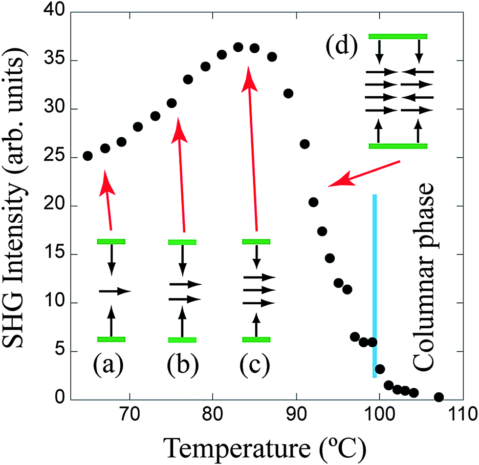 | ||
| Fig. 4 SHG intensity versus temperature of the studied compound after application of an electric field of 5 V μm−1 at 65 °C and its subsequent removal. The angle of incidence of the fundamental light was 25°. (a), (b), (c), and (d) are schematic representations of how the molecular dipole moments are oriented within the cell at several temperatures. Scheme (d) indicates the coexistence of FE and AF domains. The green segments are the cell substrates. | ||
At normal incidence the SHG intensity under electric field was null, as the sample polarization is along the light direction. In contrast, after field removal, a noticeable SHG intensity was detected at normal incidence. In general this intensity was even higher than the one detected under field at moderate incidences. Such an observation implies the existence of a non-null component of the sample polarization parallel to the substrate plane.
The temperature dependence of the SHG intensity shown in Fig. 4 can be understood assuming that the anticlinic domains induced by the electric field are FE and, in contrast, the synclinic domains, coexisting between 99 °C and 85 °C, are AF. As mentioned above, the proportion of anticlinic domains increases as the temperature decreases and, therefore, the SHG signal should increase. In addition, the coexistence of FE and AF domains above 85 °C implies that two repolarization current peaks should be detected, as reported in ref. 14. Finally, below 85 °C the sample becomes uniformly FE and therefore only one repolarization current peak remains. The above conclusions are also summarized in Table 1.
Upon cooling, the decrease of the SHG signal below the maximum as shown in Fig. 4 (85 °C) is unusual. Usually, an increase of intensity would be expected when cooling the sample as a consequence of the higher order in the phase. However, the observed behavior can be explained by a simple qualitative model. As stated above, the FE ground state must contain a polarization component in the substrate plane. This occurs because near the surfaces the polarization is perpendicular to the cell substrates and points inward the cell.19 Therefore the dipoles must rotate as we move along the cell thickness and, thus, a region where the polarization has a component parallel to the substrate is required in the middle of the cell. Without applied electric field, this region must be the main contributor to the SHG signal, especially for small angles of incidence of the fundamental beam. The size of this region increases if we assume that the surface effects are less important with increasing temperature (see schemes (a)–(c) of Fig. 4). Therefore, the SHG signal must increase upon heating, as in fact is observed. Similar profiles for the polarization inside the sample in other FE liquid crystals have been proposed in the literature.20 Usually the polarization has a splayed structure along the sample thickness and the molecular director rotates around the tilt cone. In our case, since the switching takes place around the main director of the molecule, the molecular change associated with the polarization variations must be a rotation about the same axis (there is no texture change upon field application or field removal).
4. SHG interferometry
In order to further understand the FE phase in this material, we also carried out SHG interferometry experiments. In this case, an SHG active material (a suitably oriented quartz crystal) was inserted in the light path to generate a coherent SHG wave that interferes with the one produced by the liquid crystal under study (Fig. 5). The phase shift between the two waves was controlled by placing a fused silica plate on a rotating stage between the two samples. The relative phase shift δ is produced as a consequence of the optical path difference between the fundamental and SHG waves in the silica plate. On rotating the plate the optical path difference varies, and interference fringes are obtained for the resulting SHG signal. The SHG intensity is given by| I2ω ∝ |eiδE12ω + E22ω|2. |
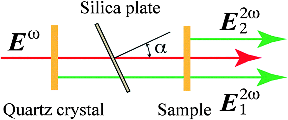 | ||
| Fig. 5 Scheme of the SHG interferometry experiment. The fundamental light and the SHG ones generated by the quartz crystal and the sample are indicated. The electric fields are Eω, E12ω and E22ω respectively. | ||
Fig. 6 shows the SHG intensity versus the incidence angle on the silica plate at 70 °C. Solid and open circles represent the measured intensities under electric fields of ±5 V μm−1 respectively. The result can be interpreted as follows: if solid circles represent the intensity corresponding to a SHG light electric field E22ω, then open circles are obtained for a field −E22ω, i.e., the inversion of the sample polarization leads to the inversion of the SHG field. Then, since both interference patterns are in anti-phase, the result in Fig. 6 implies an inversion of the polarization between both states, as expected.
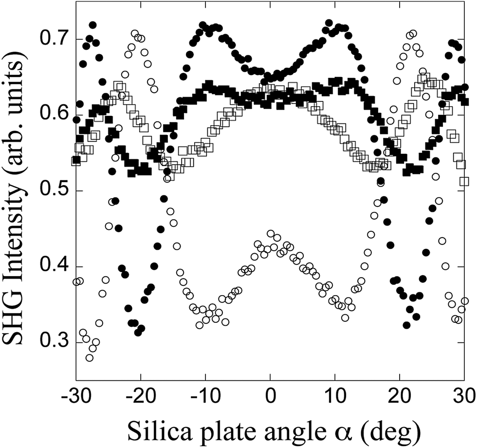 | ||
| Fig. 6 SHG interference fringes at 70 °C. The angle of incidence α on the silica plate is indicated in Fig. 5. Solid and open circles correspond to + and −5 V μm−1 respectively. Solid and open squares represent respectively the interference pattern after application of + and −5 V μm−1 followed by the field removal. The angle of incidence of the fundamental light on the sample was 25°. | ||
The fringe patterns after field removal are represented by solid and open squares (after having applied ±5 V μm−1 electric field respectively). A decrease in the visibility of the fringes is observed in both cases. This fact is due to the enhancement of the SHG signal in the sample without field, which becomes larger than the one generated by the quartz crystal. In this case the fringes are not in anti-phase, which means that the relationship between both states is not a simple polarization inversion, in qualitative agreement with schemes (a)–(c) in Fig. 4. In addition, open squares present a phase shift with respect to the corresponding situation under field (open circles). This implies that the FE state under field is not perfectly retained. This behavior is expected under the model proposed in this work for the FE ground state, in which there is an important component of the bulk polarization parallel to the substrate plane. In contrast, solid circles and solid squares present practically the same phase, which corresponds more to the ideal FE behavior. Thus, we deduce that the surface interactions do not maintain symmetrically the orientation of the dipoles after positive or negative field application, but favor the state induced by a given direction of the electric field.
5. Structural study
According to the X-ray diffraction results of Geese et al.14 the material is lamellar for all temperatures. However, the authors themselves present conclusions derived from texture analysis and electro-optical behavior that are in contradiction with the lamellar character, indicating the existence of undulated and columnar mesophases in certain temperature ranges. Then, it is reasonable to think that the resolution that could be achieved in the small angle region in the measurements of ref. 14 was not enough to detect the features characteristic of 2D structures. To investigate this point, we performed a complete structural study of the material by means of high-resolution powder diffraction measurements, using synchrotron radiation, and resonant X-ray diffraction experiments. The first type of study checked the existence of further lattice periodicities. On the other hand, resonant scattering experiments are able to detect polarization or tilt periodicities and their relationship to the translation lattice.15,215.1 Conventional powder diffraction
High-resolution powder X-ray diffraction measurements were carried out at a fixed energy of 13.5 keV on a six-circle diffractometer at SpLine beamline (BM25B), ESRF, Grenoble, France.22 A point-scintillation detector was used. The distance from the sample to the detector was about 70 cm and the experimental resolution in 2θ (θ is the Bragg angle) was better than 0.01°. Measurements were performed in Debye–Scherrer operation mode using a Lindemann capillary of diameter 0.6 mm placed in a hot stage with 0.1 °C temperature resolution.
Fig. 7 shows the X-ray results at 75 °C and 95 °C (Fig. 7a) and at 105 °C (Fig. 7b) during a heating run. In all cases there are several diffraction peaks, which indicates that the material has indeed a 2D structure at all temperatures. However, it can be seen that the peaks extend in a very narrow angular range (2θ < 0.12°). The results of Geese et al.,14 which were not able to distinguish more than one periodicity in their surface-aligned samples, can now be explained under the assumption that the set of reflections occupy a very small region of the reciprocal space around the meridian line for being resolved in their experiment. Basically, this implies a smectic structure with a long wave modulation. Data in Fig. 7(a) and (b) are compatible with a phase transition at 99 °C. No more transitions could be recognized in our data, and below 99 °C the peaks evolve continuously (Fig. 7a). Three peaks were observed in this temperature range. They are better resolved in the curve with full circles in Fig. 7a, and were interpreted on the basis of an oblique lattice as indicated in the figure. At 95 °C the X-ray data yielded three lattice parameters a = 186 Å, c = 48.6 Å and β = 84.8°. The lattice parameter a decreases on heating as the material approaches the columnar phase (see the inset in Fig. 7a). In our experiment we also observed a very slight decrease in c together with a small variation of β on heating. The spacing that results from the reciprocal c* parameter is c![[thin space (1/6-em)]](https://www.rsc.org/images/entities/char_2009.gif) sinβ = 48.4 Å, in good agreement with the (apparently smectic) spacing reported for this material in ref. 14. In summary, according to our results T-Bip-Si presents frustrated lamellar structures in the whole temperature range. Above 99 °C the frustration is stronger and results in a phase that may be classified as columnar according to ref. 14 (Fig. 7b). Below 99 °C (Fig. 7a) the frustration is weaker and yields an undulated structure with a similar diffraction pattern to that of a typical B7 phase.23 No further transitions were detected between 95 °C and 65 °C.
sinβ = 48.4 Å, in good agreement with the (apparently smectic) spacing reported for this material in ref. 14. In summary, according to our results T-Bip-Si presents frustrated lamellar structures in the whole temperature range. Above 99 °C the frustration is stronger and results in a phase that may be classified as columnar according to ref. 14 (Fig. 7b). Below 99 °C (Fig. 7a) the frustration is weaker and yields an undulated structure with a similar diffraction pattern to that of a typical B7 phase.23 No further transitions were detected between 95 °C and 65 °C.
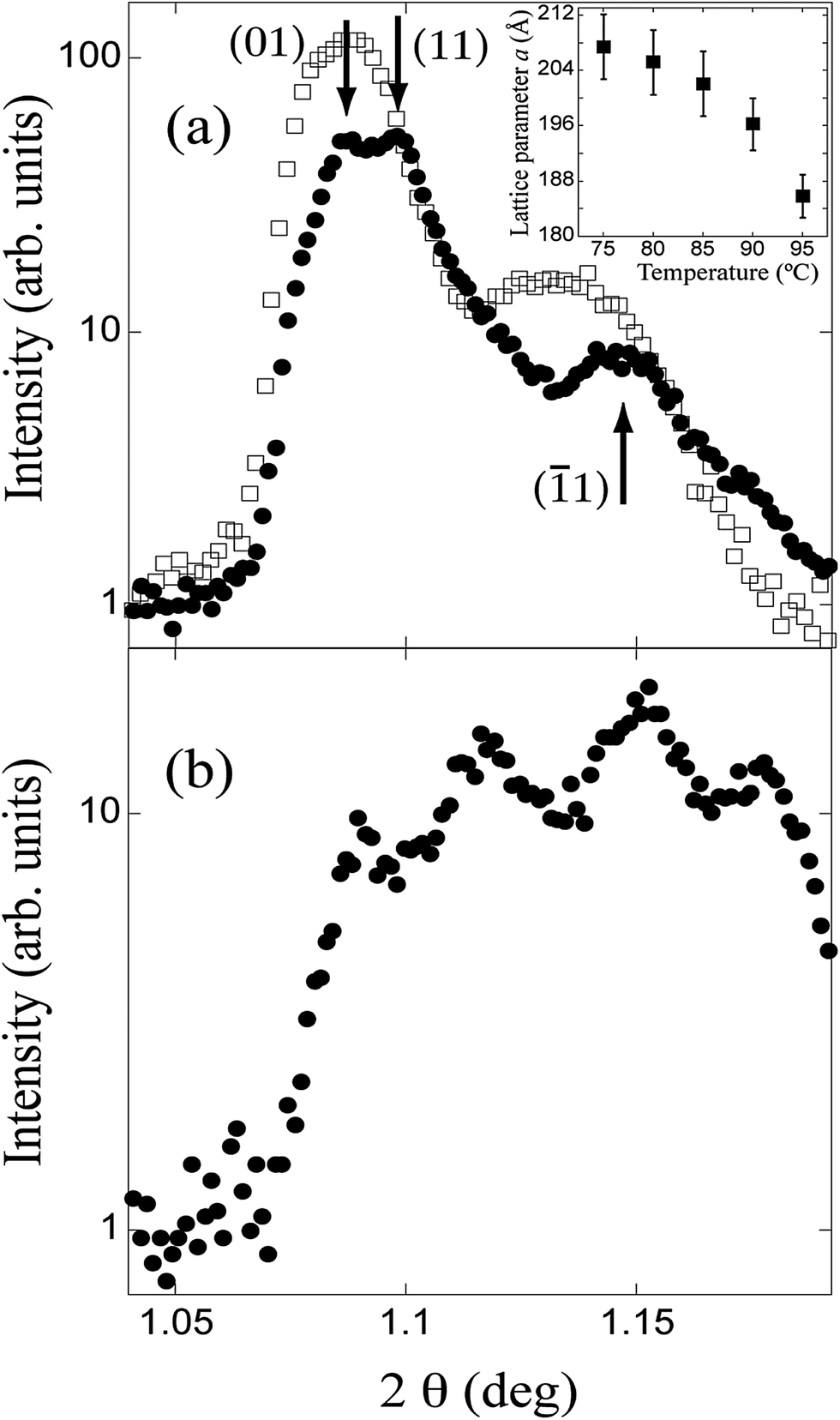 | ||
| Fig. 7 (a) X-ray diffraction patterns of T-Bip-Si at 75 °C (open squares) and 95 °C (full circles). The inset represents the temperature dependence of the lattice parameter a. (b) X-ray results for 105 °C. Intensities are plotted against 2θ, where θ is the Bragg angle. All diagrams are compatible with a 2D translation lattice. An indexation on the basis of an oblique lattice is given in (a) for the data at 95 °C. | ||
5.2 Resonant diffraction in oriented samples
As is well known, in conventional X-ray scattering in 2D columnar phases (as in our case) the diffraction pattern is the same regardless of the polarization of the different columns. The reason is that the contribution to the structure factor of any atom does not depend on its position along the column. The situation is different with resonant diffraction. In this technique the X-ray energy is tuned to the K absorption edge of a given probe atom in the molecule. As a consequence, an inner-shell electron is excited to an outer-shell empty quantum state whose symmetry depends on the arrangement of the atomic neighborhood. Thus, the atomic response can be described by a second order susceptibility tensor χ, relating the incident and diffracted fields. Then the structure factor becomes anisotropic and the systematic extinction rule related to a non-symmorphic symmetry element (in our case a glide plane relating columns with opposite polarization) does not hold for the set of resonant scatterers.24 In other words, resonant diffraction breaks the systematic extinction rule of non-symmorphic symmetry operations, revealing the extra periodicity.Resonant X-ray diffraction experiments were carried out at beamline X-19A of the National Synchrotron Light Source, Brookhaven National Laboratory. A complete description of the Huber two-circle goniometer was published previously.25 The sample was prepared by spreading the material on a glass substrate treated with hexadecyl trimethyl ammonium bromide. By rubbing in a given direction, a specimen whose smectic layers (or pseudolayers) are parallel to the substrate is obtained.15,21,26 Since the rubbing process promotes a preferred direction, a triad of axes X, Y, and Z, with Z along the glass plate normal and X along the rubbing direction can be defined (see Fig. 8a). It is well known that in lamellar phases the rubbing direction fixes the tilt plane.15,21,26 Consequently, it can be expected that in 2D structures it is the 2D translation lattice plane that is fixed by rubbing, i.e., both in the lamellar and columnar cases polar and rubbing directions are perpendicular to each other. The sample was mounted on a rotation platform with the rotation axis along the Z axis inside a two-stage oven having a 20 mK temperature resolution. It was oriented in such a way that the X, Z plane was the incidence plane (Fig. 8a). Due to geometrical constraints (e.g., those imposed by the windows of the sample chamber and vacuum assembly of the arms), the range of accessible reciprocal space extends only to a thin region surrounding the QZ direction. The beam dimensions were set to 0.2 mm vertical by 0.5 mm horizontal using the adjustable slit assembly at the input arm. An experimental resolution ΔQZ ≈ 1.5 × 10⁻³ Å−1 was estimated from the goniometer geometry and slits' apertures.
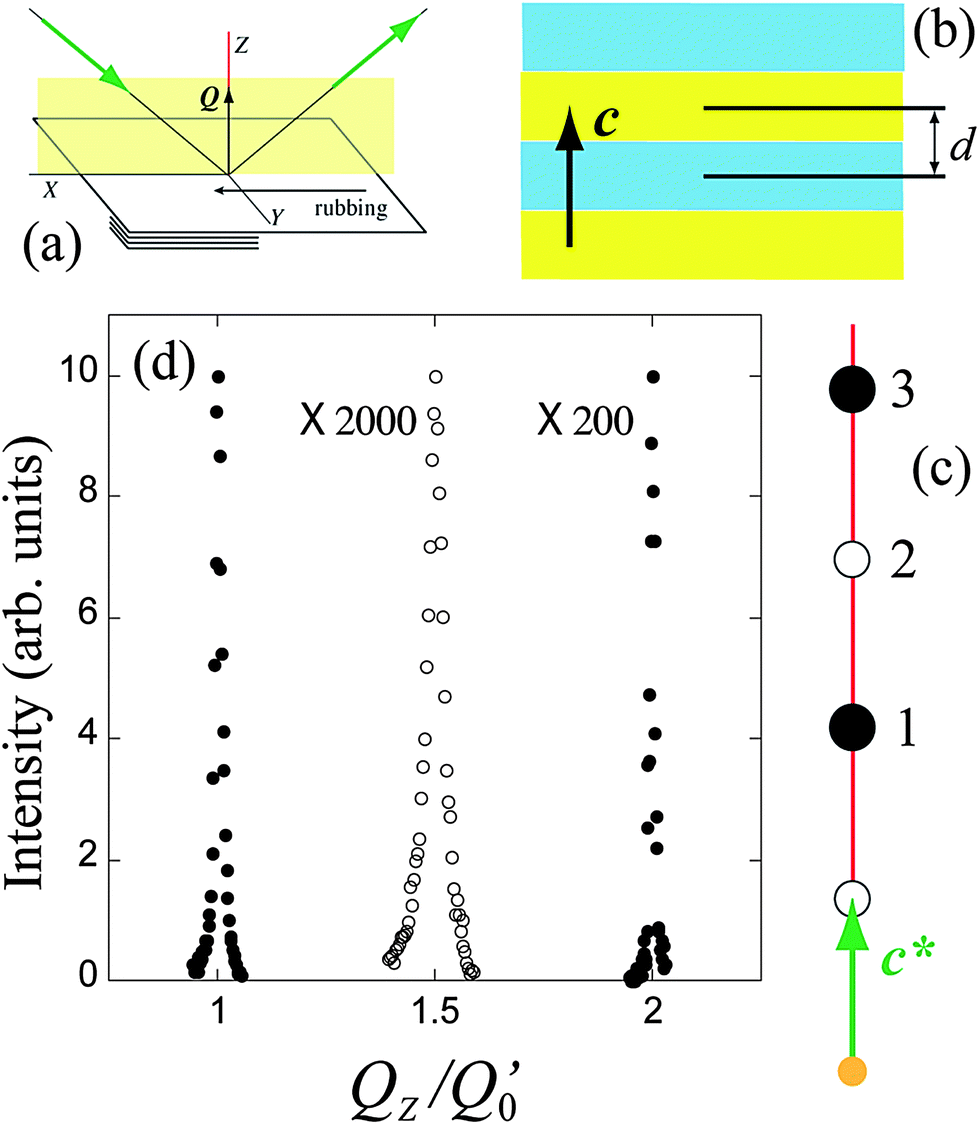 | ||
| Fig. 8 (a) Geometry of the diffraction experiment: X is along the rubbing direction, Y is the polar direction, and Z is along the glass plate normal. Polarization periodicity of the antiferroelectric smectic structure (b) together with a scheme of the resonant diffraction diagram (c). Different colors represent opposite polarization directions. (d) X-ray diffraction pattern showing the normal Bragg reflections designated as 1 and 3 in (c) (full circles), and the resonant reflection 2 (open circles). The intensities of 2 and 3 were expanded as indicated, and the background was subtracted. Q′0 = 2π/d, where d is the smectic spacing as indicated in (b). | ||
Once prepared, the sample was introduced in the oven at a temperature of about 75 °C. Then, after tuning the beam energy to the sulfur K edge (2.473 keV) the alignment of the goniometer was optimized by scanning the reciprocal space along the QZ direction, i.e., a standard (θ, 2θ) diffraction experiment where periodicities along Z are detected. Two strong nonresonant Bragg reflections (one being the second harmonic of the other) were detected, together with a clear resonant peak at the middle position between them, which was used to optimize further the beam energy to E0 = 2.476 keV (Fig. 8d). We checked that the resonant peak vanishes for energy shifts |E − E0| > 10 eV. This is compatible with a bilayer smectic phase (Fig. 8b). As is well known, beyond the detection of resonant reflections, relevant structural information is obtained from their polarization analysis.15,21,25,26 Unfortunately, such studies were not possible in T-Bip-Si because the sample was damaged by the direct beam during the time required for a complete polarization analysis. Interestingly, the observed resonant peak became weaker and disappeared when the material was heated above a given temperature (about 90 °C) while both nonresonant reflections remained with the same intensity (only a slight variation in their positions was observed). No resonant peak was detected on further heating to the highest temperature mesophase. Then, on cooling back the sample, instead of the original resonant reflection, a double peak, whose intensity was about 15% of the original one, appeared centered at the middle position of the normal Bragg reflections (see Fig. 9). On further heating and cooling runs this double peak was observed at approximately the same temperature range as the original single peak. Since there was no further evidence of the original resonant reflection we concluded that the sample preparation procedure promoted a bilayer smectic structure that relaxed to a more stable phase (evidently different from the bilayer smectic) on heating the material. The above phenomenology was observed in the same form in different samples.
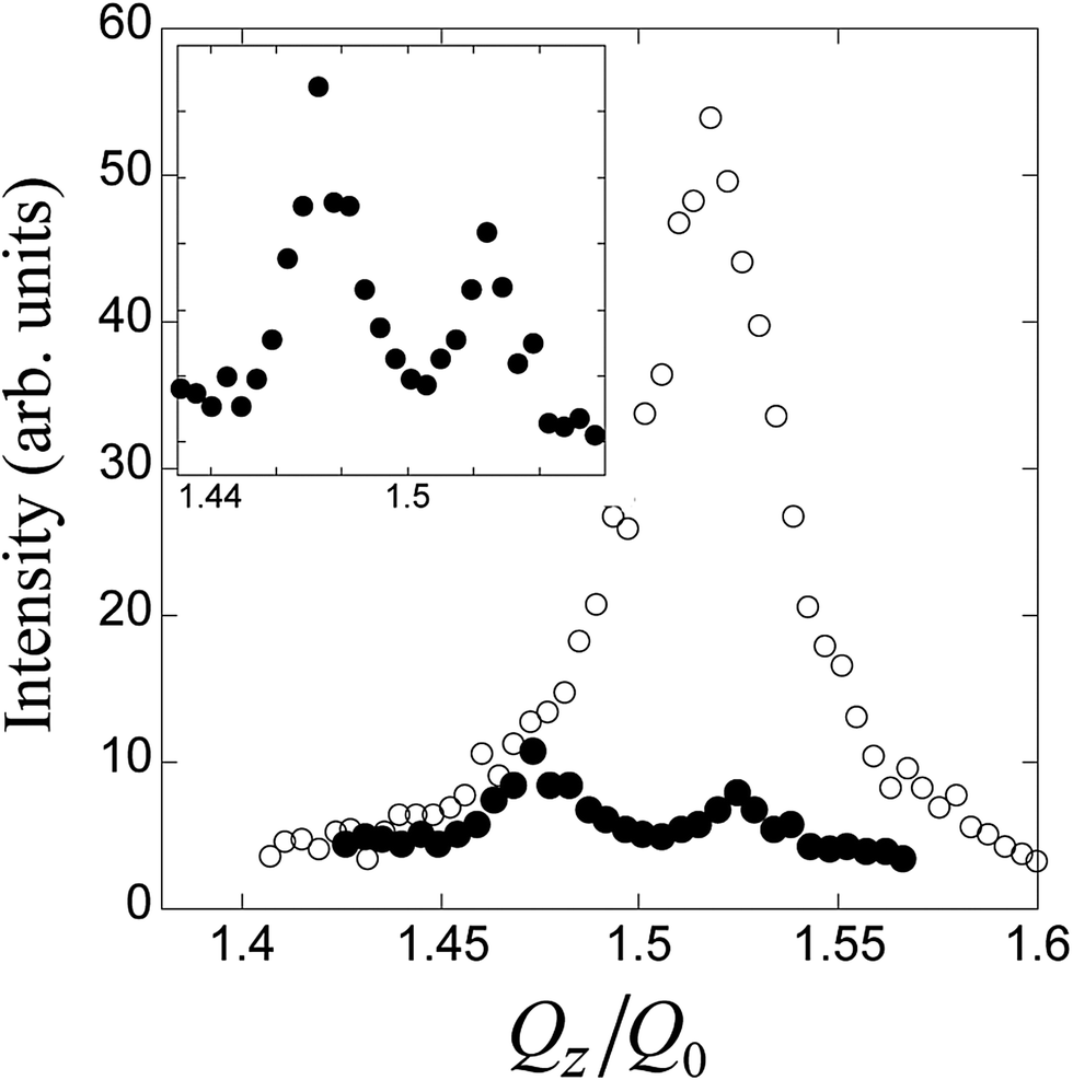 | ||
| Fig. 9 Open circles: resonant reflection observed in a first heating run after sample preparation (T = 78 °C). Full circles: a typical double resonant peak observed in the same temperature range after heating the sample above 90 °C and cooling down again. The inset shows this reflection in more detail. QZ is the reciprocal vector along the scanning direction, and Q0 is the position of the corresponding first order normal Bragg reflection after the temperature treatment. Note that Q0 and Q′0 (see Fig. 8) are slightly different since they correspond to different structures (undulated and smectic respectively). Both curves were normalized with the criterion of equalling their backgrounds. | ||
A simple explanation for the existence of a weak double resonant reflection is to attribute it to the tails of other resonant reflections that are not contained along the QZ line. The existence of the double peak is consistent with a 2D translation lattice that is necessarily oblique, in accordance with the indexation of the powder diagram in Fig. 7. Fig. 10 shows a structural model that agrees with this interpretation. According to this model the double peak is due to reflections 1 and 2. Different colors in Fig. 10 represent opposite polarization directions along bR, and the red line in the diffraction diagram is the scanning direction, i.e., the QZ direction. The polarization periodicity is the same as that reported in a previous study for another bent-core liquid crystal.21,27 Nevertheless, in the present case, blue and yellow pseudolayers are not parallel to the sample surface and, therefore, the surface is not homogeneously polarized but contains a periodic sequence of opposite polarizations. Accordingly, no resonant reflections are contained on the red line in agreement with the color sequence along this direction.28
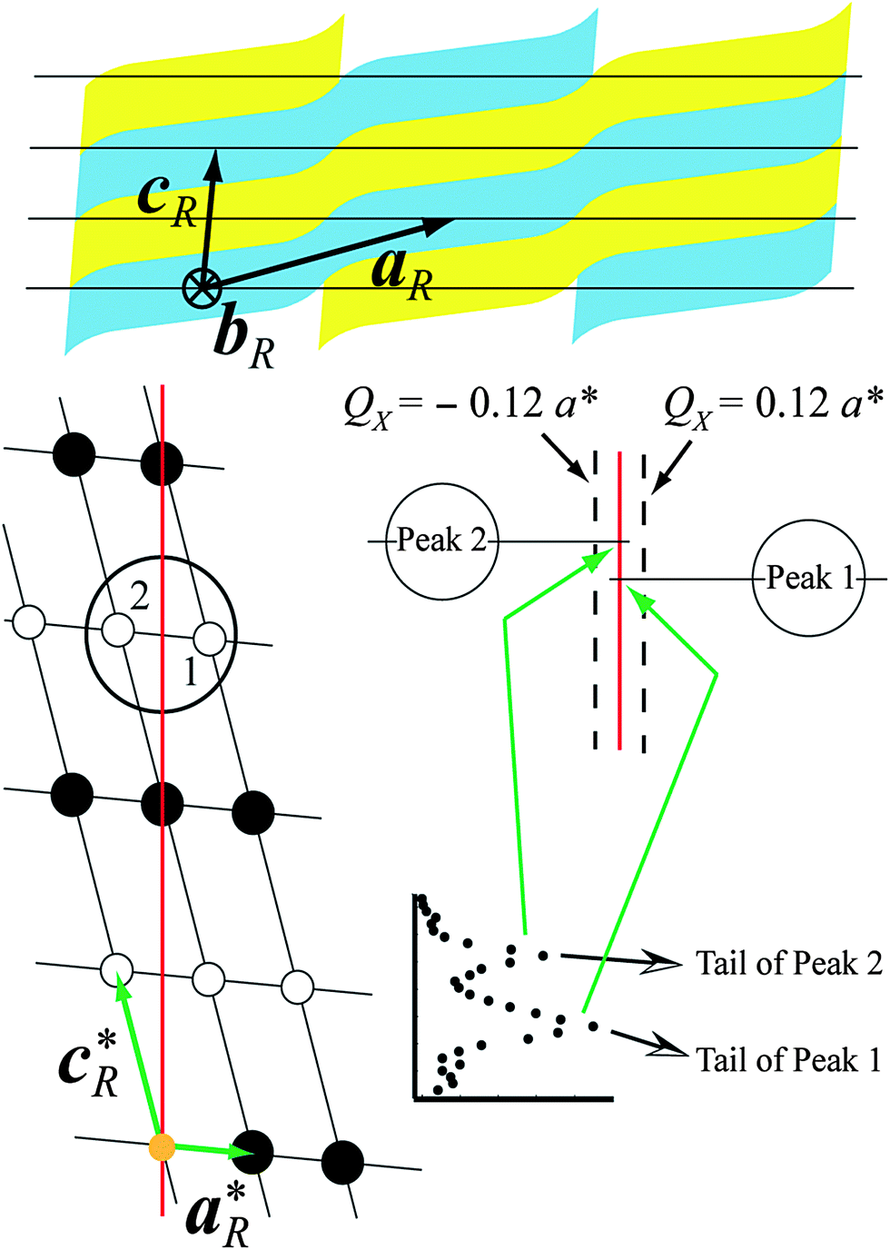 | ||
| Fig. 10 The structural model proposed together with the corresponding diffraction diagram. Open and full circles represent resonant and nonresonant reflections respectively. Direct and reciprocal lattice parameters are indicated. The reciprocal space was scanned along the red and dashed lines (see text). The region inside the circle is schematically detailed at the right side to clarify the origin of the double resonant peak. | ||
In order to test the above hypothesis, the reciprocal space was scanned along directions parallel to QZ but contained on the X, Z plane (dashed lines in Fig. 10). The results are presented in Fig. 11. As can be seen, single peaks (more intense than the double one) are clearly identified with different positions along the scanning direction in agreement with the proposed model. Due to geometrical restrictions with our experimental setup, the maximum of every single peak could not be reached. However, the structural model represented in Fig. 10 accounts properly for the results obtained in powder and resonant scattering experiments.
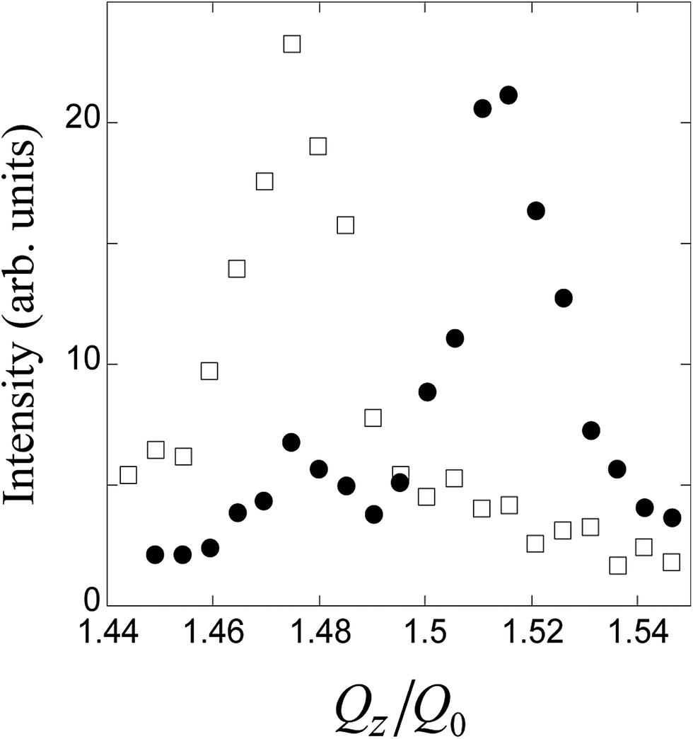 | ||
| Fig. 11 Resonant reflections observed when the reciprocal space was scanned along the lines defined by (QX = 0.12a*, QZ) (open squares) and (QX = −0.12a*, QZ) (full circles). These are the directions represented as dashed lines in Fig. 10. Q0 is the position of the corresponding first order normal Bragg reflection. The intensities were normalized with the criterion of equalling the backgrounds. | ||
Another question is why resonant reflections are not observed above a given temperature (∼90 °C) in the low temperature phase. In fact, peaks were not even detected along directions as shown by the dashed lines in Fig. 10. A simple hypothesis can be proposed regarding this point: it was observed that the birefringence of the virgin material decreases on heating, implying a more disordered structure with a higher angular spread of molecular positions about their long axes. Then, the contrast between yellow and blue blocks in Fig. 10 would be smaller, which leads to a decrease of the resonant peaks. In particular, a null contrast could imply a transition to a superparaelectric structure, where neighboring layers are completely decoupled. Beside this, a further contribution for the disappearance of the peaks would be the decrease of the lattice parameter a on heating (inset in Fig. 7a), which expands the distance between the resonant reflections and the QZ line.
6. Conclusions
In summary, we have studied a thiophene based bent-core mesogen with silyl groups at the end chains by means of SHG and X-ray diffraction. The experimental results have allowed us to clarify certain issues apparently paradoxical regarding the structure and polarity expected for the material. Some of the results found in previous reports are also revised and refined. The main conclusions are as follows:The virgin material is AF throughout the temperature range. This means that though the bulky terminal chains suppress fluctuations between adjacent layers, the removal of this entropic penalty is not enough to warrant true ferroelectricity. On the other hand, neighboring layers are not as decoupled as a superparaelectric material would require. In the superparaelectric case no SHG is expected and resonant reflections should also be absent. The observed resonant reflections clearly indicate a bilayer structure, in agreement with the AF character.
Once an electric field is applied, the sample surfaces can stabilize a FE structure upon field removal. We have found that this surface-stabilized ferroelectricity is maintained, at least to a certain extent, up to 99 °C, where a transition to a columnar AF phase takes place. The degree of stabilization of the FE character varies with temperature. For low temperatures (<85 °C) it is almost perfect, as SHG results indicate. On raising temperature, there is a coexistence of FE and AF domains. The sequence of mesophases that T-Bip-Si presents under different external conditions is summarized in Table 2. The difference with the phase sequence depicted in Fig. 2 (ref. 14) is evident and not only makes explicit the fact that the material behaves differently after field treatment.
| Mesophase sequence | |
|---|---|
| Virgin material (no field treatment) | USmCSPA 99 °C; ColobPA 118 °C; Iso |
| Material after field treatment | SmCAPFE 85 °C; SmCAPFE + USmCSPA 99 °C; ColobPA 118 °C; Iso |
The undisturbed state of the material is never strictly smectic, but undulated, with a long modulation period, and columnar (below or above 99 °C respectively). This 2D characteristic agrees with the idea that molecules with oligosiloxanes at both terminal ends frustrate the smectic phases, since there is a mismatch between the transverse cross-sections of the bulky silyl groups and those of the molecular cores.11,13 The same steric effect, which tends to separate the aromatic cores, also favors the reorientation of the molecules around their molecular long axes in a switching process, as experimentally observed in T-Bip-Si.
Author contributions
K.G. and C.T. synthesized the material and performed a preliminary phase characterization; S.R.C., G.S.E., J.O., J.E., and C.L.F. carried out texture and electro-optical analysis, and the SHG studies. They also carried out the powder X-ray diffraction measurements at the ESRF; V.P., P.B., LiDong P., Z.Q.L., B.K.M., J.O., C.L.F., and R.P. performed resonant X-ray diffraction measurements at the NSLS. The project at the NSLS was coordinated by P.B. The paper was written by C.L.F., J.O., and J.E. with some contributions from C.T., C.C.H., R.P., and P.B.Acknowledgements
This research was supported by MICINN-FEDER of Spain-UE (Project MAT2012-38538-C03-02) and the Basque Country Government (Project GI/IT-449-10). The authors acknowledge the CSIC for the provision of synchrotron radiation facility at ESRF. The use of the National Synchrotron Light Source, Brookhaven National Laboratory, was supported by the U.S. Department of Energy, Office of Science, Office of Basic Energy Sciences, under Contract no. DE-AC02-98CH10886. S.R.C. and G.S.E. thank the MEC of Spain and the Basque Country University for grants. VP acknowledges financial support from Cnano-GSO for synchrotron studies.References
- T. Niori, F. Sekine, J. Watanabe, T. Furukawa and H. Takezoe, J. Mater. Chem., 1996, 6, 1231 RSC.
- D. R. Link, G. Natale, R. Shao, J. E. Maclennan, N. A. Clark, E. Korblova and D. M. Walba, Science, 1997, 278, 1924 CrossRef CAS PubMed.
- A. Jakli, L.-C. Chien, D. Krüerke, H. Sawade and G. Heppke, Liq. Cryst., 2002, 29, 377 CrossRef CAS; Y. Shimbo, Y. Takanishi, K. Ishikawa, E. Gorecka, D. Pociecha, J. Mieczkowski, K. Gomola and H. Takezoe, Jpn. J. Appl. Phys., 2006, 45, L282 CrossRef; I. Alonso, J. Martinez-Perdiguero, J. Ortega, C. L. Folcia and J. Etxebarria, Liq. Cryst., 2007, 34, 655 CrossRef.
- I. C. Pintre, N. Gimeno, J. L. Serrano, M. B. Ros, I. Alonso, C. L. Folcia, J. Ortega and J. Etxebarria, J. Mater. Chem., 2007, 17, 2219 RSC; I. C. Pintre, J. L. Serrano, M. B. Ros, J. Martínez-Perdiguero, I. Alonso, J. Ortega, C. L. Folcia, J. Etxebarria, R. Alicante and B. Villacampa, J. Mater. Chem., 2010, 20, 2965 RSC; Y. Zhang and J. Etxebarria, in Liquid Crystals Beyond Displays, ed. Q. Li, Wiley, Hoboken, NJ, 2012, ch. 4, pp. 111–156 Search PubMed.
- J. Harden, B. Mbanga, N. Eber, K. Fodor-Csorba, S. Sprung, J. T. Gleeson and A. Jakli, Phys. Rev. Lett., 2006, 97, 157802 CrossRef CAS PubMed; J. Harden, M. Chambers, R. Verduzco, P. Luchette, J. T. Gleeson, S. Sprunt and A. Jakli, Appl. Phys. Lett., 2010, 96, 102907 CrossRef.
- G. Pelzl, S. Diele and W. Weissflog, Adv. Mater., 1999, 11, 707 CrossRef CAS; R. Amaranatha Reddy and C. Tschierske, J. Mater. Chem., 2006, 16, 907 RSC; H. Takezoe and Y. Takanishi, Jpn. J. Appl. Phys., 2006, 45, 597 CrossRef.
- J. Etxebarria and M. B. Ros, J. Mater. Chem., 2008, 18, 2919 RSC.
- D. M. Walba, E. Körblova, R. Shao, J. E. Maclennan, D. R. Link, M. A. Glaser and N. A. Clark, Science, 2000, 288, 2181 CrossRef CAS PubMed.
- G. Dantlgraber, A. Eremin, S. Diele, A. Hauser, H. Kresse, G. Pelzl and C. Tschierske, Angew. Chem., Int. Ed., 2002, 41, 2408 CrossRef CAS.
- C. Keith, R. Amaranatha Reddy, H. Hahn, H. Lang and C. Tschierske, Chem. Commun., 2004, 1898 RSC.
- C. Keith, R. Amaranatha Reddy, A. Hauser, U. Baumeister and C. Tschierske, J. Am. Chem. Soc., 2006, 128, 3051 CrossRef CAS PubMed.
- G. Dantlgraber, U. Baumeister, S. Diele, H. Kresse, B. Lühmann, H. Lang and C. Tschierske, J. Am. Chem. Soc., 2002, 124, 14852 CrossRef CAS PubMed; H. Hahn, H. Lang, C. Keith, R. Amaranatha Reddy and C. Tschierske, Adv. Mater., 2006, 18, 2629 CrossRef; W.-H. Chen, W.-T. Chuang, U.-S. Jeng, H.-S. Sheu and H.-C. Lin, J. Am. Chem. Soc., 2011, 133, 15674 CrossRef PubMed.
- C. Keith, R. Amaranatha Reddy, M. Prehm, U. Baumeister, H. Kresse, J. L. Chao, H. Hahn, H. Lang and C. Tschierske, Chem.–Eur. J., 2007, 13, 2556 CrossRef CAS PubMed.
- K. Geese, M. Prehm and C. Tschierske, J. Mater. Chem., 2010, 20, 9658 RSC.
- P. Barois, H. Gleeson, C. C. Huang and R. Pindak, Eur. Phys. J.: Spec. Top., 2012, 208, 333 CrossRef CAS.
- N. Pereda, C. L. Folcia, J. Etxebarria, J. Ortega and M. B. Ros, Liq. Cryst., 1998, 24, 451 CrossRef CAS.
- R. Amaranatha Reddy, G. Dantlgraber, U. Baumeister and C. Tschierske, Angew. Chem., Int. Ed., 2006, 45, 1928 CrossRef CAS PubMed; C. Keith, G. Dantlgraber, U. Baumeister and C. Tschierske, Chem. Mater., 2007, 19, 694 CrossRef; Y. Zang, M. J. O'Callaghan, U. Baumeister and C. Tschierske, Angew. Chem., Int. Ed., 2008, 47, 6892 CrossRef PubMed; Y. Zang, U. Baumeister, C. Tschierske, M. J. O'Callaghan and C. Walker, Chem. Mater., 2010, 22, 2869 CrossRef.
- R. Amaranatha Reddy, U. Baumeister, C. Keith and C. Tschierske, J. Mater. Chem., 2007, 17, 62 RSC; C. Keith, G. Dantlgraber, R. Amaranatha Reddy, U. Baumeister, M. Prehm, H. Hahn, H. Lang and C. Tschierske, J. Mater. Chem., 2007, 17, 3796 RSC; R. Amaranatha Reddy, U. Baumeister, H. Hahn, H. Lang and C. Tschierske, Soft Matter, 2007, 3, 558 RSC; R. Amaranatha Reddy, U. Baumeister and C. Tschierske, Chem. Commun., 2009, 4236 RSC.
- F. Araoka, H. Hoshi and H. Takezoe, Phys. Rev. E: Stat., Nonlinear, Soft Matter Phys., 2004, 69, 051704 CrossRef.
- M. Nakata, D. R. Link, F. Araoka, J. Thisayukta, Y. Takanishi, K. Ishikawa, J. Watanabe and H. Takezoe, Liq. Cryst., 2001, 28, 1301 CrossRef CAS.
- C. L. Folcia, J. Ortega, J. Etxebarria, L. D. Pan, S. Wang, C. C. Huang, V. Ponsinet, P. Barois, R. Pindak and N. Gimeno, Phys. Rev. E: Stat., Nonlinear, Soft Matter Phys., 2011, 84, 010701(R) CrossRef.
- G. R. Castro, J. Synchrotron Radiat., 1998, 5, 657 CrossRef CAS PubMed.
- D. A. Coleman, J. Fernsler, N. Chattham, M. Nakata, Y. Takanishi, E. Körblova, D. R. Link, R. F. Shao, W. G. Jang, J. E. Maclennan, O. Mondainn-Mova, C. Boyer, W. Weissflog, G. Pelzl, L. C. Chien, J. Zasadzinski, J. Watanabe, D. M. Walba, H. Takezoe and N. A. Clark, Science, 2003, 301, 1204 CrossRef CAS PubMed.
- V. E. Dmitrienko, Acta Crystallogr., Sect. A: Found. Crystallogr., 1983, 39, 29 CrossRef.
- P. Mach, R. Pindak, A.-M. Levelut, P. Barois, H. T. Nguyen, H. Baltes, M. Hird, K. Toyne, A. Seed, J. W. Goodby, C. C. Huang and L. Furenlid, Phys. Rev. E: Stat., Nonlinear, Soft Matter Phys., 1999, 60, 6793 CrossRef CAS.
- P. Fernandes, P. Barois, S. T. Wang, Z. Q. Liu, B. K. McCoy, C. C. Huang, R. Pindak, W. Caliebe and H. T. Hguyen, Phys. Rev. Lett., 2007, 99, 227801 CrossRef CAS PubMed.
- J. Martinez-Perdiguero, J. Etxebarria, C. L. Folcia, J. Ortega, N. Gimeno and M. B. Ros, Phys. Rev. E: Stat., Nonlinear, Soft Matter Phys., 2010, 82, 041706 CrossRef CAS.
- Note that reflections on the QZ line are generated by the planes parallel to the surface (horizontal lines in Fig. 10). Resonant reflections would appear if neighboring planes contained a different sequence of colors (except for an arbitrary shift), which is not the case for the proposed structure.
| This journal is © The Royal Society of Chemistry 2014 |