DOI:
10.1039/C4RA13555E
(Paper)
RSC Adv., 2014,
4, 63866-63874
Pyrene nanoparticles as a novel FRET probe for detection of rhodamine 6G: spectroscopic ruler for textile effluent†
Received
31st October 2014
, Accepted 14th November 2014
First published on 14th November 2014
Abstract
An aqueous suspension of pyrene nanoparticles (PyNPs) stabilized by sodium lauryl sulfate exhibit red shifted aggregation induced enhanced emission (AIEE) in the spectral region where Rhodamine 6G (R6G) absorbs strongly. Dynamic light scattering results of the aqueous suspension show a narrow particle size distribution with an average size of 38 nm and the zeta potential of −22 mV predicted a high degree of stability and surface charge modification of the nanoparticles. The negative zeta potential allowed cationic R6G to adsorb on the oppositely charged surface of the nanoparticles and both the molecules bind within the close distance required for efficient fluorescence resonance energy transfer (FRET) to take place from PyNPs to R6G. Systematic FRET experiments performed by measuring quenching of fluorescence of PyNPs with successive addition of R6G solution exploited the use of the PyNPs as a novel probe first time for the detection and estimation of R6G from textile effluents with a Limit of Detection (LOD) equal to 8.905 × 10⁻⁶ mol L−1 by fluorimetric measurements. The quenching results obtained at different constant temperatures were found to fit the well-known Stern–Volmer relation and were used further to estimate photokinetic and thermodynamic parameters such as quenching rate constant, enthalpy change (ΔH), Gibbs free energy change (ΔG) and entropy change (ΔS). The mechanism of binding and quenching of fluorescence of PyNPs by R6G is proposed based on the thermodynamic parameter, the energy transfer efficiency, critical energy transfer distance (R0) and distance of approach between donor–acceptor molecules (r). The fluorescence quenching results are used further to develop analytical methods for estimation of R6G from industrial textile effluents.
1. Introduction
Rhodamine 6G (R6G) is among the oldest and most commonly used synthetic dyes, which are widely used as color additives in cosmetic, food and pharmaceutical industries, and also as a colorant in textiles and plastic industries.1–3 However, irritation to the skin, eyes and respiratory track is a serious health concern. Moreover, the carcinogenicity, reproductive and developmental toxicity toward humans and animals have also been experimentally proven.4,5 Many investigations have focused on the removal of these synthetic dyes from wastewater treatments or from highly colored waste water containing hazardous industrial chemical effluents.6–13 Few methods have been used for detection and estimation of dyes.14 A direct fluorimetric method needs to separate the analyte from interfering constituents in the samples and have absorption in the region of the analyte molecule. In contrast the fluorescence quenching methods based on FRET have higher sensitivity and more simple detection and do not require separation of analyte molecules from other interfering constituents.15,16 FRET is a nonradiative process whereby excited donors (the fluorescent probe) transfer energy to a ground state acceptor (the analyte molecule) and results in quenching of the fluorescence of the probe.17,18 The FRET process does not involve an intermediate photon.19 Use of a fluorescent dye as a probe to sense other dyes suffers from the problem of self-absorption.20 The absorption and emission spectral regions of most dyes exhibit overlap which introduces spectral interference and selectivity problems.21 The use of a water soluble probe emitting in the spectral region where R6G has strong absorption can induce excitation in the dye molecule by FRET.22 It is thought that the nanoparticles of polynuclear aromatic hydrocarbons like anthracene, pyrene, perylene, those absorbing in the ultraviolet region and exhibiting red shifted AIEE,23 can show sufficient overlap with the absorption spectrum of R6G to meet the requirement for efficient FRET. The fluorescence spectrum of an aqueous suspension of pyrene nanoparticles prepared by the reprecipitation method in the laboratory was found to have appreciable overlap with the absorption spectrum of R6G. Moreover, the large Stokes shift observed between the enhanced emission spectrum and the absorption spectrum of pyrene nanoparticles prevents self-absorption.23 The present paper reports preparation of monodispersed PyNPs under controlled conditions which would exhibit large Stokes shifted enhanced emission due to specific nanoclusters formed by π–π stacking and fluorescence quenching studies of pyrene nanoparticles by the systematic addition of R6G solution. A simple, more sensitive and selective fluorimetric method is developed for estimation of R6G from industrial samples.
2. Experimental
2.1 Apparatus and reagent
The size of the pyrene nanoparticles was measured using a Malvern Zetasizer (nano ZS-90) equipped with a 4 mW, 633 nm He–Ne Laser (U.K.) at 25 °C under a fixed angle of 90 °C in disposable polystyrene cuvettes. The morphology of PyNPs was assessed by Transmission Electron Microscope (TEM), (PHILIPS CM-200 operating voltages: 20–200 kV resolution: 2.4 Å) and with a scanning electron microscope (SEM), (JEON-6360 Japan), operated at an accelerating voltage of 5 kV. UV-visible absorption spectra of the aqueous suspension of pyrene nanoparticles with and without analyte were measured using a UV-visible-NIR spectrophotometer (Shimadzu, UV-3600, Japan). The excitation and fluorescence spectra were recorded with a (JASCO, Japan, and Model FP-8300) by monitoring emission and excitation wavelength at values obtained from their respective spectra. We use Horiba's Jobin-Yv on- IBH time resolved fluorescence spectrometer. This spectrometer uses nanosecond LED's (352, 389, 472 and 584 nm). The fluorescence lifetimes are measured by time correlated single photon counting method. Lifetimes in the time scales of 500 picoseconds to 1 microsecond are measured at emission wavelength 470 nm by monitoring excitation wavelength to a value of 354 nm.
The pyrene (99%, Sigma Aldrich), and sodium dodecyl sulfate (SDS, 99%, Aldrich) were used as received for the preparation of nano clusters. Ethanol (99.9% AR S. D. Fine Chem. Ltd. India) was used as received after confirming its boiling point. Ultra high pure water (Millipore, India) was used in all quenching experiments.
2.2 Preparation of pyrene nanoparticles (PyNPs) by reprecipitation method
Pyrene Nanoparticles of pyrene was prepared by a reprecipitation method24 using SDS as a template. Solution of pyrene in ethanol (6.822 × 10⁻³ M L−1) 1000 μL was injected directly into 50 mL aqueous SDS micelle solution of concentration 7.2 × 10⁻³ M L−1 and stirred for 30 min, followed by ultrasonication for 15 minutes. It was then left standing for 30 minutes for stabilization. The use of surfactant helped to control the growth rate of nanoparticles as they surround the nanoparticles and therefore prevents the nanoparticles agglomerate to form larger nanoparticles. More fine nanoparticles are known to exhibit bright fluorescence due to increase in surface to volume ratio.
 |
| | Scheme 1 Proposed scheme for FRET between SDS capped PyNPs and its hydrophobic force of interaction with R6G. | |
3. Characterization of PyNPs
3.1 Particles size distribution and morphology of PyNPs
Fig. 1(a) shows the histogram of particle size distributions in an aqueous suspension of PyNPs recorded by the Dynamic Light Scattering (DLS). It is seen that the size distribution of the nanoparticles is remarkably narrow and the average diameter of the nanoparticles are 38 nm. The zeta potential is −22 mV. The magnitude of zeta potential is predictive of stability of the nanoparticles. The nanoparticles with zeta potential values greater than +25 mV or less that −25 mV typically have high degrees of stability.25 Hence the present reprecipitation method developed for pyrene nanoparticles by using SDS surfactant gives highly stable nanoparticles. The SEM photomicrograph of an air dried layer of PyNPs presented in Fig. 1(b) reveals distinct spheres and clearly indicates that the aggregated particles are spherical and identical in size which indicated the monodispersity. For SEM the mass-weighted particle size distribution overestimates larger and suffers from agglomeration problems of samples due to the drying step in sample preparation.26
 |
| | Fig. 1 (a) Particle size distribution histogram of pyrene nanoparticles obtained by DLS Analysis. (b) SEM photomicrograph of air dried layer of pyrene nanoparticles. | |
3.2 Photophysical properties of PyNPs
3.2.1 UV-visible and fluorescence properties of PyNPs.
The photophysical study of PyNPs has provided information in support of the formation of nanoparticles by self-assembly and has also helped to explore the suitability of nanoparticles for sensing application. Absorption spectra of aqueous suspension PyNPs (A) and ethanol solution of pyrene (B) are shown in Fig. 2. The structured absorption band of pyrene seen in the region 310–340 nm is reduced appreciably due to aggregation of molecules and a new red shifted broad band appeared with maximum at 375 nm is assigned to the nanostructure. The planar pyrene molecules in SDS stabilized self-assembled nanoclusters are held together by intermolecular π-stacking (J-aggregates) or head-to-tail structure which is confirmed by a bathochromic shift in UV-visible spectrum of pyrene nanoparticles.
 |
| | Fig. 2 Absorption spectra of pyrene nanoparticles suspension (spectrum A) and pyrene solution in ethanol (spectrum B). | |
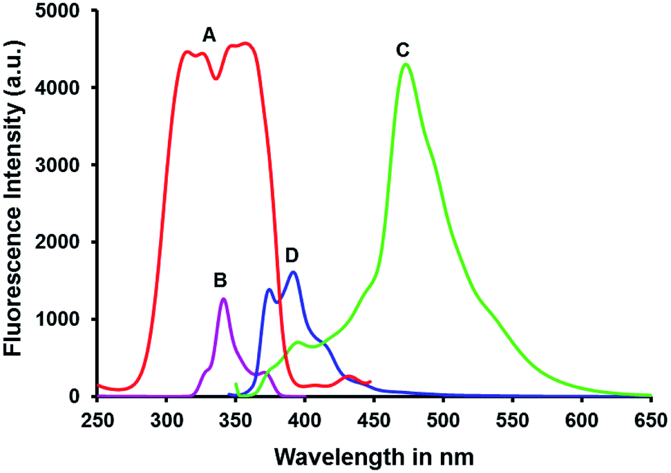
The excitation and fluorescence spectrum shown in Fig. 3 reveals that the excitation spectrum of aqueous suspension of PyNPs (spectrum A) is blue shifted broad band in comparison with the structured excitation spectrum of pyrene monomer in ethanol solution (spectrum B). The fluorescence spectrum of the nanostructure (spectrum C) is broad, structure less band with a maximum at 470 nm and red shifted from that of monomer fluorescence of pyrene in ethanol solution (spectrum D). The monomer emission disappeared totally from the emission spectrum of the nanostructure.
 |
| | Fig. 3 Excitation spectra of pyrene nanoparticles suspension (A), and dilute solution of pyrene in ethanol (A) and emission spectra of pyrene nanoparticles suspension (C), and dilute solution of pyrene in ethanol (D). | |
3.2.2 Fluorescence lifetime of PyNPs.
Fig. 4 shows the decay profile of dilute solution of pyrene in ethanol (A) and aqueous suspension of PyNPs (B). It is known that the aggregation and molecular interactions lead to a prolonged life time.23 The relatively longer life time of PyNPs (6.5 ns) than that of its solution in ethanol (2.5 ns) confirms that the pyrene molecules aggregates by self-assembly24a to form nanostructure and the characteristic enhanced emission from the excited state nanoparticles appears at 470 nm. The long lifetimes of PyNPs compared with that of monomer solution are easily understood in that the formation of aggregated nanoparticles restricts the molecular rotation and vibration of molecules and thus increases the emission lifetime of PyNPs.24b
 |
| | Fig. 4 Fluorescence decay profile of dilute solution of pyrene in ethanol (A) and pyrene nanoparticles suspension (spectrum B). | |
3.3 Stoke's shift in PyNPs
The value of Stoke's shift estimated as a difference between the excitation and fluorescence energy for PyNPs suspension is Δ![[v with combining macron]](https://www.rsc.org/images/entities/i_char_0076_0304.gif) = 6835 cm−1. This value is significantly larger than the Stoke's shift of Δ = 3644.5 cm−1 estimated for the pyrene solution in ethanol. The largest value of Stoke's shifts is an indication of the aggregation of molecules with stacking effects to form clusters. The observed large Stoke's shift in the emission of nanoparticles is attributed to the π–π interaction between closely stacked neighboring molecules which causes a gradual increase in the excitonic coupling effect by which the exciton relaxes to an energetically lower lying excited state and therefore in the case of the PyNPs the emission originates from a lower lying excited state as compared to the isolated pyrene molecules.27 This results in large Stoke's shifted emission from PyNPs.
= 6835 cm−1. This value is significantly larger than the Stoke's shift of Δ = 3644.5 cm−1 estimated for the pyrene solution in ethanol. The largest value of Stoke's shifts is an indication of the aggregation of molecules with stacking effects to form clusters. The observed large Stoke's shift in the emission of nanoparticles is attributed to the π–π interaction between closely stacked neighboring molecules which causes a gradual increase in the excitonic coupling effect by which the exciton relaxes to an energetically lower lying excited state and therefore in the case of the PyNPs the emission originates from a lower lying excited state as compared to the isolated pyrene molecules.27 This results in large Stoke's shifted emission from PyNPs.
4. Result and discussion
4.1 Fluorescence quenching experiments
Fluorescence of the probe is known to be quenched by Forster nonradiative energy transfer and by the binding of the analyte molecule with probe.28 The present pair of pyrene nanoparticles and R6G dye exhibits significant overlap between the fluorescence spectrum of pyrene nanoparticles and excitation spectrum of R6G in aqueous solution as presented in Fig. 5.
 |
| | Fig. 5 Integral overlap of the emission spectrum of pyrene nanoparticles (D) with the excitation spectrum of rhodamine 6 G (A). | |
The fluorescence spectra of aqueous suspension of PyNPs containing various amounts of R6G, monitored at excitation wavelength 354 nm are shown in Fig. 6(a). The inset of the figure shows fluorescence spectra of R6G excited at 354 nm without PyNPs which were subtracted from emission spectra of PyNPs with corresponding amount of R6G. The spectral data obtained after subtraction is shown in Fig. 6(b). It is seen that strong aggregation induced enhanced fluorescence of PyNPs at 470 nm is decreased gradually with increasing amounts of R6G, and significant new sensitized fluorescence band characteristic of R6G has appeared at 550 nm with an isoemissive point at 516 nm. This observation and significant overlap between the emission spectrum of PyNP and excitation spectrum of R6G led us to consider that the fluorescence quenching is due to resonance energy transfer from excited PyNPs to R6G which resulted in sensitization of fluorescence of R6G. There is no possibility of direct excitation of R6G at excitation on wavelength of PyNPs which is shown in Fig. S1.† The quenching experiments are repeated at three different temperatures and spectral data are analyzed by Stern–Volmer relation given by eqn (1).
| |  | (1) |
Where,
F0 and
F are the steady-state fluorescence intensities in the absence and presence of quencher, respectively,
Kq is the quenching rate constant and
τ0 is the average lifetime of the donor molecule in the absence of quencher (
τ0 = 6.5 × 10
−9 s).
KSV is the Stern–Volmer quenching constant, and [Q] is the concentration of quencher (R6G).
Fig. 7 shows Stern–Volmer plots for quenching of fluorescence of PyNPs by R6G at three different temperatures
viz. 298, 308, and 318 K. It is clear that the curves show good linear relationship, according to
eqn (1). The estimated values of photokinetics are summarized in
Table 1. The increase in
KSV and
Kq with increase in temperature indicates that the quenching interaction between PyNPs and R6G is dynamic and depends upon the diffusion of molecules.
29a,b The dynamic quenching phenomenon also support from absorption spectroscopy given in Fig. S2.
†
 |
| | Fig. 6 (a) Fluorescence quenching of pyrene nanoparticles (1.36 × 10⁻⁴ mol L−1) with varying amounts of R6G spectra A to I (0.0 to 8 × 10⁻⁶ mol L−1). [The inset of the figure shows fluorescence spectra of R6G excited at at 354 nm without PyNPs.]. (b) Fluorescence quenching spectra of PyNPs (1.36 × 10⁻⁴ mol L−1) with varying amounts of R6G obtained after subtraction of emission of R6G excited at 354 nm without PyNPs. (A to I: 0.0 to 8 × 10⁻⁶ mol L−1). | |
 |
| | Fig. 7 Stern–Volmer plots of the quenching of fluorescence of PyNPs by R6G at their different temperatures. | |
Table 1 Stern–Volmer quenching constants, binding and thermodynamic parameter of PyNPs estimated for binding interaction with R6G in aqueous solution
|
T/K |
K
sv (× 104 L mol−1) |
K
q (× 1012 L mol−1 s−1) |
K (× 104 L mol−1) |
ΔH (KJ mol−1) |
ΔG (KJ mol−1) |
ΔS (J mol−1 k−1) |
n
|
R
a
|
|
R is the correlation coefficient.
|
| 299 |
1.5 |
2.3 |
1.11 |
34.30 |
−23.12 |
192.71 |
0.9752 |
0.9986 |
| 309 |
3.0 |
8.5 |
1.49 |
−25.05 |
0.9679 |
0.9988 |
| 319 |
4.5 |
18 |
2.70 |
−26.97 |
0.9516 |
0.9963 |
4.2 Effect of coexisting cations on fluorescence quenching of PyNPs by R6G
We have to perform an interference experiment of acid violet 43 and some of the cation. To explore the selectivity of the method using pyrene nanoparticles as a probe for R6G in aqueous solution, the changes in fluorescence intensity of the probe were measured in the presence of co-existing cation such K+, Pb2+, Cd2+, Co2+, Ni2+, Fe2+, Al3+, Ca2+, Zn2+, Na+, Na2+, Sn2+, NH4+ and cationic dye (acid violet 43) solution of 5 × 10⁻⁶ mol L−1 concentrations each. The results presented in Fig. 8 (red bar histogram) reveal a remarkable fluorescence quenching response only in the composite holding R6G ion solution and no other cations caused observable quenching in these experiments. This is because of R6G specifically adsorbed on the negatively charged surface of pyrene nanoparticles than other cation resulting in fluorescence quenching. Evidently, the coexistence of this cation did not interfere with the fluorescence ratiometric detection of R6G.
 |
| | Fig. 8 Fluorescence intensity response [ΔF/F] in the presence of the R6g and several coexisting cations like K+, Pb2+, Cd2+, Co2+, Ni2+, Fe2+, Al3+, Ca2+, Zn2+, Na+, Na2+, Sn2+, NH4+ acid violet 43 solution of 5 × 10⁻⁶ mol L−1 concentrations (excitation wavelength is 354 nm). | |
4.3 Energy transfer efficiency and binding distance between PyNPs and R6G
The efficiency of energy transfer (E) and its relation with ‘r’ distance between donor and acceptor molecule and ‘R0’ the critical energy transfer efficiency equals to 50% is given by following equations,30| |  | (2) |
| | | R06 = 8.8 × 10⁻²⁵K2n−4ΦJ | (3) |
In eqn (3)K2 is the spatial orientation factor between the emission dipole of the donor and the absorption dipole of the acceptor. If both the donor and the acceptor are tumbling and free to assume any orientation, then K2 equals 2/3.31n = 1.336 (the refractive index of the medium) and Φ = 0.4765 is the fluorescence quantum yield of the donor in the absence of the acceptor. J is given by the expression,
| |  | (4) |
The value J = 9.5977 × 10⁻¹⁴ cm3 L mol−1 was evaluated from the integral region of overlap seen in Fig. 5. The value of R0 = 45.1 Å is calculated using eqn (3) and the average distance ‘r’ is 6.286 nm which is lower than the value 10 nm required for efficient energy transfer to occur between PyNPs and R6G.19 The decrease in fluorescence lifetime of PyNPs upon addition of R6G solutions supports efficient FRET from PyNPs to R6G is presented in Fig. S3.†
4.4 Thermodynamic parameters and nature of the acting forces
Further details of the molecular interaction between PyNPs and R6G molecule are given by the thermodynamic parameters (ΔH) and (ΔS) which were calculated from the Van't Hoff equation given below.| |  | (5) |
Where K is the binding constant at the corresponding temperature, and R is the gas constant.32,33 The nature of the plot of ln![[thin space (1/6-em)]](https://www.rsc.org/images/entities/char_2009.gif) K against 1/T is shown in Fig. 9. The enthalpy change (ΔH) and entropy change (ΔS) are obtained from the slope and intercept of Van't Hoff plot. Finally the Gibbs energy change (ΔG) was calculated by following thermodynamic relation.
K against 1/T is shown in Fig. 9. The enthalpy change (ΔH) and entropy change (ΔS) are obtained from the slope and intercept of Van't Hoff plot. Finally the Gibbs energy change (ΔG) was calculated by following thermodynamic relation.| | | ΔG = ΔH − TΔS = −RTlnk | (6) |
 |
| | Fig. 9 Van't Hoff plot for binding of PyNPs with R6G. | |
The estimated values of thermodynamic parameter are summarized in Table 1. Positive ΔH and ΔS value indicate the less dominant electrostatic and predominant hydrophobic force of interaction between PyNPs and R6G34 respectively. Thus the nonpolar hydrophobic group of PyNPs capped SDS may be involved in the binding with R6G. Rhodamine dyes are moderately hydrophobic cationic dye.35 In solution when both molecules are brought closer at a distance ‘r’ equal to 6.286 nm, the hydrophobic interaction becomes more probable. This is also supported by the fact that the hydrophobic forces36 are inversely proportional to the seventh power the distance r. The estimated smaller value indicates that two hydrophobic forces are appreciably large to favor the hydrophobic interaction rather than electrostatic interaction.37 The negative value of Gibbs free energy change (ΔG) indicates that the binding process is spontaneous.38
4.5 Estimation of binding parameter of PyNPs-R6G
In order to evaluate the binding constant K and the number of binding sites n, the double logarithmic plot based on following equation as shown in Fig. 10 is obtained.39| |  | (7) |
 |
| | Fig. 10 Double logarithmic plot of log10[(F0 − F)/F] versus log10[Q]. | |
From the nature of the plot shown in Fig. 10, the values of K and n are obtained from the intercept and slope of the straight line of the graph respectively. The estimated values of binding parameter are listed in Table 1. The results show that the binding constants increased with temperature, which manifests that binding, is an endothermic reaction and stable PyNPs–R6G complex is formed.40 The value of the binding site ‘n’ approximately equal to one indicates that the binding ratio is 1:1.
4.6 Mechanisms of fluorescence quenching
The Zeta potential value of −22.6 mV obtained from the electrophoretic mobility measured via phase analysis light scattering indicates that PyNPs stabilized by SDS. The addition of 4 × 10⁻⁶ mol L−1 and 8 × 10⁻⁶ mol L−1 solution of cationic R6G in the nanoparticles suspension has decreased the negative zeta potential to −19.9 mV and −15.7 mV respectively (Fig. 11) due to plausible adsorption41 of dye ions onto the surface of PyNPs. The centers of these ions are localized in between the PyNPs surface and Stern plane and thus both the PyNPs (donor) and R6G (acceptor) molecules are within a close distance of approach required for efficient transfer of energy there by exhibiting strong quenching of fluorescence of PyNPs. The aggregation of R6G with PyNPs is further supported by the size of nanoparticle42 which is seen to be increased from from 38 nm to 62 nm and to 743 nm upon addition of 4 × 10⁻⁶ mol L−1 and 8 × 10⁻⁶ mol L−1 solution respectively in Fig. 11.
 |
| | Fig. 11 Representation of average particle size and zeta potential of PyNPs measured by DLS and their variation response to addition of R6G concentration in PyNPs suspension (1.36 × 10⁻⁴ mol L−1): (a) in absence of R6G (b) and (c) in presence of 4 × 10⁻⁶ mol L−1 and 8 × 10⁻⁶ mol L−1 R6G respectively. | |
From the above observations and thermodynamic parameter study it is reasonable to conclude that the complexation through hydrophobic adsorption between PyNPs and R6G leads to fluorescence quenching. The mechanism of FRET is illustrated in Scheme 1.
4.7 Calibration curve and detection limit of the method
In order to avoid the contribution of sample matrix into the fluorescence signal of analyte molecule, calibration curve is used. The calibration graph is linear over the range of R6G is 0.0 to 8 × 10⁻⁶ mol L−1 in Fig. S4† with a correlation coefficient 0.985 obtained. The limit of detection (LOD) of the method is 8.905 × 10⁻⁶ mol L−1 which was calculated by the equation LOD = (3σ/k), where σ is the standard deviation of the y-intercepts of regression lines and k is the slope of the calibration graph.23
4.8 Analysis of R6G from textile effluent
The proposed fluorescence quenching method was applied for determination of R6G in the textile effluent sample collected from Yashwant Textile Ichalkaranji. The textile effluent was filtered through Whatmann filter paper no. 41 to remove solid impurity and diluted it and used in further analysis. Textile effluent samples were spiked with standard R6G of two concentration level and diluted within the working range with distilled water. The effluent sample was analyzed by the proposed fluorimetric method using calibration curve shown in Fig S4.† The results are summarized in Table 2. It can be seen that the value of R6G found in the two concentration level are identical with expected values. The recovery and relative standard deviation are very satisfactory.
Table 2 Determination of R6G in textile effluent from Yashwant processing Ichalkarangi by standard addition method (n = 3)
| Textile effluent sample studied |
Amount of Standard R6G added (×10−6 M L−1) |
Total R6G found (n = 3) (×10−6 M L−1) |
Recovery of R6G added (%) |
RSD (%) |
Relative error (%) |
| Yashwant Co-operative processers Ltd. Ichalkaranji Maharashtra (India) |
5.00 |
5.041 |
100.82 |
1.82 |
0.829 |
| 7.00 |
7.066 |
100.95 |
0.48 |
0.950 |
5. Conclusion
A novel FRET PyNPs probe prepared by the reprecipitation method under optimized preparation conditions. Dynamic light scattering results showed a narrow particle size distribution with average size of 38 nm. Zeta potential −22 indicated a high degree of stability PyNPs. SEM microphotography of air dried film of aqueous suspension of PyNPs showed that the nanoparticles are spherical in shape and monodispersed. The formation of nanoparticles was also evidenced by the absorption and fluorescence spectroscopy. The as prepared PyNPs emitting red shifted enhanced fluorescence showed greater spectral overlap with the absorption spectrum of R6G required for more efficient energy transfer. The value of critical energy transfer distance (R0) calculated by using Forster relation is 45.1 Å and as it is less than 50 Å, supported the spectral observation of efficient FRET from excited PyNPs to R6G. The FRET experiments performed at three different temperatures by the systematic addition of R6G into an aqueous suspension of PyNPs used to further evaluate photokinetics and thermodynamic parameter which helped in understanding mechanisms of interaction and observed quenching of fluorescence of PyNPs by R6G. Also the thermodynamic parameters and particle size along with zeta potential study supports the hydrophobic interaction between PyNPs and R6G. Finally, based on the fluorescence quenching results first fluorimetric method was developed for detection of R6G from industrial textile effluents.
Acknowledgements
The authors are thankful to the University Grants Commission (UGC), New Delhi for research funding of the project no. F. no. 41-217/2012 (SR) and a research fellowship with one of the author DPB. We are also thankful to the Department of Science and Technology (DST), New Delhi for providing funds to the Department of Chemistry, Shivaji University, Kolhapur under the FIST-Level-1 program for infrastructure improvement and UGC New Delhi for grants under the program UGC-DRS-I.
References
- L. Gagliardi, D. D. Orsi, G. Gavazzutti, G. Multari and D. Tonelli, Chromatographia, 1996, 43, 76–78 CrossRef CAS.
- S. Scalia and S. Simeoni, Chromatographia, 2001, 53, 490 CrossRef CAS.
- L. Valianou, I. Karapanagiotis and Y. Chryssoulakis, Anal. Bioanal. Chem., 2009, 395, 2175–2189 CrossRef CAS PubMed.
- R. Jain, M. Mathur, S. Sikarwar and A. Mittal, J. Environ. Manage., 2007, 85, 956–964 CrossRef CAS PubMed.
-
D. S. Mull, D. T. Liebermann, J. L. Smoot and L. H. Woosley, Application of Dye-Tracing Techniques for Determining Solute-transport Characteristics of Groundwater in Karst Terranes, EPA 904/6 88 001, US Environmental Protection Agency, Atlanta, Georgia, 1988, pp. 90–103 Search PubMed.
- O. Tunay, I. Kabdasli, G. Eremektar and D. Orhon, Water Sci. Technol., 1996, 34, 9–16 CrossRef CAS.
- A. Cassano, R. Molinari, M. Romano and E. J. Drioli, J. Membr. Sci., 2001, 181, 111 CrossRef CAS.
- N. D. Lourenco, J. M. Novais and H. M. Pinheiro, J. Biotechnol., 2001, 89, 163 CrossRef CAS PubMed.
- D. Pokhrel and T. Viraraghavan, Sci. Total Environ., 2004, 333, 37–38 CrossRef CAS PubMed.
- E. Forgacs, T. Cserháti and G. Oros, Environ. Int., 2004, 30, 953–971 CrossRef CAS PubMed.
- G. Crini, Bioresour. Technol., 2006, 97, 1061 CrossRef CAS PubMed.
- C. K. Lee, S. S. Liu, L. C. Juang, C. C. Wang, K. S. Lin and M. D. Lyu, J. Hazard. Mater., 2007, 147, 997–1005 CrossRef CAS PubMed.
- C. Fernández, M. S. Larrechi and M. P. Callao, Trends Anal. Chem., 2010, 29, 1202 CrossRef.
- C. Tsung-Ling, W. Yu-Chen and D. Wang-Hsien, J. Chin. Chem. Soc., 2011, 59, 1 Search PubMed.
- D. T. Patil, S. L. Bhattar, G. B. Kolekar and S. R. Patil, J. Solution Chem., 2011, 40, 211–223 CrossRef CAS.
- D. T. Patil, V. V. Mokashi, G. B. Kolekar and S. R. Patil, Luminescence, 2012, 28, 821–826 CrossRef PubMed.
-
J. R. Lakowicz, Principles of Fluorescence Spectroscopy, Plenum Press, New York, 3rd edn, 2006 Search PubMed.
- S. L. Bhattar, G. B. Kolekar and S. R. Patil, J. Lumin., 2008, 128, 306–310 CrossRef CAS.
- G. Chen, F. Song, X. Xiong and P. Peng, Ind. Eng. Chem. Res., 2013, 52, 11228–11245 CrossRef CAS.
-
http://scbaghdad.edu.iq/library/Physics/PhD/2013/Photobleaching%20Spectroscopic%20Studies.pdf, Republic of Iraq Ministry of Higher Education and Scientific Research, University of Baghdad College of Science.
- P. Marius, C. G. Jan, L. Philipp, G. Bernhard, U. Johannes, D. Frank, R. B. Wehrspohn and B. Benedikt, Energies, 2010, 3, 171–193 CrossRef.
-
(a) D. Zhou, L. Ying, X. Hong, E. A. Hall, C. Abell and D. Klenerman, Langmuir, 2008, 24, 1659–1664 CrossRef CAS PubMed;
(b) B. Mariana, M. A. Carlos-A, S. F. José-Paulo, C. Marie-Thérèse and M. G. José-Martinho, Polymer, 2011, 52, 5933–5946 CrossRef.
- D. P. Bhopate, G. B. Kolekar, K. M. Garadkar and S. R. Patil, Anal.
Methods, 2013, 5, 5324–5330 RSC.
-
(a) X. Zhang, X. Zhang, W. Shi, X. Meng, C. Lee and S. Lee, J. Phys. Chem. B, 2005, 109, 18777–18780 CrossRef CAS PubMed;
(b) M. Y. Berezinand and S. Achilefu, Fluorescence Lifetime Measurements and Biological Imaging, Chem. Rev., 2010, 110(5), 2641–2684 CrossRef PubMed.
-
http://cdn.shopify.com/s/files/1/0257/8237/files/nanoComposix_Guidelines_for_Zeta_Potential_Analysis_of_Nanoparticles.pdf
.
- Y. Dieckmann, H. Colfen, H. Hofmann and A. Petri-Fink, Anal. Chem., 2009, 81, 3889–3895 CrossRef CAS PubMed.
- D. P. Bhopate, G. B. Kolekar, K. M. Garadkar and S. R. Patil, Anal. Methods, 2013, 5, 5324–5330 RSC.
-
T. Foster and O. Sinanoglu, Quantum Chemistry, Academic Press, New York, 1996, vol. 3, p. 93 Search PubMed.
-
(a)
J. R. Lakowicz, Principles of Fluorescence Spectroscopy, Plenum Press, New York, 3rd edn, 2006 CrossRef;
(b) X. Zhu, A. Gong, B. Wang and S. Yu, J. Lumin., 2008, 128, 1815–1818 CrossRef CAS;
(c) U. S. Mote, S. R. Patil, S. H. Bhosale, S. H. Han and G. B. Kolekar, J. Photochem. Photobiol., B, 2011, 103, 16–21 CrossRef CAS PubMed.
- V. R. More, U. S. Mote, S. R. Patil and G. B. Kolekar, Spectrochim. Acta, Part A, 2009, 74, 771–775 CrossRef CAS PubMed.
- T. Forster, Discuss. Faraday Soc., 1959, 25, 7 RSC.
- Y. Q. Wang, B. P. Tang, H. M. Zhang, Q. H. Zhou and G. C. Zhang, J. Photochem. Photobiol., B, 2009, 94, 183–190 CrossRef CAS PubMed.
- M. Choudhury and R. Basu, J. Photochem. Photobiol., A, 1995, 85, 89–92 CrossRef CAS.
- U. S. Mote, S. L. Bhattar, S. R. Patil and G. B. Kolekar, Luminescence, 2010, 25, 1–8 CAS.
- S. Freire, J. Bordello, D. Granadero, W. Al-Soufi and M. Novo, Photochem. Photobiol. Sci., 2010, 9, 687–696 RSC.
- J. C. Micheau, G. V. Zakharovab and A. K. Chibisov, Phys. Chem. Chem. Phys., 2004, 6, 2420–2425 RSC.
- J. Shobini, A. K. Mishra, K. Sandhya and C. Nagsuma, Spectrochim. Acta, Part A, 2001, 57, 1133–1147 CrossRef.
- Y. J. Hu, Y. Liu, R. M. Zhao, J. X. Dong and S. S. Qu, J. Photochem. Photobiol., A, 2006, 179, 324–329 CrossRef CAS.
- X.-Y. Jiang, W.-X. Li and H. Cao, J. Solution Chem., 2008, 37, 1609–1623 CrossRef CAS.
- U. S. Mote, S.-H. Han, S. R. Patil and G. B. Kolekar, J. Lumin., 2010, 130, 2059–2064 CrossRef CAS.
-
(a)
http://www.diss.fuberlin.de/diss/servlets/MCRFileNodeServlet/FUDISS_derivate_000000002344/06_7Chapter7.pdf?hosts
;
(b) M. Kabiri, Z. Amiri-Tehranizadeh, A. Baratian, M. R. Saberi and J. Chamani, Molecules, 2012, 17, 3114–3147 CrossRef CAS PubMed;
(c) N. Wangoo, J. Kaushal, K. K. Bhasin, S. K. Mehta and C. R. Suri, Chem. Commun., 2010, 46, 5755–5757 RSC.
- A. H. Gore, S. B. Vatre, P. V. Anbhule, S.-H. Han, S. R. Patil and G. B. Kolekar, Analyst, 2013, 138, 1329 RSC.
Footnote |
| † Electronic supplementary information (ESI) available. See DOI: 10.1039/c4ra13555e |
|
| This journal is © The Royal Society of Chemistry 2014 |
Click here to see how this site uses Cookies. View our privacy policy here.