Song Wanga,
Bing Wang*a,
Huitong Sia,
Juanjuan Shana and
Xinlin Yang*b
aState Key Laboratory of Hollow Fiber Membrane Materials and Processes (Tianjin Polytechnic University), School of Materials Science and Engineering, Tianjin Polytechnic University, Tianjin 300387, P. R. China. E-mail: bingwang@tjpu.edu.cn
bKey Laboratory of Functional Polymer Materials, Ministry of Education, Institute of Polymer Chemistry, Collaborative Innovation Center of Chemical Science and Engineering (Tianjin), Nankai University, Tianjin 300071, P. R. China. E-mail: xlyang88@nankai.edu.cn
First published on 17th December 2014
Magnetite–β-cyclodextrin@poly(ethyleneglycol dimethacrylate-co-methacrylic acid) core–shell microspheres imprinted with baicalein (Fe3O4–β-CD@MIPs) were synthesized by ultrasonic assisted precipitation polymerization (UAPP) using methacrylic acid (MAA) as a functional monomer and EGDMA as a crosslinker in the presence of Fe3O4–β-CD nanoparticles as the template and baicalein (BAI) as the imprinting molecule. The structure, morphology and magnetic properties of the resultant Fe3O4–β-CD@MIPs have been systematically characterized by transmission electron microscopy (TEM), Fourier-transform infrared spectroscopy (FT-IR), vibrating sample magnetometry (VSM), and thermogravimetric analysis (TGA). The unique recognition ability of Fe3O4–β-CD@MIPs for BAI was evaluated by the adsorption experiments via comparing those of Fe3O4@MIPs and Fe3O4–β-CD@NIPs non-imprinted nanoparticles.
It is well-known that the pharmacologically active compounds in herbal plants have low concentrations and complex chemical constituents. It is essential to develop an effective technique for quantitative determination, recognition and purification of BAI molecules from those components in SBG. Several analytical methods, such as heat refluxing6 and adsorption chromatography separation,7 have been successfully utilized for the quantitative determination of BAI. However, these analytical procedures are always time-consuming with a high cost. A feasible way to solve these problems is via utilization of a solid adsorbent with specific recognition property, which has a great potential value for the study of biological process as well as its in-depth application. From these aspects, it is a superior choice to employ molecularly imprinted materials with specific adsorption.
Molecularly imprinted polymers (MIPs) have attracted much attention for their outstanding advantages, including unique adsorption performances, chemical resistance, mechanical stability, and relatively facile and low-cost preparation.8 As a result, MIPs have been widely used in many areas, such as adsorbents,9 membranes,10 solid-phase extraction,11 biosensors,12 isolation,13 chromatography stationary phase for efficient separation and specific recognition.14 The outstanding performance of MIPs depends on the shape memory towards the template via hydrogen bonding, and hydrophobic interactions. MIPs has been manufactured in various morphologies, including membrane, microspheres. Recently, core–shell magnetic MIPs have attracted much attention. It would be a promising solution to fully utilize the advantages of the magnetic NPs for efficient separation and enrichment, and recognition of the template molecule via a simple imposed magnetic field from a complex system. MIP coatings on magnetic Fe3O4 NPs with a core–shell structure (Fe3O4@BHb–MIPs) have been synthesized via combination of surface imprinting and sol–gel techniques for recognition and enrichment of proteins with accessible recognition sites, high capacity and favorable selectivity.15 Magnetic indole-3-acetic acid (IAA) imprinted polymer beads have been synthesized by suspension polymerization of 4-vinylpyridine (4-VPy) and a trimethylolpropane trimethacrylate (TRIM) crosslinker having an improved recognition ability to IAA with the aid of a silanized β-CD arm.16
In recent years, β-cyclodextrin (β-CD) has been widely utilized to recognize target molecules of compatible size based on its hydrophobic cavity together with a hydrophilic external surface.17 Therefore, it would be a promising solution through the preparation of MIPs with β-CD to contain hydrophobic and hydrophilic functionalities in a single material. The imprinting efficiency of traditional MIPs with an individual functional monomer, such as MAA, 4-VP, will be improved.
In the process of synthesis of MIPs, many methods have been used, such as bulk polymerization,18 suspension polymerization,19 emulsion polymerization,20 and precipitation polymerization.21 In recent years, precipitation polymerization has been developed as a heterogeneous polymerization technique for the synthesis of polymer micro-/nanospheres with uniform size, shape and various functionalities in the absence of any added surfactant or stabilizer.22 Unfortunately, the abhorrently long polymerization time is inevitable. In order to resolve the problem, ultrasound-assisted preparation polymerization23 was utilized. As a result, the polymerization time was greatly shortened. Herein, the magnetic MIP beads with a core–shell structure were designed and synthesized by the microwave-assisted precipitation polymerization of methacrylic acid functional monomer (MAA) and ethyleneglycol dimethacrylate (EGDMA) crosslinker in the presence of the silanized β-CD arm coated magnetite nanoparticle (Fe3O4) as a template. The mechanism for the construction of Fe3O4–β-CD@BAI–MIPs core–shell microspheres and the primary recognition of the MIPs for baicalein with a facile recovery via a magnetic separation process have been elucidated. The imprinting efficiency between individual functional monomer and assisted β-CD for conventional MIPs has been discussed.
For comparisons, Fe3O4@MIPs in absence of the β-CD arm and Fe3O4–β-CD@non-imprinted polymers without BAI template (Fe3O4–β-CD@NIPs) were also prepared for evaluation of the imprinting efficiency.
Q = (C0 − Ct)V/M | (1) |
Based on the above experiment, the dynamic adsorption experiment was implemented. 50 mg of the resultant beads were suspended in 5 mL 2 mmol L−1 BAI methanol solution. After different adsorption time (5–210 min), the concentration of BAI in the supernatant was detected as same as for isothermal adsorption.
Kd = Cp/Cs | (2) |
a = Kd(BAI)/Kd(reference) (reference = Qu, CAP) | (3) |
β = Qm/Qn | (4) |
The Fourier infrared spectra (FT-IR) were recorded on a TENSOR-37 spectrometer (Brucker, Germany) ranging from 4000 to 400 cm−1 with a potassium bromide pellet.
Thermogravimetric analysis (TGA) was performed on a SAT 409 PC (Netzsch, Germany) via heating from room temperature till 800 °C with a rate of 15 °C min−1 under a nitrogen flow.
The magnetic properties of the nanoparticles were measured with a vibrating sample magnetometer (VSM) (LDJ 9600-1, America) at room temperature.
The concentrations of BAI, Qu and CAP were determined by UV-vis spectroscopy on a UV spectrometer (Tu-1901, China).
The silica content of silylated β-CD was measured by Inductively Coupled Plasma (ICP, JY/ACTIVA-M, France).
The magnetite (Fe3O4) nanoparticles were prepared by the chemical co-precipitation of Fe(III)/Fe(II) (2/1 in molar ratio) cations in a basic solution (1.0 M NaOH) at 60 °C via a sol–gel process. These magnetite nanoparticles were prone to aggregate in the solvent and during the drying process due to the high specific surface area and strong interparticle interaction. This was confirmed by the FESEM micrograph in Fig. 1A with presence of the agglomerate consisting of several magnetite nanoparticles with the average diameter of 10 nm. The formation of the PEGylated Fe3O4 nanoparticles not only improved the stability of the composite nanoparticles as shown by the FESEM and TEM micrograph in Fig. 1B, but also endowed the hydrophilicity and the hydroxyl groups on the surface for the further functionalization and modification of the resultant magnetic nanoparticles.25 The mass ratio of the PEG components in the PEGylated magnetite nanoparticles was as high as 22% determined by TGA as shown in Fig. 5b.
Fig. 1 FESEM and TEM micrographs of (A) Fe3O4 nanoparticles; (B) PEGylated Fe3O4 nanoparticles; (C) Fe3O4–β-CD@BAI–MIPs. |
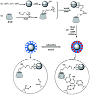
Molecularly imprinted polymers (MIPs) have special recognition performance based on the specific interaction/reaction between the functional monomer and the template molecule. Therefore, the selection of functional monomer was the most important aspect to achieve a good imprinting efficiency for the MIPs. However, the imprinting efficiency of traditional MIPs with a simple monomer system was not satisfactory enough for recognition after the selective removal of the template molecule. Herein, the β-CD, as a functional bridge between the BAI molecule and the magnetite core, was introduced to improve the imprinting efficiency during the recognition. The natural β-CD has certain limitations in practical applications,26 such as poor water-solubility and fewer effective functional sites. In order to improve the compatibility of β-CD, GPTMS was used to modify the β-CD molecule.27 The silylated β-CD was prepared via the inter-molecular condensation of the hydroxyl groups on the hydrophilic external surface of β-CD and the methoxy groups of GPTMS with presence of sodium hydride as shown in Scheme 1. The silica content in the silylated β-CD was 6.9% determined by ICP, which was consistent with the theoretical value of 7.7% according to the chemical structure in Scheme 1. The incorporation of the reactive methylsilane group via this modification provided the possibility for silylated β-CD as a functional bridge between the PEGylated magnetite core and the BAI imprinted polymer shell as shown in Scheme 1.
To elucidate the mechanism of the precipitation polymerization and the role of β-CD for the resultant Fe3O4–β-CD@BAI–MIPs, the UV-spectra were carefully and systematically investigated for BAI, MAA–BAI, BAI/β-CD, and MAA–BAI/β-CD in DMF solutions as shown in Fig. 2. The UV-spectrum of BAI in DMF has two peaks at 275 and 323 nm, which may be attributed to the hydrophilic cinnamoyl group and the hydrophobic benzoyl group, respectively. The UV-vis spectra of MAA–BAI with different ratios in DMF were illustrated in Fig. 2A. It indicates that the peak at 323 nm was gradually weakened with the molar ratio of MAA–BAI increasing from 0/1 to 4/1, while the peak intensity at 275 nm was weakened from 1.33 to 1.23 in the presence of MAA. According to the structure of BAI and the changes in the UV spectra, we deduced that there was a strong hydrogen-bonding interaction between the carboxyl groups of MAA and the hydroxyl groups in the BAI molecule as illustrated in Scheme 1(3). After the formation of hydrogen-bonding, the density of electron clouds decreased, the π-electron mobility increased, and a lower energy was required for the transition of electronic energy levels. Consequently, there was a significant weakening of the spectral band. In Fig. 2A, there was an isoabsorptive point at 350 nm. It revealed that two coordination interactions between BAI and MAA were formed in the process of self-assembly. Fig. 2B shows the UV-spectra of BAI/silylated β-CD in DMF with different ratios ranging from 1/0 to 1/4 in molar ratio. It demonstrates that the peak at 323 nm was considerably red-shifted from 323 to 329 nm with decreasing the ratio of BAI/silylated β-CD from 1/0 to 1/4, while the peak at 275 nm slightly changed. These red-shifts may originate from the efficient interaction between the hydrophobic benzoyl group and hydrophobic cavity of β-CD as described in Scheme 1(3), which reduced the energy gap for the π–π* transition to arise a better stabilization of excitons. With the BAI/silylated β-CD (1/1) as an example, the influence of the MAA on the UV-vis spectra were determined via addition of different amounts of MAA functional monomers as shown in Fig. 2C. For the MAA–BAI/silylated β-CD tri-component systems, both the peaks at 283 and 322 nm of the UV-vis spectra were red-shifted. Addition of more and more MAA in this tri-component system had a more significant influence on the absorption peak at 322 nm in UV-vis spectra as observed in Fig. 2C, which was attributed to the hydrogen-bonding interaction between MAA and BAI/β-CD complex. The UV-vis peak at 322 nm almost disappeared, which may be due to the formation of a core–corona micelle for MAA–BAI/silylated (7:1:1) tri-layer system. In such a case, the hydrophobic part may be located inside the micelle as core, which was shielded by the hydrophilic core during UV-vis irradiation. All these results demonstrated that the self-assembled MAA–BAI/silylated β-CD super-monomers were formed as shown in Scheme 1(3) via the strong hydrogen-bonding interaction between the MAA group and the phenol group of BAI together with the efficient hydrophobic interaction between the phenyl group of BAI and the hydrophobic cavity of β-CD, which would facilitate the precipitation polymerization and the improvement in recognition property of the Fe3O4–β-CD@BAI–MIPs.
Fig. 2 The UV-spectra of (A) BAI/MAA, (B) BAI/β-CD and (C) BAI/MAA/β-CD with different molar ratios in DMF solution. |
The MAA monomer, silylated β-CD, and BAI were mixed in DMF with a BAI–β-CD–MAA molar ratio of 1:1:2 in a round bottom flask with mild agitation on a SHA-B shaker overnight to form a super-MAA monomer. The Fe3O4–β-CD@BAI–MIPs were synthesized by precipitation polymerization of the super-MAA as functional monomer and EGDMA as crosslinker, in presence of PEGylated nanoparticles as the core with the aid of ultra-sonic irradiation in DMF/deionized water solvent. The FESEM micrograph of the resultant Fe3O4–β-CD@BAI–MIPs in Fig. 1C shows that these microparticles have a spherical shape and rough surface with the average diameter of 300 nm. The inserted TEM micrograph in Fig. 1C indicates that Fe3O4–β-CD@BAI–MIPs have a typical core–shell structure with a deep contrast inner core containing Fe3O4 nanoparticles and a slight contrast of MIP polymer shell. For comparisons, the molecularly imprinted polymer beads in absence of β-CD arm (Fe3O4@BAI–MIPs) were similarly prepared by precipitation polymerization of MAA functional monomer and EGDMA crosslinker in the presence of BAI as the template molecule and PEGylated Fe3O4 nanoparticles as the core. The molecularly non-imprinted polymer beads in absence of the BAI template (Fe3O4–β-CD@NIPs) were prepared by precipitation polymerization of MAA functional monomer and EGDMA crosslinker with presence of PEGylated Fe3O4 nanoparticles as the core.
The chemical structures of Fe3O4–β-CD@BAI–MIPs were confirmed by the FT-IR spectra in Fig. 3. The presence of a peak at 2929 cm−1 in the FT-IR spectrum of Fe3O4–β-CD@BAI–MIPs (Fig. 3a) proved that the β-CD was incorporated in the imprinted polymer beads, which was consistent with the imprinted polymer using acryloyl-β-CD and acrylamide as functional monomers in the literature.28 The presence of the peaks at 1728 cm−1 attributed to the stretching vibration of the carbonyl components from PMAA as well as PEGDMA network were clearly observed in Fig. 3a, c and d for all these three polymer beads. The formation of P(EGDMA-co-MAA) shell-layer over Fe3O4nanoparticles for Fe3O4–β-CD@BAI–MIPs and Fe3O4–β-CD@NIPs was attributed to the reaction between the tri-methoxy group from GPTMS and the hydroxyl groups of PEGylated magnetic nanoparticles, which was confirmed by Ji et al.29 The P(EGDMA-co-MAA) shell-network in Fe3O4@BAI–MIPs was formed by ultrasonic assisted precipitation polymerization with the aid of efficient hydrogen-bonding interactions between the carboxyl group of PMAA and the hydroxyl group of the PEGylated Fe3O4 nanoparticles, which was similar to the construction of P(DVB-co-AA)@PAA@P(DVB-co-AA) (DVB: divinylbenzene) tri-layer microspheres in our previous work.30 These peaks ranging from 1630 to 1450 cm−1 in Fig. 3a proved the incorporation of the BAI template molecules in Fe3O4–β-CD@BAI–MIPs, which was contributed from the stretching vibration of C–C bonds in BAI species. The strength for this wide peak was considerably decreased in Fig. 3b after the extraction of the BAI with a mixed solvent of methanol and acetic acid (8/2, v/v) using the Soxhlet extraction technique, which confirmed the selective removal of the BAI templates from Fe3O4–β-CD@BAI–MIPs through the damage of hydrogen bonding interaction between the monomer and BAI.
Fig. 3 FT-IR spectra: (a) Fe3O4–β-CD@BAI–MIPs; (b) Fe3O4–β-CD@MIPs; (c) Fe3O4@MIPs; (d) Fe3O4–β-CD@NIPs. |
The magnetic properties of the Fe3O4, Fe3O4@BAI–MIPs, Fe3O4–β-CD@BAI–MIPs, and Fe3O4–β-CD@BAI–NIPs were determined by VSM at room temperature, as shown in Fig. 4. The saturation magnetization value of the particles was 15.99, 3.67, 2.43, and 2.0 emu g−1, respectively. The saturation magnetization values for the functionalized nanoparticles were significantly smaller than that for magnetite core nanoparticles, which was due to the considerable mass decrease of the neat magnetite species after the functionalization. No magnetic hysteresis loops were observed from the field dependent magnetization plots in Fig. 4 for all these samples. In other words, the inexistence of remanence when magnetic field was removed, implies that all the magnetic particles retained paramagnetic properties originating from the magnetite core nanoparticles at room temperature. With the existence of an external magnetic field, the black suspension with these magnetic nanoparticles became transparent and clear within a short time. All these beads were attached to the walls of the vial as shown by the inserted digital photo in Fig. 4. As a result, it would be possible to facilely separate these magnetic particles during the recovery of BAI recognition for these molecularly imprinted beads.
Fig. 4 Magnetic properties by VSM technique: (a) Fe3O4 nanoparticles; (b) Fe3O4@MIPs; (c) Fe3O4–β-CD@BAI–MIPs; (d) Fe3O4–β-CD@NIPs. |
The amount of the inorganic components in Fe3O4, PEGylated Fe3O4, Fe3O4@BAI–MIPs, Fe3O4–β-CD@BAI–MIPs and Fe3O4–β-CD@NIPs were determined by TGA as shown in Fig. 5. These results demonstrated that the magnetite contents were 29.5 mass% for Fe3O4@MIPs, 19.3 mass% in Fe3O4–β-CD@BAI–MIPs and 16.1 mass% for Fe3O4–β-CD@NIPs, as the residual silica from β-CD was very low after pyrolysis of these hybrid beads. The significant decrease of magnetite content was consistent with the decrease of the saturation magnetization value from 15.99 emu g−1 to 2.43 emu g−1 for these particles.
Fig. 5 TGA curves: (a) Fe3O4 nanoparticles; (b) PEGylated Fe3O4 nanoparticles; (c) Fe3O4@BAI–MIPs; (d) Fe3O4–β-CD@BAI–MIPs; (e) Fe3O4–β-CD@NIPs. |
Fig. 6 Adsorption isotherms of BAI on functional beads: (a) Fe3O4–β-CD@MIPs; (b) Fe3O4@MIPs; (c) Fe3O4–β-CD@NIPs. |
A BAI standard solution with the concentration of 2.0 mmol L−1 was used to evaluate the dynamic adsorption for rebinding capacity of the resultant polymer beads. The adsorption dynamic curves of Fe3O4–β-CD@MIPs, Fe3O4@MIPs and Fe3O4–β-CD@NIPs are shown in Fig. 7. It can be observed that the adsorption capacities of all these three beads increased very quickly during the first 20 min and then leveled off between 30 and 120 min. The adsorption equilibrium state was reached after 120 min. The results indicated that the adsorption capacities of Fe3O4–β-CD@MIPs (130 μmol g−1) and Fe3O4@MIPs (100 μmol g−1) were significantly larger than that of Fe3O4–β-CD@NIPs (50 μmol g−1). These were mainly attributed to the presence of the specific binding cavities, which were afforded by the selective removal of the templated BAI molecules from Fe3O4–β-CD@BAI–MIPs and Fe3O4@BAI–MIPs via Soxhlet extraction. Compared to Fe3O4@MIPs, Fe3O4–β-CD@MIPs had a higher adsorption capacity via the incorporation of the β-CD arm in this system, which implied that more imprinted sites were well maintained during the extraction of BAI molecules due to the synergic effect of the silylated β-CD arm.
Fig. 7 Dynamic adsorption of BAI on functional polymer beads: (a) Fe3O4–β-CD@MIPs; (b) Fe3O4@MIPs; (c) Fe3O4–β-CD@NIPs. |
The adsorption kinetics of MIPs can be described via utilization of the internal diffusion model and the pseudo-first-order and pseudo second-order kinetic models.31,32 Through fitting the experimental data from Fig. 6, the pseudo second-order model is more consistent with the kinetics of the adsorption characteristic of MIPs than those of the pseudo first-order according to the internal diffusion model. The kinetic data were calculated by the following formula according to the second-order model:
t/Qt = 1/(KsQe2) + (1/Qe)t | (5) |
The dynamic binding data were summarized in Table 1. These results indicated that the binding process could be the rate-limiting step for the whole adsorption process for BAI.
Beads | Equation | Qe | Qt | Ks | R2 |
---|---|---|---|---|---|
Fe3O4–β-CD@MIPs | t/Qt = 0.0073t + 0.3441 | 137.3 | 133.5 | 0.00015 | 0.9949 |
Fe3O4@MIPs | t/Qt = 0.0091t + 0.0698 | 110.2 | 108.5 | 0.00119 | 0.9967 |
Fe3O4–β-CD@NIPs | t/Qt = 0.0146t + 0.3186 | 68.6 | 64.2 | 0.00067 | 0.9990 |
The thermodynamic adsorption of Fe3O4–β-CD@MIPs was investigated under the temperature ranging from 293 to 333 K, which was treated by the thermodynamic equation:
lnKD = −ΔrGm/RT = −ΔrHm/RT + ΔrSm/R | (6) |
KD = Qe/Ce | (7) |
The ΔrHm was −28.1 kJ mol−1, which was obtained from the straight slope via plotting lnKD and 103/T (Fig. 8). This suggested that the process of imprinting adsorption was mainly via a physicochemical adsorption mechanism. The hydrogen-bonding interaction between the resultant polymers and BAI played as a driving force for the construction of Fe3O4–β-CD@MIPs.
Fig. 8 The relationship between the dissociated equilibrium constant and temperature for the adsorption of BAI on Fe3O4–β-CD@MIPs. |
The selectivity of Fe3O4–β-CD@MIPs, Fe3O4@MIPs, and Fe3O4–β-CD@NIPs was investigated by UV measurement of the residual amounts of BAI, Qu and non-analogue CAP in the upper liquid after the separation of the adsorbed beads via external magnetic field from the adsorption system. The adsorption was performed by adding independently these polymer beads into the solution of BAI, Qu and CAP with mild agitation on a SHA-B shaker for 20 h, respectively. The relevant data are summarized in Table 2. The selectivity factor of Fe3O4–β-CD@MIPs for Qu and CAP was 3.85 and 4.88, respectively. These results demonstrated that Fe3O4–β-CD@MIPs exhibited a higher selectivity for the adsorption of BAI templated molecules than those of Fe3O4@MIPs (2.56 and 2.94) and Fe3O4–β-CD@NIPs (1.16 and 1.20) for the reference molecules of Qu and CAP. The highest selectivity of Fe3O4–β-CD@MIPs was mainly contributed from the molecular size recognition via the hydrogen-bonding interaction between the BAI molecules and the carboxyl groups of PMAA with the synergic effect of β-CD arm in Fe3O4–β-CD@MIPs compared to that in Fe3O4@MIPs. The higher selectivity of BAI to CAP (4.85, 2.94) than that of BAI to Qu (3.85, 2.56) for the BAI-imprinted Fe3O4–β-CD@MIPs and Fe3O4@MIPs as chemical structure of Qu was more similar to that of the BAI molecule. The selectivity of Fe3O4–β-CD@NIPs (1.16 and 1.20 to Qu and CAP) was very low as there were not specific cavities/sites left in the polymer network for the selective adsorption of BAI molecules with a very low saturate adsorption capacity (50 μmol g−1).
Substance | Fe3O4–β-CD@MIPs | Fe3O4@MIPs | Fe3O4–β-CD@NIPs | ||||||||
---|---|---|---|---|---|---|---|---|---|---|---|
Q | Kd | α | β | Q | Kd | α | β | Q | Kd | α | |
BAI | 133.5 | 201 | 1.00 | 2.08 | 108 | 118.5 | 1.0 | 1.69 | 64 | 47.31 | 1.00 |
Qu | 68.5 | 52 | 3.85 | 1.18 | 63 | 46.3 | 2.56 | 1.10 | 58 | 40.65 | 1.16 |
CAP | 58.1 | 401 | 4.88 | 1.03 | 57 | 40.3 | 2.94 | 1.02 | 56 | 39.31 | 1.20 |
This journal is © The Royal Society of Chemistry 2015 |