
Ag/ZnO nanoparticles thin films as visible light photocatalysts
Jamshaid Rashida,
M. A. Barakat*abc,
Numan Salahd and
Sami S. Habibd
aDepartment of Environmental Sciences, Faculty of Meteorology, Environment and Arid Land Agriculture, King Abdulaziz University, Jeddah, Saudi Arabia. E-mail: mabarakat@gmail.com; Fax: +966 02 6952364; Tel: +966 02 6400000/64821
bCentral Metallurgical R & D Institute, Helwan 11421, Cairo, Egypt
cCenter of Excellence in Environmental Studies (CEES), King Abdulaziz University, Jeddah, Saudi Arabia
dCenter of Nanotechnology, King Abdulaziz University, Jeddah, Saudi Arabia
First published on 28th October 2014
Abstract
Silver (Ag) and zinc oxide (ZnO) nanoparticles were simultaneously deposited on a glass substrate using the radio frequency (RF) sputtering technique at different substrate temperatures. Detailed characterization of the co-sputtered Ag/ZnO thin films was performed by X-ray diffraction (XRD), field emission scanning electron microscopy (FESEM) and X-ray photoelectron spectroscopy (XPS). The as synthesized thin films were tested using UV-Vis diffuse reflectance spectroscopy to evaluate their optical properties. The obtained SEM results show a uniform dispersion of Ag nanoparticles within the ZnO matrix. These nanoparticles have average particle size of 20 nm. The optical band gap value was calculated from UV transmission spectra of Ag/ZnO thin films deposited at various substrate temperatures. This value was observed to be in the visible light range (i.e., 2.7–3.1 eV), which is much smaller than that of pure ZnO (3.37 eV). The photocatalytic activity of the produced thin films was evaluated through visible light photodegradation of 2-chlorophenol (2-CP) which has been used as a pollutant model in water. The synthesized thin films showed enhanced visible light photocatalytic efficiency towards 2-CP degradation at elevated substrate temperature and retained their catalytic efficiency with only 8% loss in efficiency after four reuse cycles. Kinetic parameters involved in the degradation process were investigated by applying a pseudo-second-order kinetic model.
Introduction
Zinc oxide (ZnO) is a wide band gap semiconductor (3.37 eV), environmentally friendly in nature and potentially can be used in photo-induced redox reactions. These properties make it suitable for various applications including solar energy conversion, optoelectronics, UV transparent coatings and photocatalysis.1,2 The electronic structure of ZnO is characterized by a filled valance band and an empty conduction band with a band gap energy comparable to TiO2. Surface oxygen defects acting as electron trap sites and efficient hydroxyl ions generation in aqueous medium and high surface reactivity make ZnO a competitive photocatalyst.3,4 For instance, in some studies ZnO has been reported to be more efficient and economically valuable than titanium oxide (TiO2) for photodegradation of azodyes.5,6Photocatalysis as an advanced oxidation processes (AOPs), has gained considerable attention towards complete, non-selective destruction of organic contaminants such as 2-chlorophenol (2-CP) in industrial wastewater.7 However the energy band gap of ZnO (3.37 eV for pure crystals) limits its use in large scale application as a photocatalyst; requiring ultraviolet (UV) radiation for electronic excitation to its conduction band. The UV irradiation comprises only about 5% of natural solar spectrum while visible light makes approximately 43% of the incident solar light.8 Laboratory and industrial scale use of UV radiation is very expensive while prolong exposure to UV radiation may have chronic health risks including premature skin aging, suppressed immune system, damage to the eyes and skin cancer.9 High degree of recombination of photogenerated species (electron–hole pairs e−/h+) also impedes its practical use of ZnO photocatalysis.10,11 Furthermore, recovery of used powder photocatalyst from the solution is another problem which is generally associated with significant loss of activity due to catalyst agglomeration.
To circumvent these shortfalls, addition of a suitable electron scavenger and immobilization of the photocatalyst as thin films over transparent supports could be a suitable solution. Addition of metal impurities into ZnO has been reported to enhance its photocatalytic performance.12–14 The presence of metal Ag in relatively low concentrations (less than 2%) in photocatalyst systems has been reported to enhance photocatalytic activity for degradation of organic contaminants with increase in specific surface area of the catalyst.15 The surface plasmon resonance (SPR) properties of silver are also known to assist in visible light absorption and subsequent electron–hole pair generation for the decontamination of pollutants in water.16,17
ZnO thin films can be easily deposited on a variety of substrates including, sapphire, silicon, GaAs and glass,18–21 by a number of physical deposition techniques that may include chemical vapor deposition (CVD),22 radio frequency-magnetron sputtering (RF-MS),23 pulsed laser deposition (PLD)24 and plasma-assisted molecular beam epitaxy (P-MBE).25 RF sputtering is comparatively flexible technique to achieve high deposition rate ensuring adherence for a large variety of compounds while maintaining the optical and electronic properties of transparent thin films intact.26 This work reports the enhancement effect of RF co-sputtering of Ag with ZnO on visible light photocatalytic activity of Ag/ZnO nanoparticles thin films. The thin films were prepared at different low substrate temperatures. Their characterization and application as photocatalyst were studied for degradation of 2-CP in synthetic wastewater under visible irradiation.
Materials and methods
Materials
UV grade glass slides were used as substrates for deposition of Ag and ZnO nanostructure. Acetone and ethyl alcohol were used as cleaning solvents for the glass slides. Then the substrates were dried in argon flow and mounted on the aluminum custom made slide holders of the deposition system. Standard grade 2-Chlorophenol (Merck) was utilized as a pollutant in simulated wastewater for the photodegradation experiments. Deionized water was used for solution preparation. All other chemicals were also analytical grade.Fabrication of Ag/ZnO thin films
Simultaneous radio frequency (RF) magnetron sputtering (DC/RF Magnetron Sputter System, Syskey Technologies, Taiwan) of Ag and ZnO was performed to deposit Ag/ZnO thin films on glass substrate. Before deposition the glass slides were thoroughly cleaned with acetone and dried with pressurized nitrogen. High purity (99.999%) targets for both sintered ZnO (3 × 0.06 inch) and Ag (3 × 0.236 inch) metal were used for the deposition studies. The plasma was generated inside the sputtering chamber by applying a RF power of 200 W for ZnO and 30 W for Ag. The base pressure in chamber was adjusted to 1 × 10⁻⁶ torr before introducing Argon gas. The operating pressure of 5 × 10⁻³ torr was maintained. Thin films of average thickness 110 nm were deposited by co-sputtering for 300 s. Three different thin films were prepared by varying the substrate temperatures i.e., room temperature (RT), 50 and 100 °C.Materials characterization
The synthesized thin films of Ag/ZnO co-sputtered nanoparticles were characterized by X-ray diffractometer (Ultima-IV; Rigaku, Japan) with Cu Kα radiation λ = 1.5418 Å wavelength having 40 kV accelerating voltage and 30 mA current. Surface elemental analysis was performed by X-ray photoelectron spectroscopy (XPS); (PHI 5000VersaProbe II), while the morphology was studied by a field emission scanning electron microscopy (FESEM) (JSM-7500 F; JEOL-Japan) using parallel beam geometry and a multi-purpose thin film attachment. Thickness of the thin films was determined by cross-sectional SEM observations. Optical transmittance of Ag/ZnO co-sputtered nanoparticles thin films in the range of 300 to 800 nm was recorded by UV-Vis-NIR spectrophotometer (PerkinElmer UV/Vis/NIR; Lambda 750).Photocatalytic experiments
The photocatalytic performance of the synthesized thin films was evaluated in Luzchem photochemical reactor (LZC4) system. For a typical experiment the photocatalyst thin film was immersed in a cylindrical rector cell (250 ml capacity) provided with air circulation to ensure aerobic conditions required for photocatalysis. The reaction contents were irradiated with 112 W cool white visible light lamps from top and sides with spectral range from 400 to 700 nm and spectral irradiance of 17.45 mW cm−2 monitored by Luzchem power monitor at a distance of 12 cm from the light source for 3 h irradiation time. For analysis 2.5 ml samples were drawn after every 30 min interval and analyzed by using UV-visible spectrophotometer (HACH LANGEDR6000) at 274 nm. The degradation efficiency was calculated, after analysis, using the following relation:| Degradation% = {(Co − Ct)/Co} × 100 | (1) |
Results and discussion
Morphology, structural and optical properties of Ag/ZnO nanoparticles thin films
The X-ray diffraction patterns of Ag/ZnO co-sputtered thin films deposited at different substrate temperatures along with pure ZnO thin film are provided in Fig. 1. The thin films display main peaks of hexagonal ZnO identified at 2θ values of 31.51 (1 0 0), 34.15 (0 0 2) and 64.7 (1 0 3) while the peak at 38.66 (1 1 1) indicates the presence of Ag in Ag/ZnO thin films. An apparent increase in ZnO peak intensity with (0 0 2) preferential orientation was observed at elevated substrate temperatures indicating that adequate increase in substrate temperature may facilitate atomic diffusion to stable lattice sites. As reported by other studies27–29 addition of Ag to ZnO resulted in decrease in peak intensities however no shift in the (0 0 2) diffraction peak was observed when compared to pure ZnO indicating that Ag was not incorporated as Ag ions in the Zn2+ (72 pm) substitution sites contrary to results reported in other studies30. For Ag nanoparticles indexing process of X-ray diffraction pattern was done and Miller Indices (h k l) were assigned to each peak. The detail of the diffraction peaks for Ag is provided in Table 1. A number of strong Bragg reflections corresponding to the (111), (200), (220) and (311) reflections of fcc silver were observed at 2θ values of 38.10, 44.37, 64.18 and 77.55 respectively. The XRD study confirms that the resultant particles are face centered cubic nanoparticles of Ag metal (PDF card no: 00-001-116). | ||
| Fig. 1 XRD patterns of ZnO and Ag/ZnO co-sputtered thin films. | ||
| 2Theta | d-value | h k l | Intensity |
|---|---|---|---|
| 38.10 | 2.360 | 1 1 1 | 100 |
| 44.37 | 2.040 | 2 0 0 | 53.0 |
| 64.18 | 1.450 | 2 2 0 | 27.0 |
| 77.55 | 1.230 | 3 1 1 | 53.0 |
The average crystallite size of ZnO nanoparticles in Ag/ZnO thin films were estimated by XRD data to be in the range of 3 to 9 nm. Compared to pure ZnO (5–15 nm) the decrease in the crystallite size may be attributed to the oxygen vacancies leading to lattice deformation and limiting crystallite growth.31,32 No significant change in d space values was observed that suggests that silver co-deposition did not induce any variation in average unit cell dimensions.
X-ray photoelectron spectroscopy was performed to investigate the surface compositions and chemical states of the species existing in the Ag/ZnO thin films (Fig. 2). Oxygen 1s state has been reported to be characterized by three binding energy components denoted by the low, intermediate and high binding energy peaks located at 530.15, 531.25 and 532.40 eV respectively.33,34 The low binding energy state are characteristic of intrinsic O2− ions present in regular ZnO lattice sites, middle one represent O2− ions in oxygen deficient sites while the high binding energy peak attributes to the loosely bound surface oxygen form surface bound H2O, O2 or CO3− species. From Fig. 2(a), the low and intermediate energy peaks of O1s can be identified with peak values 530.2 and 531.76 eV. It is clear that the low binding energy peak identifying oxygen atoms in fully oxidized stoichiometric species is comparatively weaker than the intermediate binding energy peak corresponding to the oxygen species in the oxygen deficient regions. This demonstrates most of the oxygen exists within the interstices and oxygen deficient sites in Ag/ZnO thin film representing poor stoichiometry and crystal quality.
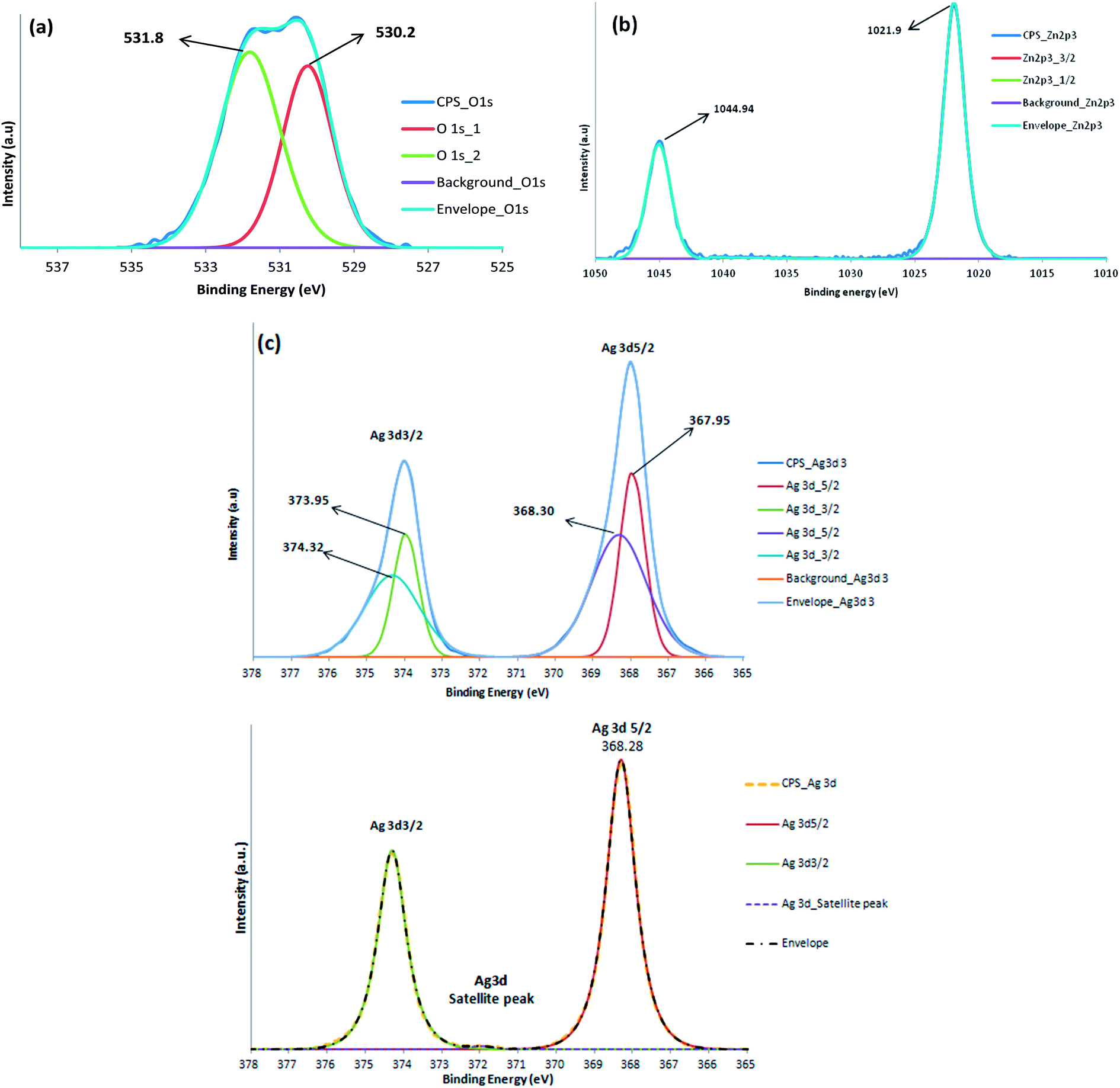 | ||
| Fig. 2 XPS spectra of (a) O 1 s, (b) Zn 2p and (c) Ag 3d regions of Ag/ZnO thin film compared with Pure Ag 3d Spectra. | ||
The typical XPS analysis of Ag/ZnO thin film in Fig. 2(b) shows high symmetry corresponding to the position of Zn 2p1/2 and Zn 2p3/2 peaks centered at binding energy values of 1044.94 eV and 1021.9 eV, respectively according to the regular lattice sites in crystalline ZnO. The calculated difference in binding energy (23.04 eV) corresponds to the characteristic value for ZnO and no binding energy peak at 1021.50 eV for metallic Zn confirm the existence of Zn in oxidized states only.35 The deconvolution of the Ag 3d XPS peaks for and evaluation of the spectra was performed using the CasaXPS software package Version 2.3.16. The Shirley method and the Gaussian–Lorentzain (GL) function were used for the background subtraction and peak fitting procedures, respectively. The XPS curves of Ag 3d region are shown in Fig. 2(c) with doublet Ag 3d5/2 and Ag 3d3/2 peaks with an intensity ratio of 3–2, located at 368.14 and 374.10 eV respectively. The Ag 3d peaks of Ag/ZnO show asymmetry on the higher binding energy side, which is indicative of the presence of Ag in more than one chemical state. The comparison of Ag 3d peak of Ag/ZnO with the pure metallic silver sample shows the characteristic of metallic silver36–38 with the Ag 3d peak appearing at a binding energy of 368.5 eV and separation between Ag 3d5/2 and Ag 3d3/2 doublet peaks of 6 eV. Based on this, the highest binding energy components in these two samples have been assigned to metallic silver. The two component peaks however appearing at lower binding energy values of 367.9 and 373.95 may be assigned to mono and bi-valent Ag–O possibly resulting from Ag oxidation at elevated temperature.39,40
Surface morphology has important significance to evaluate the thin film microstructure. SEM images of Ag/ZnO co-sputtered thin films deposited on glass substrate at room temperature (RT), 50 °C and 100 °C are illustrated in Fig. 3 (images a, b and c, respectively). The SEM images show uniform distribution of Ag nanoparticles uniformly distributed within the ZnO nanoparticles layer with typical Ag grain size within 9–20 nm. The particle size measured from SEM however appeared to be slightly greater than that calculated from XRD results therefore it can be assumed that the particles appearing in SEM are result of aggregation of small crystallites. Excess silver nanoparticles also appear as white clusters as reported by other studies by assembling at grain boundaries and aggregating into cluster form41,42 as confirmed from the XRD data of Ag (111) plane. The morphology of Ag nanoparticles also changed from flake like to cylindrical particles with the increase in substrate temperature from RT to 100 °C as illustrated in Fig. 3(a–c). Increase in particle size of Ag with elevated deposition temperature can be explained by the difference in energy of Ag nanoparticles. At lower temperatures (RT–50 °C), the energy of Ag nanoparticles is too small to move freely; therefore they cannot form large Ag grains. At temperatures of 100–200 °C, Ag atoms could absorb enough energy to move freely. So they cling together and form large grains. The largest grains with the size of ∼30 nm can be observed at the growth temperature of 100 °C.43 A high resolution cross sectional image of thin film deposited at 100 °C showed in Fig. 3(d) provides with the approximate thickness of the thin film.
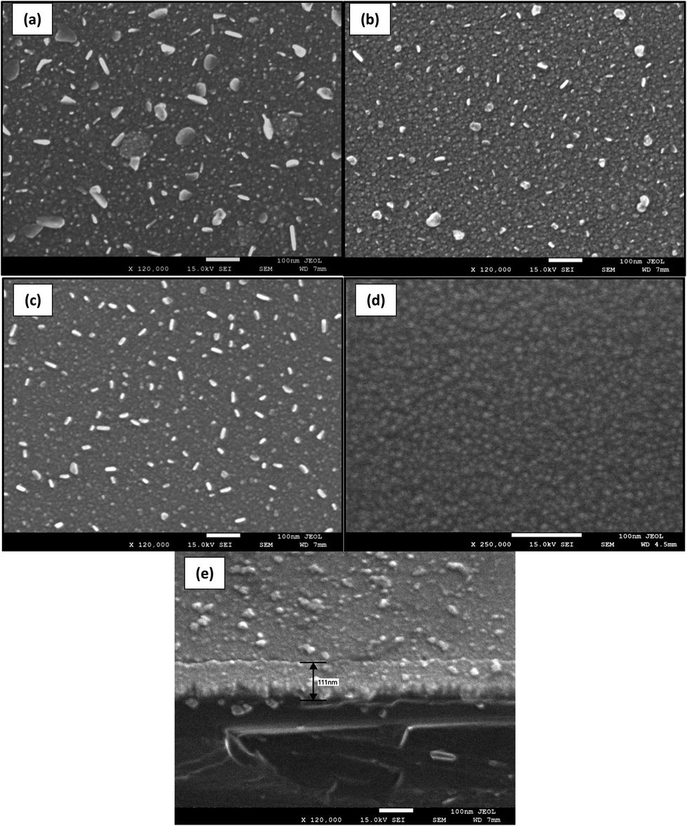 | ||
| Fig. 3 SEM images of Ag/ZnO thin film co-sputtered at different temperatures (a) Room temp. (b) 50 °C, (c) 100 °C and (d) Pure ZnO (e) cross sectional view of Ag/ZnO thin film. | ||
The EDS spectra provides with the elemental composition of the nanoparticles of deposited thin film. The EDS analysis recorded at various sites on the thin film indicated that the thin film mainly comprised of Zn, Ag and O as shown in Fig. 4. The percent atomic composition was found to vary at different positions, indicating non uniform deposition of Ag over the nanoparticles thin film. This may partially be due to the fact that EDS spectrum only provides the element's relative abundance in the area swept by the electron gun.44 For comparison the EDS spectra of flaked and non flaked areas are provided in Fig. 4(a and b). The UV-visible transmission spectra of Ag/ZnO at various substrate temperatures measured by UV/Vis/NIR spectrometer showed high transmittance in the visible region; however the percentage transmittance decreased by moving to the shorter wavelengths of visible region as a result of Ag loading. The average transmittance increased from 50% to 78% with increase in substrate temperature from RT to 100 °C. Increase in crystallinity and change in morphology of Ag nanoparticles at higher substrate temperatures as suggested by the SEM results may be responsible for high transmittance values. These results were in coincidence with the study conducted by Sahu30 however other studies have reported an opposite trend45 suggesting that difference in the Ag content in the synthesized thin films may be responsible for such behavior of the thin films.
 | ||
| Fig. 4 Representative EDS of Ag/ZnO nanoparticles (a) unflaked and (b) flaked areas. | ||
The direct band gap of the Ag/ZnO thin films was determined through Tauc's relationship46 as shown in eqn (2):
| αhν = A (hν − Eg)n | (2) |
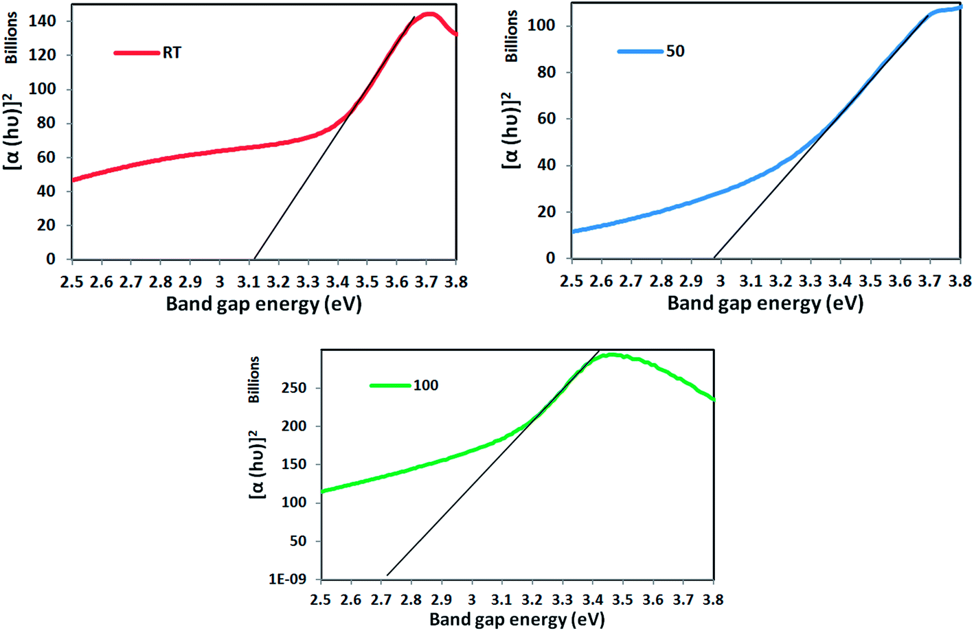 | ||
| Fig. 5 Plot of band gap energy of Ag/ZnO co-sputtered thin films at different substrate temperatures. | ||
Photocatalysis
Photocatalytic degradation of 2-CP using synthesized thin film was performed under visible light and varied experimental conditions to determine the optimum reaction parameters. In each experiment, the thin films were suspended and dipped in 250 ml 2-CP solution having certain initial concentration, at normal solution pH and constant aeration to maintain aerobic conditions; the reaction contents were subjected to 112 W (λmax > 350 nm) visible light irradiation for 3 h.To assess the effect of Ag addition on the photocatalytic activity, 10 mg L−1 of 2-CP solution was subjected to visible light irradiation in the presence of both Ag/ZnO and pure ZnO thin films (prepared under same conditions). Results obtained showed that Ag/ZnO thin film had superior photocatalytic activity as compared to pure ZnO (Fig. 6). Degradation efficiency of 54.1% was achieved with Ag/ZnO as compared to 20.9% with pure ZnO after 3 h of irradiation. The enhancement in photocatalytic activity can be attributed to the Ag nanoparticles present on the surface of the Ag/ZnO thin film which act as electron sink. The metallic silver Ag nanoparticles act as electron scavengers facilitating the transfer of photogenerated ZnO conduction band electrons to dioxygen as a means of increasing the efficiency of the photodegradation of 2-CP compared to pure ZnO alone. The Ag nanoparticles also hinder the recombination of photogenerated electrons–hole species thus making available ZnO surface for the photo-induced oxidation reactions thus promoting the photocatalytic activity.49–51 Furthermore, the narrowing of the band gap as a result of Ag addition may also be a significant contributor towards enhancement of the photocatalytic efficiency.52
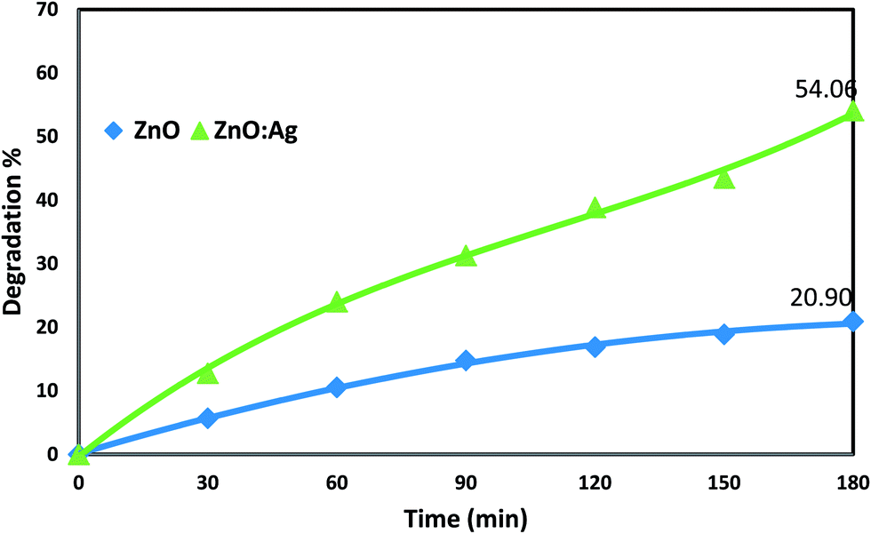 | ||
| Fig. 6 Effect of Ag addition to ZnO thin film. (2-CP Conc. = 10 mg L−1; Visible lamp = 112 W; Irradiation time = 3 h, pH = 6.2). | ||
Ag/ZnO thin films deposited at different substrate temperatures (25, 50 and 100 °C) were tested to evaluate the effect of substrate temperature on the photocatalytic efficiency. As shown in Fig. 7, at the initial 2-CP concentration of 25 mg L−1, the photocatalytic degradation efficiency increased from 30 to 43.3%with increase in the substrate temperature from 25 to 100 °C after 3 h. The increase in the crystallinity of Ag/ZnO nanoparticles and the change in morphology of Ag at higher substrate temperature may be collectively responsible for the narrowing of energy band gap from 3.1 to 2.7 eV (Fig. 5) which was evidenced as increased photocatalytic efficiency. For further experiments Ag/ZnO thin films deposited at substrate temperature 100 °C were used as photocatalyst.
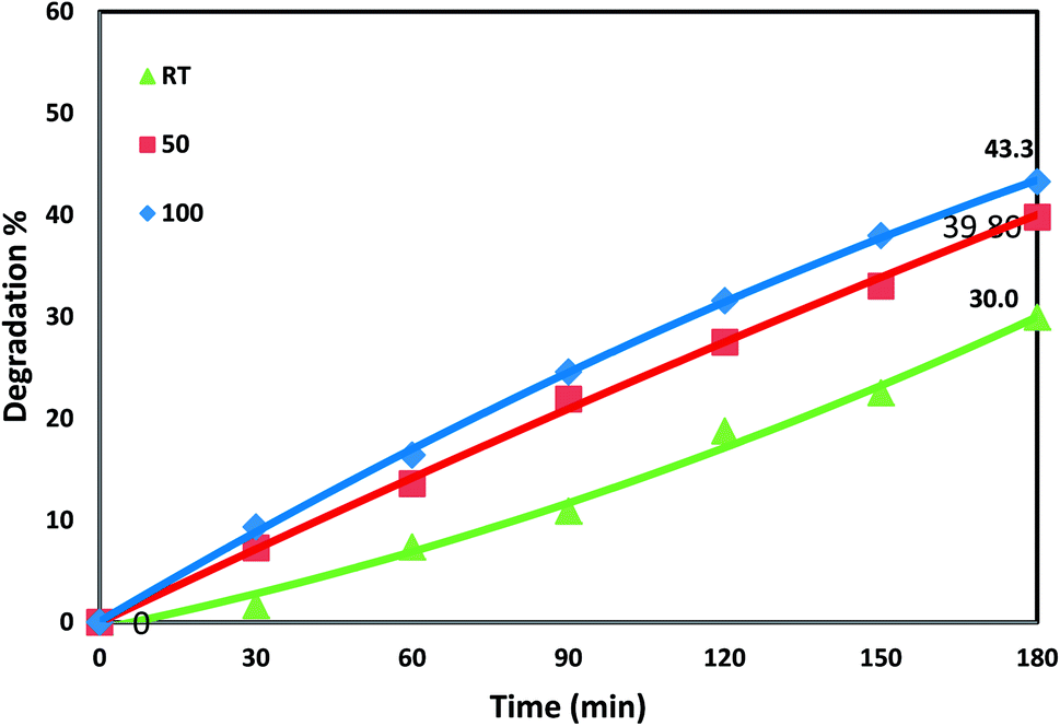 | ||
| Fig. 7 Effect of substrate temperature on photocatalytic efficiency. (2-CP Conc. = 25 mg L−1; Visible lamp = 54 W; Irradiation time = 3 h, pH = 6.2). | ||
To evaluate the impact of initial pollutant concentration on the rate of photocatalytic degradation, initial 2-CP concentration range of 5 to 50 mg L−1 was investigated. As Shown in Fig. 8 the photocatalytic efficiency of the thin film decreased with an increase in the 2-CP concentration. However, complete degradation of 2-Cp was not achieved within 3 h. Once the active sites on the surface of the thin films become completely occupied any further increase in pollutant molecules per unit volume appears to slow down the photocatalytic degradation rate. Also increase in number of pollutant molecules may prolong the photon path length resulting in decrease in photocatalytic efficiency.53
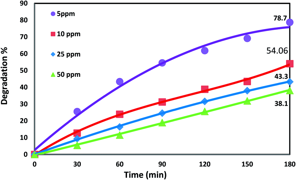 | ||
| Fig. 8 Pollutant concentration effect on photocatalysis. (Visible lamp = 112 W; Irradiation time = 3 h, solution pH = 6.2). | ||
The stability and reusability of thin films was assessed by subjecting the thin film deposited at 100 °C to three complete photocatalytic cycles under optimized conditions (Fig. 9). The obtained results indicate that the thin films are very stable in aqueous media and retain high photocatalytic activity upon subsequent reuse and showed only 8.7% decrease in efficiency after 4th use.
 | ||
| Fig. 9 Effect of catalyst reuse. (2-CP Conc. = 10 ppm, Visible lamp = 54 W; Irradiation time = 3 h, solution pH = 6.2). | ||
Photocatalysis of organic pollutants in aqueous medium is governed by pseudo-first-order reaction kinetics. The kinetic equation for such reactions can be represented by the following relation:
| ln(Co/Ct) = kapp × t | (3) |
| Catalyst Sample (Fabrication temp. °C) | kaap (min−1) | R2 |
|---|---|---|
| 25 | 0.0022 | 0.984 |
| 50 | 0.0029 | 0.9968 |
| 100 | 0.0032 | 0.9992 |
Conclusion
Ag/ZnO nanoparticles thin films were prepared by radiofrequency (RF) co-sputtering technique. The detailed investigations of the structural properties and the interaction of Ag with ZnO nanoparticles were performed by using XRD, SEM, EDS, scanning XPS probing, and UV-visible-NIR spectrometry. The SEM images of the thin films deposited at various temperatures showed that the Ag nanoparticles have variable morphology and exist as randomly anchored clusters within the ZnO matrix. The synthesized thin films showed enhanced visible light photocatalytic activity towards degradation of 2-CP compared to pure ZnO which can be attributed to the decreased band gap of Ag/ZnO and electron scavenging property of surface Ag. The synthesized thin films showed increased efficiency with elevated substrate temperature and high stability after multiple uses. Therefore, Ag/ZnO co-sputtered thin films can be considered a good candidate for photocatalytic applications for wastewater treatment.Acknowledgements
The authors are grateful for the financial support from King Abdulaziz City for Science and Technology (KACST), Saudi Arabia.References
- M. Caglar, S. Ilican, Y. Caglar and F. Yakuphanoglu, Appl. Surf. Sci., 2009, 255, 4491–4496 CrossRef CAS PubMed.
- Y. Li, W. Xie, X. Hu, G. Shen, X. Zhou, Y. Xiang, X. Zhao and P. Fang, Langmuir, 2010, 26, 591–597 CrossRef CAS PubMed.
- Y. H. Jang, S. T. Kochuveedu, M. Cha, Y. J. Jang, J. Y. Lee, J. Lee, J. Lee, J. Kim, D. Y. Ryu and D. H. Kim, J. Colloid Interface Sci., 2010, 345, 125–130 CrossRef CAS PubMed.
- A. M. Ali, E. A. C. Emanuelsson and D. A. Patterson, Appl. Catal., B, 2010, 97, 168–181 CrossRef CAS PubMed.
- I. Udom, K. Manoj, E. Stefanakos, A. F. Hepp and D. Y. Goswami, Mater. Sci. Semicond. Process., 2013, 16, 2070–2083 CrossRef CAS PubMed.
- N. Daneshvar, D. Salari and A. R. Khataee, J. Photochem. Photobiol., A, 2004, 162, 317–322 CrossRef CAS.
- S. Song, C. Wang, F. Hong, Z. He, Q. Cai and J. Chen, Appl. Surf. Sci., 2011, 257, 3427–3432 CrossRef CAS PubMed.
- S. Malato, P. Fernandez-Ibanez, M. I. Maldonado, J. Blanco and W. Gernjak, Catal. Today, 2009, 147, 1–59 CrossRef CAS PubMed.
- Y. Matsumura and H. N. Ananthaswamy, Toxicol. Appl. Pharmacol., 2004, 195, 298–308 CrossRef CAS PubMed.
- L. R. Zheng, Y. H. Zheng, C. Q. Chen, Y. Y. Zhan, X. Y. Lin, Q. Zheng, K. M. Wei and J. F. Zhu, Inorg. Chem., 2009, 48, 1819–1825 CrossRef CAS PubMed.
- C. Wang, X. M. Wang, B. Q. Xu, J. C. Zhao, B. X. Mai, P. A. Peng, G. Y. Sheng and J. M. Fu, J. Photochem. Photobiol., A, 2004, 168, 47–52 CrossRef CAS PubMed.
- S. Gao, X. Jia, S. Yang, Z. Li and K. Jiang, J. Solid State Chem., 2011, 184, 764–769 CrossRef CAS PubMed.
- B. Subash, B. Krishnakumar, B. Sreedhar, M. Swaminathan and M. Shanthi, Superlattices Microstruct., 2013, 54, 155–171 CrossRef CAS PubMed.
- T. Jia, W. Wang, F. Long, Z. Fu, H. Wang and Q. Zhang, J. Alloys Compd., 2009, 484, 410–415 CrossRef CAS PubMed.
- Y. Zheng, C. Chen, Y. Zhan, X. Lin, Q. Zheng, K. Wei and J. Zhu, J. Phys. Chem. C, 2008, 112, 10773–10777 CAS.
- P. Christopher, H. Xin and S. Linic, Nat. Chem., 2011, 3, 1–6 CrossRef PubMed.
- W. Hou, Z. Liu, P. Pavaskar, W. H. Hung and S. B. Cronin, J. Catal., 2011, 277, 149–153 CrossRef CAS PubMed.
- S. Bethke, H. Pan and B. W. Wessels, Appl. Phys. Lett., 1988, 52, 138–140 CrossRef CAS PubMed.
- B. Guo, Z. R. Qiu and K. S. Wong, Appl. Phys. Lett., 2003, 82, 2290–2292 CrossRef CAS PubMed.
- F. K. Shan, B. I. Kim, G. X. Liu, Z. F. Liu, J. Y. Sohn, W. J. Lee, B. C. Shin and Y. S. Yu, J. Appl. Phys., 2004, 95, 4772–4776 CrossRef CAS PubMed.
- H. T. Ng, B. Chen, J. Li, J. Han and M. Meyyappan, Appl. Phys. Lett., 2003, 82, 2023–2025 CrossRef CAS PubMed.
- A. Hongsingthong, I. A. Yunaz, S. Miyajima and M. Konagai, Sol. Energy Mater. Sol. Cells, 2011, 95, 171–174 CrossRef CAS PubMed.
- N. Ekem, S. Korkmaz, S. Pat, M. Z. Balbag, E. N. Cetin and M. Ozmumcu, Int. J. Hydrogen Energy, 2009, 34, 5218–5222 CrossRef CAS PubMed.
- S. Lemlikchi, S. Abdelli-Messaci, S. Lafane, T. Kerdja, A. Guittoum and M. Saad, Appl. Surf. Sci., 2010, 256, 5650–5655 CrossRef CAS PubMed.
- B. Zhang, B. Yao, S. Wang, Y. Li, C. Shan, J. Zhang, B. Li, Z. Zhang and D. Shen, J. Alloys Compd., 2010, 503, 155–158 CrossRef CAS PubMed.
- T. J. Coutts, D. L. Young and X. Li, MRS Bull., 2000, 25, 58–65 CrossRef CAS.
- K. Liu, B. Yang, H. Yan, Z. Fu, M. Wen, Y. Chen and J. Zuo, J. Lumin., 2009, 129, 969–972 CrossRef CAS PubMed.
- X. B. Wang, C. Song, K. W. Geng, F. Zeng and F. Pan, J. Phys. D: Appl. Phys., 2006, 39, 4992–4996 CrossRef CAS.
- R. Wang, J. H. Xin, Y. Yang, H. Liu, L. Xu and J. Hu, Appl. Surf. Sci., 2004, 227, 312–317 CrossRef CAS PubMed.
- D. R. Sahu, Microelectron. J., 2007, 38, 1252–1256 CrossRef CAS PubMed.
- P. M. R. Kumar, K. P. S. Kartha and V. Kumar, J. Appl. Phys., 2005, 98, 023509 CrossRef PubMed.
- C. E. Benouis, M. Benhaliliba, A. Sanchez Juarez, M. S. Aida, F. Chami and F. Yakuphanoglu, J. Alloys Compd., 2010, 490, 62–67 CrossRef CAS PubMed.
- M. N. Islam, T. B. Ghosh, K. L. Chopra and H. N. Acharya, Thin Solid Films, 1996, 280, 20–25 CrossRef CAS.
- S. Major, S. Kumar, M. Bhatnagar and K. L. Chopra, Appl. Phys. Lett., 1986, 40, 394–396 CrossRef PubMed.
- Y. Chen, X. L. Xu, G. H. Zhang, H. Xue and S. Y. Ma, Phys. B, 2009, 404, 3645–3649 CrossRef CAS PubMed.
- J. Xu, X. Han, H. Liu and Y. Hu, Colloids Surf., A, 2006, 273, 179–183 CrossRef CAS PubMed.
- J. Xu, Y. Chang, Y. Zhang, S. Ma, Y. Qu and C. Xu, Appl. Surf. Sci., 2008, 255, 1996–1999 CrossRef CAS PubMed.
- J. F. Moulder, W. F. Stickle, P. E. Sobol and K. D. Bomben, Handbook of X-ray Photoelectron Spectroscopy, Perkin-Elmer Corporation Physical Electronics Division, 2nd edn, 1992 Search PubMed.
- J. F. Weaver and G. B. Hoflund, J. Phys. Chem., 1994, 98, 8519–8524 CrossRef CAS.
- K. Kowal, K. W. Król, M. Kopaczynska, E. Dworniczek, R. Franiczek, M. Wawrzynska, M. Vargova, M. Zahoran, E. Rakovsky, P. Kus, G. Plesch, A. Plecenik, F. Laffir, S. A. M. Tofail and H. Podbielska, J. Colloid Interface Sci., 2011, 362, 50–57 CrossRef CAS PubMed.
- J. C. Li, Q. Cao and X. Y. Hou, J. Appl. Phys., 2013, 113, 203518 CrossRef PubMed.
- D. Y. Wang, J. Zhou and G. Z. Liu, J. Alloys Compd., 2009, 481, 802–805 CrossRef CAS PubMed.
- L. X. Kun, L. Q. Shan, L. D. Chun, X. Y. Dong and X. X. Jun, Sci. China, Ser. E: Technol. Sci., 2009, 52, 2779–2784 CrossRef PubMed.
- F. Albano, Y. S. Lin, D. Blaauw, D. M. Sylvester, K. D. Wise and A. M. Sastry, J. Power Sources, 2008, 185, 1524–1532 CrossRef CAS PubMed.
- S. H. Jeong, B. N. Park, S. B. Lee and J. H. Boo, Surf. Coat. Technol., 2007, 201, 5318–5322 CrossRef CAS PubMed.
- J. Tauc, R. Grigorvici and A. Vancu, Phys. Status Solidi B, 1996, 15, 627–637 CrossRef.
- K. P. Liu, B. F. Yang, H. W. Yan, Z. P. Fu, M. W. Wen, Y. J. Chen and J. Zuo, Appl. Surf. Sci., 2008, 255, 2052 CrossRef CAS PubMed.
- D. R. Sahu, S. Y. Lin and J. L. Huang, Appl. Surf. Sci., 2007, 253, 4886–4890 CrossRef CAS PubMed.
- C. Gu and C. Shannon, J. Mol. Catal. A: Chem., 2007, 262, 185–189 CrossRef CAS PubMed.
- M. Ahmad, E. Ahmed, Z. L. Hong, N. R. Khalid, W. Ahmed and A. Elhissi, J. Alloys Compd., 2013, 577, 717–727 CrossRef CAS PubMed.
- Y. H. Zheng, C. Q. Chen, Y. Y. Zhan, X. Y. Lin, Q. Zheng, K. M. Wei and J. F. Zhu, J. Phys. Chem. C, 2008, 112, 10773–10777 CAS.
- C. S. Hong, H. H. Park, J. Moon and H. H. Park, Thin Solid Films, 2006, 515, 957–960 CrossRef CAS PubMed.
- J. C. Sin, S. M. Lam, A. R. Mohamed and K. T. Lee, Int. J. Photoenergy, 2012, 2012, 1–23 CrossRef.
| This journal is © The Royal Society of Chemistry 2014 |