DOI:
10.1039/C4RA08258C
(Paper)
RSC Adv., 2014,
4, 56883-56891
Preparation and characterization of TiO2–Graphene@Fe3O4 magnetic composite and its application in the removal of trace amounts of microcystin-LR†
Received
7th August 2014
, Accepted 14th October 2014
First published on 14th October 2014
Abstract
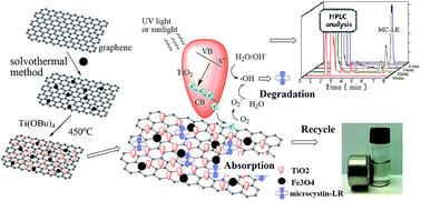
Microcystins (MCs), a family of potent cyclic heptapeptides, are produced by cyanobacteria blooms in eutrophic water and can cause acute and chronic toxicity and even mortality to animals and humans. Previous MC removal strategies concerned only highly contaminated water, in which the concentration of the pollutant was considerably larger than that in the natural world. Herein, we developed a ternary composite of TiO2-coated magnetic graphene and used it as an adsorbent and photocatalyst to efficiently remove microcystin-LR (MC-LR) from water. The two-dimensional sheets of graphene were decorated with a large quantity of spherical Fe3O4 nanoparticles (10–20 nm) and then coated with crystallized TiO2. These TiO2–graphene@Fe3O4 composites exhibited a high magnetic response to the external magnetic field. And the huge surface of the graphene dramatically boosted the adsorbability and charge mobility, which lowered the recombination rate of electron–hole pairs, and hence systematically enhanced photocatalytic activity. The combination of adsorption and photodegradation endowed the composite with a better performance in the removal of trace amounts of MC-LR than the commercial photocatalyst, Degussa P25. The concentration of MC-LR can be lowered to less than 1 μg L−1 (a provisional safety guideline by the World Health Organization) from 500 μg L−1 under UV light in 30 min. The loading of TiO2–graphene@Fe3O4, the pH, and the UV energy were also optimized. Moreover, the stable removal capability of TiO2–graphene@Fe3O4 was confirmed over multiple cycles. Finally, the removal performance was also evaluated under natural light illumination in real surface water samples. This work paves the way for the development of more efficient and easily separable purifiers for the removal of pollutants and toxins from contaminated water.
1. Introduction
With the intensification of anthropogenic activities, especially untreated industrial sewage and agricultural runoff, eutrophication has become a major threat to global aquatic ecosystems.1 One of many negative consequences is rapid increase in the occurrence of cyanobacteria blooms, which can potentially produce harmful substances, such as cyanotoxins, with adverse health effects on living biota.2,3 Microcystins (MCs), a family of potent cyclic heptapeptides, are the most common cyanotoxins in eutrophic water, particularly their derivatives known as microcystin-LR (MC-LR).4 These toxins can damage the nervous system or liver and thus cause acute and chronic toxicity or even mortality to humans and animals who come into contact with MC-contaminated water.5 Because of the potential toxicity of MC-LR, the World Health Organization (WHO) has proposed a provisional safety guideline of 1 μg L−1 in drinking water.6 Various methods have been used to remove MCs, but conventional water treatment processes (e.g., coagulation, flocculation and filtration) have proven to be unreliable or less effective for eliminating these toxins due to their chemical stability and resistance to a range of pHs.7,8 Advanced treatment processes, including ultra- and nano-filtration9 activated carbon adsorption,10 pre-ozonation, photo-irradiation, and chemical processes that use oxidising agents such as ozone,11 potassium permanganate,12 and chlorine,13 can successfully remove MCs, but they are either not cost-effective or have a risk of negative side effects.14
Advanced oxidation processes (AOPs) comprise a reasonable alternative for removing many organic contaminants from water and air,15 especially the heterogeneous photocatalysis degradation mediated by titanium dioxide (TiO2).16–20 Anatase TiO2 has attracted significant attention owing to its high efficiency, non-toxicity, low cost, physical and chemistry stability, and environmentally “green” characteristics.21–24 Generally speaking, anatase TiO2 generates electron–hole pairs when exposed to ultraviolet (UV) irradiation, and then the positive holes and the electrons react with adsorbed H2O and O2 to form hydroxyl radicals(˙OH) and superoxide radicals(˙O2), which have highly reactive activities, resulting in degradation of most organic compounds, including MCs25–27 (Scheme 1). However, the application of pure TiO2 is limited, owing to its low quantum efficiency,28 poor adsorptive power,29 narrow light absorbance range30 and difficult separation from water.31
 |
| | Scheme 1 The synthesis route of TiO2–graphene@Fe3O4 composite. | |
In the last decade, various strategies have been developed to suppress the recombination of photogenerated electron–hole pairs in TiO2. In organic pollutant degradation using TiO2 as the photocatalyst, the substrate can be adsorbed on the inert support for TiO2 loading, and the photodecomposition rate of TiO2 is affected by the adsorption capability of the support;32 and some toxins or dyes were also absorbed rather than decomposed.33–36 The difference between adsorption sites and photoactive sites on the TiO2 surface and the importance of surface migration of substrates has been illustrated. Graphene, due to its atomic sheet of sp2-bonded carbon atoms arranged in a honeycomb crystal lattice,37 has several excellent attributes, such as large theoretical specific surface area,38 high transparency,39 unique electrical conductivity, high mechanical flexibility, and – especially – its adsorption activity.40,41 In particular, graphene oxide (GO), as a novel cousin of graphene, has oxygen-containing groups that enable GO to exhibit excellent hydrophilic properties and rich intercalation chemistry,42,43 making it a highly functional substrate with abundant anchoring sites for efficiently binding with TiO2 photosensitizers.44 The graphene-based TiO2 photocatalysts exhibit systematically enhanced photocatalytic activity: photogenerated electrons are scavenged and subsequently transported by GO after excitation in the conduction band of TiO2 by UV illumination,45 generating a greater density of electron–hole pairs for high quantum efficiency.46 Furthermore, the huge surface area of graphene dramatically boosts adsorbability, and hence systematically enhances photocatalytic activity.47 Recent reports indicated that hybridizing TiO2 nanoparticles with graphene showed a higher photocatalytic activity than Degussa P25 or pure TiO2.5,46–53
Magnetic graphene, the graphene decorated with magnetic iron oxide nanoparticles (NPs), has a desirable rapid magnetic response54 and has been widely applied in the biomedical field, including uses in bioseparation, medical diagnosis, and magnetically targeted drug delivery, as well as in magnetic energy storage, fluids and catalysis.55 However, up to now, no report has concentrated on applications in the photocatalysis field, although it will potentially improve the separation, recovery and reuse of the photocatalyst, and optimize photodegration.56–59
We developed a ternary composite of TiO2-coated magnetic graphene (TiO2–graphene@Fe3O4) and used it as an efficient and recyclable agent to remove trace amounts of hazardous compound microcystin-LR from aqueous solutions. Due to its adsorptive and photocatalytic properties, TiO2–graphene@Fe3O4 was found to have superior removal performance in comparison with commercial Degussa P25. Moreover, the composite's removal performance was also effective in multiple recycling. It is expected that the proposed magnetic photocatalyst will pave the way for the development of more efficient and easily separable agents to deal with pollutants and toxins in contaminated water.
2. Experimental
2.1 Materials
All reagents were at least of analytical grade and used without further purification. Graphene oxide (GO) was provided by Nanjing XFNANO Materials Tech Co Ltd, China. Tetrabutyl titanate (TBOT) was purchased from Heowns Business License Co Ltd. Iron(III) chloride hexahydrate (FeCl3·6H2O), sodium acetate, ethylene glycol, ethylene diamine, and anhydrous ethanol were all purchased from Tianjin Chemical Reagent Company (Tianjin, China). Degussa P25 (Degussa AG, Germany) was used as a standard material for comparison purposes. Microcystin-LR (≥95% by HPLC) was purchased from Enzo Life Science Co Ltd and stored at −20 °C. Solutions with an initial concentration of 50 μg L−1 were prepared in methanol and employed in all experiments. Trifluoroacetic acid (TFA, 99.5+%, HPLC grade) was purchased from Alfa Aesar. HPLC-grade methanol and acetonitrile (JT Baker) were used in chromatographic analyses. Highly purified water was prepared with a Milli-Q water purification system (Millipore, Milford, MA).
2.2 Preparation of graphene@Fe3O4 composite by solvothermal reaction
The magnetic graphene was prepared according to a modified solvothermal reaction.55 In a typical procedure, graphene oxide (50 mg) was dissolved into ethylene glycol (25 mL), followed by addition of FeCl3·6H2O (0.5 g), NaAc (1.5 g, as a protective agent) and ethylene diamine (5 mL) to form a homogeneous solution under ultrasonic dispersion. The mixture was transferred to a Teflon-lined stainless steel autoclave for solvothermal reaction at 200 °C for 16 h. After cool-down to room temperature, the black product was collected by a magnet and washed three times with highly purified water and three times with ethanol, and oven-dried at 50 °C for further use.
2.3 Preparation of functionalized TiO2–graphene@Fe3O4 composite
The TiO2–graphene@Fe3O4 composite was synthesized by directly coating the TiO2 layer on the surface of graphene@Fe3O4.60 Briefly, the as-prepared magnetic graphene (50 mg) was dispersed in alcohol/water (v/v, 140 mL/10 mL) mixture under ultrasonic for 10 min, and heated to 70 °C. Ti(BuO)4 (3 mL) and H2SO4 (1 mL) were then added and the solution was mechanically stirred for 12 h at the same temperature. The product, amorphous TiO2–graphene@Fe3O4, was washed three times with ethanol and three times with water and finally recovered by magnetic separation. Finally, the dried particles were calcined at 450 °C for 2 h in air atmosphere to improve the crystallinity of TiO2.
2.4 MC-LR removal experiments
MC-LR removal experiments were performed in a glass vessel reactor containing 20.0 mL MC-LR solution with an initial concentration of 500.0 μg L−1. The pH of the solution was adjusted to 6.0 with TFA; a known concentration (0.50 g L−1) of TiO2–graphene@Fe3O4 and commercial Degussa P25 were dispersed under ultrasonication for 1 min, respectively. For comparison, the same mixture was stirred in the dark. The photodegradation was induced by a 125 W mercury vapour lamp positioned 15 cm from the solution (under constant stirring) in a dark box. A series of aqueous solutions (0.50 mL) were sampled using a magnet to remove the suspended catalyst particles at specific time intervals.
2.5 HPLC analytical procedures
The concentration of MC-LR was measured by high-performance liquid chromatography (HPLC). The obtained samples were analyzed on an Agilent Series 1100 system equipped with a photodiode array detector (PDA) set at 238 nm. The HPLC was carried out under the reverse-phase condition,19,48 and a C18 column (4.6 mm × 150 mm, 5 μm particle size) was used. The mobile phase in isocratic method, a mixture of 0.05% (v/v) TFA in acetonitrile solution and 0.05% (v/v) TFA in MilliQ water at 40
:
60 ratio, was used in the analysis at a flow rate of 1 mL min−1. The injection volume was 50 μL and the temperature of the column was set at 40 °C. Under these conditions, the MC-LR eluted at about 6.7 min. The concentration change of MC-LR follows Lambert–Beer's law (Fig. S1; see ESI†).
2.6 Characterization
The morphology and structure of the synthesized magnetic composites were evaluated using a Tecnai G2T2 S-TWIN transmission electron microscope (TEM). Samples for TEM were prepared by placing a drop of dilute nanoparticles in the ethanol solvent on a copper grid. The infrared spectra were recorded on a Nicolet AVATAR-360 Fourier transform infrared (FTIR) spectrometer. After vacuum drying, the samples were thoroughly mixed with KBr (the weight ratio of sample/KBr was 1%) in a mortar, and then the fine powder was pressed into a pellet. Raman spectra were measured by an FT-Raman microspectrometer (Bruker RFS100/S, Germany) with a 1046 nm laser with the laser power at 40 mW. Identification of the crystalline phase was performed on a Rigaku D/max/2500v/pc (Japan) X-ray diffractometer with a Cu-Ka source. The 2θ angles probed were from 3° to 80° at a rate of 4° min−1. The magnetic properties were analyzed with a vibrating sample magnetometer (VSM) (LDJ 9600-1, USA). The X-ray photoelectron spectra were obtained on a Shimadzu (Japan) Kratos AXIS Ultra DLD X-ray photoelectron spectrometer (XPS) with Mg-Ka anode (15 kV, 400 W) at a takeoff angle of 45°. The source X-rays were not filtered and the instrument was calibrated against the C1s band at 285 eV.
3. Result and discussion
3.1 Preparation and characterization of TiO2–graphene@Fe3O4 composite
The synthesis route to prepare TiO2–graphene@Fe3O4 composite is illustrated in Scheme 1. First, the graphene@Fe3O4 composites were prepared via a one-step hydrothermal method as described above. The TEM image of graphene@Fe3O4 (Fig. 1a) showed that two-dimensional graphene oxide (GO) sheets were decorated with a large quantity of spherical Fe3O4 NPs. The Fe3O4 NPs have an average diameter of approximately 10–20 nm and are well distributed on the surface of GO without serious aggregation. The outline of GO and Fe3O4 nanoparticles can be clearly observed. Then the TiO2 layer was directly coated on the surface of graphene@Fe3O4 and the synthesized TiO2–graphene@Fe3O4 were calcined at 450 °C for 2 h to improve TiO2 crystallinity. The TEM image of TiO2–graphene@Fe3O4 composites (Fig. 1b) clearly showed that TiO2 crystals were successfully coated on the surface of graphene@Fe3O4, and due to the good chemical and physical stability of graphene, the product maintained a flake-like structure. Futhermore, compared with Fig. 1a and b shows that the graphene became thicker, and the diameter of Fe3O4 inserted in the composite increased from 20 nm to 50 nm, indicating that TiO2 had been coated on the graphene@Fe3O4.
 |
| | Fig. 1 TEM images of the graphene@Fe3O4 (a) and the TiO2–graphene@Fe3O4 composite after calcination at 450 °C for 2 h (b). HRTEM images of TiO2–graphene@Fe3O4 (c and d). | |
To evaluate the crystalline phase of TiO2–graphene@Fe3O4, the HRTEM micrograph with the lattice fringe information is displayed in Fig. 1c and d. Fig. 1c shows a well-defined crystallinity of TiO2 with lattice spacings of 0.352 nm and 0.471 nm, which matched well with the (101) and (001) crystallographic planes of anatase TiO2. In addition, the crystal lattice fringe with a spacing of 0.253 nm (Fig. 1d) can be assigned to the (311) plane of Fe3O4. These TEM and HRTEM images were consistent with the XRD results below and offered sufficient evidence that Fe3O4 and anatase TiO2 NPs were successfully coated on the GO.
The XRD patterns of the TiO2–graphene@Fe3O4 annealed at 450 °C for 2 h (c) in an air atmosphere, as well as an unannealed sample (b) and uncoated graphene@Fe3O4 (a), are shown in Fig. 2. It was observed in each sample that there are six characteristic peaks for Fe3O4 (2θ = 30.2°, 35.5°, 43.1°, 53.5°, 57.1°, and 62.7°), and the peak positions at the corresponding 2θ values were indexed as (220), (311), (400), (422), (511), and (440), respectively, which matched well with the database of Fe3O4 face center-cubic phases in the JCPDS-International Center for Diffraction Data (JCPDS Card 19-629) file. The peak intensities of Fe3O4 in Fig. 2b have partially decreased due to the absorption of X-rays through the TiO2 shell, indicating amorphous forms of TiO2. After annealing at 450 °C, anatase TiO2 (2θ = 25.3°) is dominant, which is the main crystal face (101) of anatase, with other diffraction peaks at 37.8°, 48.1°, 54.7° and 68.8°. These diffraction peaks (marked with #) were readily indexed to typical anatase TiO2 crystals (JCPDS no. 21-1272), and the Miller indexes of these diffraction peaks were calculated to be (004), (200), (211) and (116), respectively; the other diffraction peaks corresponding to the Fe3O4 remained almost the same as those without treatment (Fig. 2a). This means that the graphene@TiO2 seems to be quite well crystallized, and only TiO2 in the anatase phase is identified, with the interface structure between anatase and magnetite remaining thermodynamically stable.
 |
| | Fig. 2 The XRD pattern of uncoated graphene@Fe3O4 (a), unannealed TiO2–graphene@Fe3O4 (b), and TiO2–graphene@Fe3O4 annealed at 450 °C for 2 h in air atmosphere (c). | |
FTIR spectra of graphene@Fe3O4 (a) and TiO2–graphene@Fe3O4 (b) in the range of 4000–450 cm−1 were recorded. As exemplified in Fig. 3a, the band at 1187.5 cm−1, 1083 cm−1 and 877.7 cm−1 was due to C–O stretching of the epoxy structures and –COOH in GO, while the bands of 1557.5 cm−1 were assigned to C
C stretching vibrations of graphene. The peak at 579.3 cm−1 corresponds to the Fe–O bond61 stretching vibration on the graphene@Fe3O4. After introducing TiO2 to graphene@Fe3O4 (Fig. 3b), the intense peaks at low frequency (about 572.6 cm−1) were assigned to the combined effects of Ti–O–C and Ti–O–Ti vibrations, confirming the impregnation of TiO2 into GO,62 while the peaks at around 2337 cm−1 may originate from dioxide in the air.63 Compared with Fig. 3a, the peaks at 877.7 cm−1, 1083.0 cm−1 and 1187.5 cm−1 disappeared, implying that the –COOH and epoxy structures had been replaced by TiO2. Moreover, the peak at 3422.7 cm−1 was ascribed to the O–H stretching frequency from the surface hydroxyl group, and the peak at 1629.8 cm−1 was attributed to the skeletal vibration of GO sheets, indicative of the GO structure remaining during calcination treatment.
 |
| | Fig. 3 FTIR spectra of GO@Fe3O4 (a) and TiO2–graphene@Fe3O4 (b) composite. | |
The FT-Raman spectra of the Fe3O4 NPs, graphene oxide, reduced graphene and graphene@Fe3O4 composite were displayed in Fig. S2 and S3 (see ESI†). The weak signal of Fe3O4 can hardly be detected, but the variation of location and intensity ratio of D band and G band on the Raman spectra demonstrated the inset of Fe3O4 on the surface of graphene. The Raman spectra of the as-prepared TiO2–graphene@Fe3O4 composites was shown in Fig. 4. A well-resolved TiO2 Raman peak, clearly seen at about 145.68 cm−1, was attributed to the main Eg vibration mode of anatase TiO2; with other vibration peaks at 397.93 cm−1 (B1g), 516.89 cm−1 (A1g), and 638.62 cm−1 (Eg), it verified the existence of anatase TiO2 coated on the composite.64 The result obtained from the Raman spectra was consistent with XRD results shown in Fig. 2b. Additionally, two specific peaks—1336.84 cm−1 (D band) and 1593.40 cm−1 (G band)—were observed in the spectra (see inset in Fig. 4), which proved the existence of the graphene substrate in both samples. Compared with that of graphene@Fe3O4, the marked increase of the D/G intensity ratio for the TiO2–graphene@Fe3O4 composite indicated the growth of anatase crystallites.65
 |
| | Fig. 4 FT-Raman spectra of TiO2–graphene@Fe3O4 composite (a) and graphene@Fe3O4 (b). The inset shows amplification from 1800 to 1200 cm−1. | |
XPS was employed to ascertain the composition of the as-prepared TiO2 immobilized magnetic composites. Wide-scan XPS spectra (Fig. 5a) showed the existence of element C, O and Ti appearing on the surface of TiO2–graphene@Fe3O4 composite. The binding energies of Ti 2p3/2 and O 1 s were 456.8 and 531.7 eV, respectively, which are identical to those for pure TiO2. The weak intensity of the binding energy at 710.20 eV for Fe 2p provided further evidence that all Fe3O4 nanoparticles in the composite were covered with a shell of titanium oxide. This result was in agreement with the above TEM and XRD analyses. The two bands centered at binding energies of 462.7 and 456.8 eV in the composite were attributed to the Ti(2p1/2) and Ti(2p3/2) spin–orbital splitting photoelectrons in Ti(IV), respectively (Fig. 5b). However, the energy bonds of the Ti(IV) were perturbed by the presence of graphene@Fe3O4 (shift to low binding energy), which also suggested the substitutions of the Ti–O–Ti by Ti–O–C bonds in the titania lattice.66 The C1s spectra can be deconvoluted into four individual component peaks, which come from different groups and overlap with each other. The C1s core-level spectrum of TiO2–graphene@Fe3O4 composites was curved into four peak components located at 282.95, 286.65, 284.55, and 281.85 eV (Fig. 5c), which were assigned to the C
C in graphene, C–O, sp3 hybridized C–C,67,68 and C–Ti, respectively. It was noted that the deconvoluted peak at 281.85 eV assigned to the Ti–C bond and the characteristic peak at 286.65 eV attributed to the Ti–O–C bond were consistent with Ti 2p spectra in Fig. 5b, indicating the TiO2 anchored and extended on the surface of graphene sheets.69 All the results demonstrated the existence of both TiO2 and magnetic graphene in the composite and the intense interaction between the two components.
 |
| | Fig. 5 XPS wide-scan spectra (a), high-resolution Ti (b) and C1s core-level spectra (c) of TiO2–graphene@Fe3O4 composites. | |
The magnetic hysteresis loops of the as-synthesized graphene@Fe3O4 and annealed TiO2–graphene@Fe3O4 are displayed in Fig. 6. The saturation magnetization (Ms) values of the graphene@Fe3O4 and annealed TiO2–graphene@Fe3O4 were 53.7 and 17.9 emu g−1, respectively. The Ms of the graphene@Fe3O4 nanoparticles was significantly higher than that of TiO2-modified magnetite composite, which was attributed to introduction of the anatase TiO2 layer, which has a very small magnetic susceptibility. However, it is important that the remaining magnetism of the photocatalyst was good enough to easily recover the composites from a suspension by applying an external magnetic field, as shown in Fig. 6b. Therefore, the material can be almost completely recovered with minimum loss through many cycles without diminishing magnetic and photoactivity properties. These properties make the material highly desirable for the treatment of contaminated water.
 |
| | Fig. 6 Magnetic hysteresis loops of graphene@Fe3O4 (a) and annealed TiO2–graphene@Fe3O4 (b). Inset: magnetic separation–redispersion of TiO2–graphene@Fe3O4 composite. | |
3.2 Removal of MC-LR compounds by TiO2–graphene@Fe3O4
The performance of TiO2–graphene@Fe3O4 composites for removing toxins was measured with MC-LR as a model reaction under UV light irradiation; results are shown in Fig. 7. A control experiment in the absence of the catalyst was conducted under weakly acidic conditions (pH = 6) to indicate that the contribution of UV photolysis was negligible, since no degradation was observed. It was clear that TiO2–graphene@Fe3O4 under UV (black dots) exhibited a more effective performance in removing MC-LR than the commercial Degussa P25 (white squares). In all cases, after 30 min UV irradiation, the concentration of MC-LR in the solution with TiO2–graphene@Fe3O4 was reduced to zero (for representative HPLC chromatograms, see Fig. S4†), while 50% MC-LR still remained in the solution with Degussa P25. Experiments performed in the dark (black squares) showed that without photodegradation, TiO2–graphene@Fe3O4 adsorbed about 60% toxins and came to equilibrium within 30 min. These results indicated that the combination of adsorption and photodegradation of TiO2–graphene@Fe3O4 is effective to fully remove trace amounts of MC-LR.
 |
| | Fig. 7 Removal of MC-LR under 125 W UV-A (λmax = 365 nm) irradiation in presence of different nanostructured materials, 0.5 g L−1. Initial MC-LR concentration: 500 μg L−1 (pH = 6). | |
The adsorption and photocatalytic activity of various concentrations of TiO2–graphene@Fe3O4 for MC-LR removal from aqueous solutions were investigated (Fig. 8). In the first 30 min, we compared the adsorption ability of every sample in the absence of light; and after that, with the UV light turned on, a great reduction in MC-LR concentration was observed during the next 40 min of irradiation time. As expected, changing the TiO2–graphene@Fe3O4 loading from 0.1 to 0.5 g L−1 led to a remarkable increase in both adsorption capacity (from 30% to 60%) and the final removal efficiency (from 65% to 100%) of the toxins. A further increase in the loading of ingredients to 1 g L−1 (see white dots) only raised adsorption loading, but showed nearly the same efficiency in MC-LR photocatalytic degradation, thus proving that 0.5 g L−1 was the most cost-effective concentration for removal.
 |
| | Fig. 8 Removal of MC-LR with different loadings of TiO2–graphene@Fe3O4 photocatalytic under 125 W UV light (λ = 365 nm). Initial MC-LR concentration: 500 μg L−1 (pH = 6). | |
As solution pH has an important effect on the characteristics of catalyst surface electric charges,70 experiments to investigate the adsorption and degradation of MC-LR solutions with pH values ranging from 3 to 11 were also carried out. The initial and final pH values were recorded and no change of pH was observed because of change in composition. At first, all mixtures were incubated in the dark for 30 min (to achieve adsorption equilibrium) for investigation of absorption capacity. From the adsorption data (blue column) shown in Fig. 9, maximum adsorption was observed at pH 6. When the pH of the solution was increased or reduced, the adsorption capacity of TiO2–graphene@Fe3O4 composite for MC-LR declined. Similar results were observed in previous studies of graphene adsorption, and the reason may be ascribed to the electrostatic repulsion of the composite and ionizable groups carried by MC-LR.71 After 30 min of UV irradiation, the MC-LR in solution of pH 6 was completely removed, while the concentration of MC-LR in other solutions with different pHs also decreased to varying degrees (see gray column in Fig. 9). These results demonstrated that a weakly acidic condition (pH = 6) was more favorable for treatment of MC-LR with the TiO2–graphene@Fe3O4.
 |
| | Fig. 9 Normalized concentration of MC-LR in solution with 0.5 g L−1 photocatalyst after 30 minutes of adsorption(blue column) and following 30 minutes of 125 W UV irradiation(gray column) with solution pH values ranging from 3 to 11. Initial MC-LR concentration: 500 μg L−1. | |
Three UV sources and natural sunlight have been studied for MC-LR degradation via the TiO2–graphene@Fe3O4 photocatalyst. As shown in Fig. 10, compared to the other two UV sources (15 W and 500 W), 125 W UV light seemed slightly better due to its high efficiency, cost-effectiveness and safety. In addition, sunlight was also an optimum choice to initiate the photodegradation in view of its environment-friendly property, but it was not quantitative and stable enough for experimental analysis. Thus, we chose 125 W UV light as the light source for subsequent study, and to use sunlight as a natural light source to treat the real-world sample for proofs of practical application.
 |
| | Fig. 10 Removal of MC-LR with 0.5 g L−1 TiO2–graphene@Fe3O4 photocatalyst under 15 W, 125 W, and 500 W UV light, and natural sunlight. Initial MC-LR concentration: 500 μg L−1 (pH = 6). | |
The reuse ability of the magnetic composites TiO2–graphene@Fe3O4 was subsequently investigated at the optimal loading. In each cycle, we recycled the composites with a magnet, and washed and dried them three times. Prior to every reuse, we weighed the recollected composites to maintain consistency, so that the result only pertained to removal performance of the reused magnetic composite. Results summarized in the recycling experiment (Fig. 11A) showed that the ultimate removal performance had no obvious change in spite of the fact that it took a relatively longer time to reduce the concentration to zero. This result demonstrated that the composites could be reused repeatedly and that they maintained removal performance over multiple cycles. To further prove the reuse of TiO2–graphene@Fe3O4, the XRD patterns of TiO2–graphene@Fe3O4 before and after five cycles of removal of MC-LR are shown in Fig. 11B. Peak positions were identical and only slight decline of peak intensities of anatase TiO2 was observed, which further demonstrated that the composites possessed reusable and sustainable removal performance during multiple removal processes.
 |
| | Fig. 11 (A) Reuse of TiO2–graphene@Fe3O4 for removal of MC-LR under UV light. Initial MC-LR concentration: 500 μg L−1; (pH = 6). (B) XRD pattern of TiO2–graphene@Fe3O4 before (a) and after (b) five cycles of removal reaction of MC-LR. | |
3.3 Practical application
To evaluate the toxin removal performance of the TiO2–graphene@Fe3O4 composite in the presence of natural constituents of water, experiments with surface water were carried out, using natural sunlight irradiation. Surface waters were collected from the river on Nankai campus, and the basic physicochemical parameters of the water are shown in Table 1. In order to simulate an approximate average value of the MC-LR concentration commonly found in water blooms of cyanobacteria, an initial concentration of 50 μg L−1 was used as a spiked concentration of MC-LR. Irradiation of samples was carried out with sunlight for 1 h, and TiO2–graphene@Fe3O4 (10 mg L−1) was used as the purifier. HPLC chromatograms of MC-LR before and after the removal reaction are shown in Fig. 12, and complete removal revealed that TiO2–graphene@Fe3O4 had an effective removal performance vis-a-vis MC-LR, even in water that contains natural organic matter and other constituents.
Table 1 Physicochemical properties of samples collected from river on Nankai campus
| Physiochemical properties |
Sample 1 |
Sample 2 |
| pH |
7.45 |
7.34 |
| Turbidity (NTU) |
1.2 |
1.1 |
| Dissolved oxygen (%) |
123 |
114 |
| Total oxygen carbon (mg L−1) |
1.8 |
2.3 |
| Chloride (mg L−1) |
9.16 |
9.05 |
| Sulphate (mg L−1) |
13.5 |
12.8 |
 |
| | Fig. 12 HPLC chromatograms of MC-LR in natural water samples before (a) and after (b) the removal reaction of TiO2–graphene@Fe3O4 with natural sunlight. | |
4. Conclusions
In summary, we developed a ternary recyclable TiO2–coated magnetic graphene composite and used it as a combination of adsorbent and photocatalyst to remove trace microcystin-LR toxins. The two-dimensional sheets were decorated with a large quantity of spherical Fe3O4 NPs (average 10–20 nm and well distributed) and well coated with crystallized TiO2. The new composite exhibited a high magnetic response to the external magnetic field, high adsorption capability and excellent photocatalytic activity. Due to the combination of adsorption and photodegradation, 0.5 g L−1 of TiO2–graphene@Fe3O4, composite has a better performance in removing trace MC-LR than Degussa P25 at pH 6 under UV irradiation. The concentration of MC-LR could be reduced to 1 μg L−1 from an original 500 μg mL−1 within 30 min. Moreover, the magnetically separable TiO2–graphene@Fe3O4 exhibited remarkable recycling activity: it can be easily withdrawn from the mixed solution by a small magnet in only 10 s, and be separated from solution in subsequent runs with only slight loss. The reused composite also maintained its final removal performance over several cycles. Futhermore, real samples tested clearly indicated that TiO2–graphene@Fe3O4 was very efficient and effective at removing trace MC-LR even in real aquatic systems, and under natural sunlight. This study showed that the newly synthesized TiO2–graphene@Fe3O4 has a promising future for practical environmental protection applications.
Acknowledgements
The authors are grateful to the National Basic Research Program of China (2012CB910601), National Natural Science Foundation of China (21275080, 21475067), and Research Fund for Doctoral Program of Higher Education of China (20120031110007).
Notes and references
- M. A. Shannon, P. W. Bohn, M. Elimelech, J. G. Georgiadis, B. J. Marinas and A. M. Mayes, Nature, 2008, 452, 301–310 CrossRef CAS PubMed.
- W. W. Carmichael, Adv. Bot. Res., 1997, 27, 211–256 CAS.
- J. Pietsch, K. Bornmann and W. Schmidt, Acta Hydrochim. Hydrobiol., 2002, 30, 7–15 CrossRef CAS.
- K. Sivonen, Phycologia, 1996, 35, 12 CrossRef.
- T. Fotiou, T. M. Triantis, T. Kaloudis, L. M. Pastrana-Martínez, V. Likodimos, P. Falaras, A. M. T. Silva and A. Hiskia, Ind. Eng. Chem. Res., 2013, 52, 13991–14000 CrossRef CAS.
- WHO, Cyanobacterial toxins: microcystin-LR. Guidelines for Drinking-Water Quality, World Health Organization, Geneva, 1998, pp. 95–110 Search PubMed.
- T. P. J. Kull, P. H. Backlund, K. M. Karlsson and J. A. O. Meriluoto, Environ. Sci. Technol., 2004, 38, 6025–6031 CrossRef CAS.
- A. M. Keijola, K. Himberg and A. L. Esala, Toxic. Assess., 1988, 3, 643–656 CrossRef CAS.
- A. J. Gijsbertsen-Abrahamse, W. Schmidt and S. G. J. Heijman, J. Membr. Sci., 2006, 276, 252–259 CrossRef CAS PubMed.
- H. L. Chiang, K. H. Lin, S. Y. Chen, C. G. Choa and S. D. Pan, Dyes Pigm., 2007, 75, 52–59 CrossRef CAS PubMed.
- S. Brooke, G. Newcombe, B. Nicholson and G. Klass, Toxicon, 2006, 48, 1054–1059 CrossRef CAS PubMed.
- E. Rodríguez, M. E. Majado, J. Meriluoto and J. L. Acero, Water Res., 2007, 41, 102–110 CrossRef PubMed.
- E. M. Rodríguez, J. L. Acero, L. Spoof and J. Meriluoto, Water Res., 2008, 42, 1744–1752 CrossRef PubMed.
- L. C. V. Jacobs, P. Peralta-Zamora, F. R. Campos and R. Pontarolo, Chemosphere, 2013, 90, 1552–1557 CrossRef CAS PubMed.
- V. K. Sharma, T. M. Triantis, M. G. Antoniou, X. He, M. Pelaez, C. Han, W. Song, K. E. O'Shea, A. A. Cruz, T. Kaloudis, A. Hiskia and D. D. Dionysiou, Sep. Purif. Technol., 2012, 91, 3–17 CrossRef CAS PubMed.
- M. G. Antoniou, J. A. Shoemaker, D. D. Dionysiou and A. A. Cruz, Toxicon, 2008, 51, 1103–1118 CrossRef CAS PubMed.
- H. Choi, E. Stathatos and D. D. Dionysiou, Desalination, 2007, 202, 199–206 CrossRef CAS PubMed.
- W. Y. Dong, Y. J. Sun, Q. W. Ma, L. Zhu, W. M. Hua, X. C. Lu, G. S. Zhuang, S. C. Zhang, Z. G. Guo and D. Y. Zhao, J. Hazard. Mater., 2012, 229–230, 307–320 CrossRef CAS PubMed.
- M. G. Antoniou, J. A. Shoemaker, A. A. Cruz and D. D. Dionysiou, Environ. Sci. Technol., 2008, 42, 8877–8883 CrossRef CAS.
- X. Chen and S. S. Mao, Chem. Rev., 2007, 107, 2891–2959 CrossRef CAS PubMed.
- A. L. Linsebigler, G. Q. Lu and J. T. Jr. Yates, Chem. Rev., 1995, 95, 735–758 CrossRef CAS.
- J. C. Liu, H. G. Bai, Y. J. Wang, Z. Y. Liu, X. W. Zhang and D. D. Sun, Adv. Funct. Mater., 2010, 20, 4175–4181 CrossRef CAS.
- K. Fuku, T. Kamegawa, K. Mori and H. Yamashita, Asian J. Chem., 2012, 7, 1366–1371 CrossRef CAS PubMed.
- B. S. Huang and M. Y. Wey, Int. J. Hydrogen Energy, 2011, 36, 9479–9486 CrossRef CAS PubMed.
- C. M. Maurino and E. Pelizzetti, Res. Chem. Intermed., 1997, 33, 319–332 CrossRef PubMed.
- P. C. Ma, W. Jiang, F. H. Wang, F. S. Li, P. Shen, M. D. Chen, Y. J. Wang, J. Liu and P. Y. Li, J. Alloys Compd., 2013, 578, 501–506 CrossRef CAS PubMed.
- I. Liu, L. A. Lawton and P. K. J. Robertson, Environ. Sci. Technol., 2003, 37, 3214–3219 CrossRef CAS.
- X. F. Yan, J. L. Qing, Y. Li, R. X. Zhang and H. Tang, J. Hazard. Mater., 2013, 261, 342–350 CrossRef PubMed.
- J. Zhao, T. Wu, K. Wu, K. Oikawa, H. Hidaka and N. Serpone, Environ. Sci. Technol., 1998, 32, 2394–2400 CrossRef CAS.
- A. Di Paola, E. García-López, G. Marcì and L. Palmisano, J. Hazard. Mater., 2012, 211–212, 3–29 CrossRef CAS PubMed.
- I. Sopyan, M. Watanabe and S. Murasawa, J. Photochem. Photobiol., A, 1996, 98, 79–86 CrossRef CAS.
- N. Takeda, T. Torimoto, S. Sampath, S. Kuwabata and H. Yoneyama, J. Phys. Chem., 1995, 99, 9986–9991 CrossRef CAS.
- K. L. Lv and Y. M. Xu, J. Phys. Chem. B, 2006, 110, 6204–6212 CrossRef CAS PubMed.
- Q. Sun and Y. M. Xu, J. Phys. Chem. C, 2009, 113, 12387–12394 CAS.
- Y. M. Xu, K. L. Lv, Z. G. Xiong, W. H. Leng, W. P. Du, D. Liu and X. J. Xue, J. Phys. Chem. C, 2007, 111, 19024–19032 CAS.
- Q. Sun and Y. M. Xu, J. Phys. Chem. C, 2010, 114, 18911–18918 CAS.
- M. Ishigami, J. H. Chen, W. G. Cullen, M. S. Fuhrer and E. D. Williams, Nano Lett., 1643, 7, 2007 Search PubMed.
- M. J. McAllister, J. L. Li, D. H. Adamson, H. C. Schniepp, A. A. Abdala, J. Liu, M. Herrera-Alonso, D. L. Milius, R. Car, R. K. Prudhomme and I. A. Aksay, Chem. Mater., 2007, 19, 4396–4404 CrossRef CAS.
- R. R. Nair, P. Blake, A. N. Grigorenko, K. S. Novoselov, T. J. Booth, T. Stauber, N. M. R. Peres and A. K. Geim, Science, 2008, 320, 1308–1314 CrossRef CAS PubMed.
- P. V. Kamat, J. Phys. Chem. Lett., 2011, 2, 242–251 CrossRef CAS.
- A. K. Geim, Science, 2009, 324, 1530–1534 CrossRef CAS PubMed.
- Z. H. Liu, Z. M. Wang, X. J. Yang and K. Ooi, Langmuir, 2002, 18, 4926–4932 CrossRef CAS.
- J. Y. Xu, Y. Hu, L. Song, Q. G. Wang and W. C. Fan, Mater. Res. Bull., 2001, 36, 1833–1836 CrossRef CAS.
- S. Morales-Torres, L. M. Pastrana-Martinez, J. L. Figueiredo, A. M. Faria and J. L. Silva, Environ. Sci. Pollut. Res., 2012, 19, 3676 CrossRef CAS PubMed.
- G. Williams, B. Seger and P. V. Kamat, ACS Nano, 2008, 2, 1487–1491 CrossRef CAS PubMed.
- B. S. Huang, F. Y. Chang and M. Y. Wey, J. Mol. Catal. A: Chem., 2005, 194, 235 Search PubMed.
- W. Low and V. Boonamnuayvitaya, J. Environ. Manage., 2013, 127, 142–149 CrossRef CAS PubMed.
- H. Choi, M. G. Antoniou, M. Pelaez, A. A. de la Cruz, J. A. Shoemaker and D. D. Dionysiou, Environ. Sci. Technol., 2007, 41, 7530–7535 CrossRef CAS.
- H. Zhang, X. J. Lv, Y. M. Li, Y. Wang and J. H. Li, ACS Nano, 2010, 4, 380–386 CrossRef CAS PubMed.
- K. F. Zhou, Y. H. Zhu, X. L. Yang, X. Jiang and C. Z. Li, New J. Chem., 2011, 35, 353–359 RSC.
- X. Y. Zhang, H. P. Li, X. L. Cui and Y. Lin, J. Mater. Chem., 2010, 20, 2801–2806 RSC.
- L. M. Pastrana-Martínez, S. Morales-Torres, V. Likodimos, J. L. Figueiredo, J. L. Faria, P. Falaras and A. M. T. Silva, Appl. Catal., B, 2012, 123–124, 241–256 CrossRef PubMed.
- Y. L. Min, K. Zhang, W. Zhao, F. C. Zheng, Y. C. Chen and Y. G. Zhang, Chem. Eng. J., 2012, 193–194, 203–210 CrossRef CAS PubMed.
- X. Sun, J. P. He, G. X. Li, J. Tang, T. Wang, Y. X. Guo and H. R. Xue, J. Mater. Chem. C, 2013, 1, 765–777 RSC.
- Y. L. Liang, L. X. Chen, X. W. He and Y. K. Zhang, RSC Adv., 2014, 4, 18132–18135 RSC.
- W. Fu, H. Yang, L. Chang, B. Hari, M. Li and G. Zou, Surf. A: Phys. Eng. Aspects, 2006, 289, 47–52 CrossRef CAS PubMed.
- W. Fu, H. Yang, M. Li, L. Chang, Q. Yu, J. Xu and G. Zou, Mater. Lett., 2006, 60, 2723–2727 CrossRef CAS PubMed.
- S. Shylesh, V. Schünemann and W. R. Thiel, Angew. Chem., Int. Ed., 2010, 49, 3428–3459 CrossRef CAS PubMed.
- A.-H. Lu, E. L. Salabas and F. Schüth, Angew. Chem., Int. Ed., 2007, 46, 1222–1244 CrossRef CAS PubMed.
- Q. H. Min, X. X. Zhang, H. Y. Zhang, F. Zhou and J. J. Zhu, Chem. Commun., 2011, 47, 11709–11711 RSC.
- H. Zhou, C. Zhang, X. Wang, H. Li and Z. Du, Synth. Met., 2011, 161, 2199–2205 CrossRef CAS PubMed.
- Y. Y. Gao, X. P. Pu, D. F. Zhang, G. Q. Ding, X. Shao and M. Jing, Carbon, 2012, 50, 4093–4101 CrossRef CAS PubMed.
- D. W. T. Griffith, N. M. Deutscher, C. Caldow, G. Kettlewell, M. Riggenbach and S. Hammer, Atmos. Meas. Tech., 2012, 5, 2481–2498 CrossRef CAS.
- Y. L. Min, K. Zhang, W. Zhao, F. C. Zheng, Y. C. Chen and Y. G. Zhang, Chem. Eng. J., 2012, 193–194, 203–210 CrossRef CAS PubMed.
- F. Tuinstra and J. L. Koenig, J. Chem. Phys., 1970, 53, 1126–1130 CrossRef CAS PubMed.
- G. Williams, B. Sege and P. V. Kamat, ACS Nano, 2008, 2, 1487–1491 CrossRef CAS PubMed.
- O. Akhavan, ACS Nano, 2010, 4, 4174–4180 CrossRef CAS PubMed.
- S. W. Liu, C. Liu, W. G. Wang, B. Cheng and J. G. Yu, Nanoscale, 2012, 4, 3193–3200 RSC.
- T. Ohsaka, F. Izumi and Y. Fujiki, J. Raman Spectrosc., 1978, 7, 321–324 CrossRef.
- Y. L. Su, Y. R. Deng, L. Zhao and Y. X. Du, Chin. Sci. Bull., 2013, 58, 1156–1161 CrossRef CAS PubMed.
- S. Pavagadhi, A. L. L. Tang, M. Sathishkumar, K. P. Loh and R. Balasubramainian, Water Res., 2013, 47, 4621–4629 CrossRef CAS PubMed.
Footnote |
| † Electronic supplementary information (ESI) available. See DOI: 10.1039/c4ra08258c |
|
| This journal is © The Royal Society of Chemistry 2014 |
Click here to see how this site uses Cookies. View our privacy policy here.