DOI:
10.1039/C4RA11959B
(Paper)
RSC Adv., 2014,
4, 64267-64276
Effect of collagen sponge incorporating Macrotyloma uniflorum extract on full-thickness wound healing by down-regulation of matrix metalloproteinases and inflammatory markers†
Received
8th October 2014
, Accepted 12th November 2014
First published on 12th November 2014
Abstract
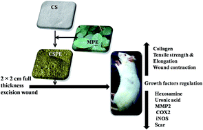
Collagen sponge (CS) was prepared using fish scales, which are a biological waste product in the marine food industry. CS was prepared so as to incorporate separately the drug mupirocin (thus designated CSM) and Macrotyloma uniflorum plant extract (CSPE). CS, CSM and CSPE were applied to the experimental wounds of rats and the healing pattern was observed using various biological and physicochemical techniques. CSPE enhanced wound healing and was involved in the up-regulation of growth factors such as vascular endothelial growth factor (VEGF), fibroblast growth factor (FGF), epidermal growth factor (EGF) and transforming growth factor β (TGF-β). Increased levels of hydroxyproline, hexosamine and uronic acid were observed in the CSPE-treated group compared with the other groups. Treatment with CSPE reduced inflammation, the expression of matrix metalloproteinases (MMPs) and scar formation, thereby contributing to faster wound healing.
1. Introduction
Collagen is an abundant and ubiquitous protein, representing nearly 30% of the total protein present in the body of animals.1 As a result of its low immunogenicity, collagen has been widely used in many pharmaceutical applications, e.g. in wound dressings, shields, injectable dispersions, sponges, moisturisers and as microparticles.2,3 Collagen induces the development of fibroblasts by its three-dimensional structure, which is essential for the formation of granular tissue, cell–cell and cell–matrix interactions, the deposition of new fibres and re-epithelialization.4 Collagen is now available in many different forms, e.g. sheets/films, gels, sponges and sprays. Of these, collagen sponge is preferred for burn/wound dressings as a result of its stability, porosity, the adsorption of large quantities of tissue exudate, its adherence to wet wounds, the maintenance of a moist environment and the enhancement of the formation of new granulation tissue and epithelium on the wound surface.5
Wound healing is a complex process, executed and regulated by a complex signalling network consisting of several growth factors, cytokines and chemokines. These are essential components for the coordination of multiple cell types during the wound-healing process. Among the known growth factors, vascular endothelial growth factor (VEGF) is thought to be the most ubiquitous, efficient and long-lasting in stimulating wound healing.6 Healing without scar formation is achieved by growth factors such as transforming growth factor β (TGF-β), fibroblast growth factors (FGFs) and epidermal growth factors (EGFs). These factors play a fundamental part in wound healing by recruiting fibroblasts to the site of injury and thereby stimulating connective tissues such as collagen and fibronectin.7
Matrix metalloproteinases (MMPs) are capable of degrading the structural components of extracellular matrix (ECM). MMPs participate in many biological processes, namely development, regeneration, morphogenesis and wound healing.8 Inflammatory mediators such as cyclooxygenase-2 (COX-2) and inducible nitric oxide synthases (iNOS) are reported to be predominantly expressed during the early inflammatory process and are reduced as the healing process continues. They are induced by different factors, including cytokines, growth factors and inflammatory stimuli.9,10
Collagen in combination with Macrotyloma uniflorum plant extract (MPE) enhances wound healing, as the plant extract has antibacterial and anti-inflammatory properties.11 The aim of the study reported here was to evaluate the feasibility of using collagen sponge impregnated with MPE to promote in vivo wound healing and its effect on growth factors, the expression of MMPs, COX-2, iNOs and various biochemical parameters.
2. Materials and methods
2.1 Preparation of the dressing material
Fish-scale collagen was isolated and MPE was obtained using previously published procedures.12 The collagen scaffolds were prepared using a slight modification of a previously published method.13 Briefly, a 1% wt/vol collagen solution in 0.5 M acetic acid (20 ml) was prepared and continuously stirred under IKA T25 using a homogenizer at 13500 rev min−1 to generate a uniform foam. A drop of Triton X-100 was added to the mixture as a frothing agent and 0.25% glutaraldehyde (0.25 ml) solution was added as a cross-linking agent. The collagen foam was poured into Teflon trays and frozen at −80 °C for 24 h, followed by freeze-drying for 48 h using a lyophilizer (Operon Co., Korea). The completely dry scaffolds were stored at 4 °C in airtight plastic containers and are referred to as collagen sponge (CS). To prepare CS incorporating the drug mupirocin (CSM), 20 mg of mupirocin were added to 20 mg of collagen solution (1% wt/vol in 0.5 M acetic acid) and the sponge was prepared following the same method as for the pure CS. To prepare the CS with MPE (CSPE), 2 ml (23 mg dry weight) of MPE were added to 20 ml of collagen solution (1% wt/vol in 0.5 M acetic acid) and the sponge was prepared following the same method as for pure CS. The characterization and in vitro studies of collagen foam impregnated with MPE and mupirocin have been reported previously.12 All the prepared materials were sterilized using ethylene oxide.14
2.2 In vivo studies
All the experiments were performed according to the Institutional Animal Care and Use Committee Approval and Guidelines (466/01a/CPCSEA). Male albino Wistar rats weighing between 180 g and 200 g were divided into four groups containing six animals in each groups (control, CS, CSM and CSPE dressings) for more details refer ESI (Table S1†). The rats in each group were acclimatized for 1 week before the study and were later housed individually under a 12 h light/dark cycle at 25 ± 1 °C. They were provided with standard rodent feed from M s−1 Hindustan Level Ltd Feeds (Mumbai, India) and water ad libitum.
2.3 Surgical procedure and dressing
After an intraperitoneal injection of standard anaesthesia (ketamine 50 mg kg−1 body weight and xylazine 10 mg kg−1 body weight), the dorsal surfaces of the rats below the cervical region were shaved and the skin disinfected with 70% ethanol. A 2 × 2 cm full thickness excision wound was created using a scalpel blade by excising the dorsal skin. The wound area was photographed and the initial wound area was traced using a transparent sheet. In the group 1 animals (control group), the wounds were dressed with sterile cotton gauze. group 2 animals were dressed with the CS scaffold, group 3 animals with the CSM scaffold and group 4 animals with the CSPE scaffold and the wounds were covered with absorbent gauze to hold the material on the wound area. The dressings were changed periodically at an interval of 4 days with the respective dressing materials. The wounds were cleaned with sterile distilled water before dressing. For the control group, the wound was treated with sterile saline in addition to distilled water to facilitate the injury-free removal of the dressing. The material was removed gently under moist conditions using a sterile pair of tweezers. Wound tissue was removed by killing six rats from each of the four groups on the 4th, 8th, 12th, 16th and 21st day after the creation of the wound and the granulation tissue formed was collected and stored at −80 °C until analysis. The progress of wound healing in the rats was evaluated by periodic monitoring of the wound contraction area and immunohistological and biochemical studies.
2.4 Planimetry: rate of contraction and period of re-epithelialization
Visual proof of the pattern of wound healing was recorded by taking digital photographs on the 0th, 4th, 8th, 12th and 16th day after wound creation. The time taken for full re-epithelialization of the wound biopsy samples was noted; the rate of contraction and the surface area were measured using a standard planimetric method by tracing the wound on a transparent graph sheet. The percentage of wound contraction was calculated using the formula:
where n = number of days (4th, 8th, 12th and 16th day). The results were analysed by one-way ANOVA at a 5% error level. The tensile strength of the incision wound tissues was measured at the end of the experiments.
2.5 Tensile strength measurements
The tensile strength measurements of the untreated and treated wound tissues collected at the end of the experiments were examined as described previously.15 The harvested tissues were trimmed into strips 20 mm long and 2 mm wide with the area of the original wound lying lengthwise in the centre of the sample. Mechanical properties such as the tensile strength (MPa) and percentage of elongation at break (%) were measured using a universal testing machine (Instron model 4501).
2.6 Biochemical analyses of the excision wounds
The excised tissue was biochemically analysed to estimate the total amount of collagen by estimating the collagen (hydroxyproline) content in defatted dried granulation tissue using the method of Woessner.16 Hexosamine was determined by the method of Elson and Morgan17 and the uronic acid content was determined using the method of Schiller et al.15 Protein was determined by the method of Lowry et al.18 Each of these experiments was performed on treated and untreated wound tissues and each was repeated six times.
2.7 Histology and immunohistochemical study of granulation tissue
The regenerated skin tissues from the wound site were periodically collected along with the healthy skin from a 2 mm strip surrounding the wound on the 4th, 8th, 12th and 16th day after wound creation following euthanasia. The samples were fixed in 10% buffered formalin, dehydrated with a graded ethanol series and embedded in paraffin blocks. The samples were sliced into 4 μm thick sections and stained with hematoxylin and eosin (H&E; Fisher Scientific) to examine the regeneration of the epidermis and dermis. The immunohistochemical expression of COX-2 and iNOs was determined as reported by Kalayarasan et al.19 Briefly, paraffin-embedded tissue sections (4 μm thickness) were rehydrated first with xylene and then in a series of ethanol solutions. The sections were then blocked with 5% BSA in pH 7.4 Tris-buffered saline (TBS) for 2 h, followed by immunostaining with a primary antibody (rabbit polyclonal IgG to rat iNOS), diluted 1:500 with 5% BSA in TBS and kept overnight incubation at 4 °C. After washing the slides three times with TBS, the sections were incubated with goat anti-rabbit secondary antibody (diluted 1:2000) with 5% BSA in TBS and incubated for 2 h at room temperature, followed by washing the sections with TBS and incubating for 5–10 min in a solution of 0.02% diaminobenzidine containing 0.01% hydrogen peroxide. The sections were counter-stained using hematoxylin; the sections were examined under a microscope and photomicrographed (DM5000, Leica, Germany).
2.8 Gelatin zymography
Total protein from the rat wound tissues was extracted at different sampling times and the extracted protein samples (n = 3 per treatment group) were pooled and tested for gelatinase activities as described previously.20 Protein samples were assessed for MMP-2 and MMP-9 by sodium dodecyl sulfate polyacrylamide gel electrophoresis (SDS-PAGE) with 10 mg of non-reduced sample loaded per lane of a 7% SDS polyacrylamide gel containing 0.5 mg ml−1 gelatin. They were finally stained with 1% acetic acid and 30% methanol. The MMPs were seen as a clear band corresponding to the zone of digestion of the gelatin substrate.
2.9 Reverse transcriptase PCR (RT-PCR)
Total RNA was isolated from the regenerated skin collected on the 4th, 8th and 12th day after wound creation; the samples were trimmed of any visible fat and healthy skin. Total RNA was isolated using TRIZOL reagent (Bangalore Genei) according to the manufacturer's instructions. A 3 μg mass of total RNA from the samples was used in the RT-PCR reaction. The PCR reaction was performed in a thermal cycler (Eppendorf, Germany) under the following conditions. Reverse transcription was performed at 50 °C for 50 min and activation at 95 °C for 15 min. The cDNA (2 μl) was amplified by PCR with the required primers. The primer sequences were designed using Primer-BLAST (http://www.ncbi.nlm.nih.gov/tools/primer-blast/) and were synthesized by Xcelris Labs Ltd (Bodakder, Ahmedabad, India). All the primer pairs were analysed by Primer-BLAST to ensure specificity for the intended target gene within the human genome. VEGF, forward 5′-AGAGTGGGAGGGAAGCTCTTAG-3′, reverse 5′-CGGGATTTCTTGCGCTTTCG-3′ (511); EGF, forward 5′-TGGAAAAGATGGCTGCCACTGGGTC-3′, reverse 5′-GTGTTCCTCTAGGACCACAAACCA-3′ (430); FGF, forward 5′-CAGGAGTACTGCAGAGCGAC-3′, reverse 5′-TCCGGTTTTGGTGCTGATGT-3′ (239); and TGF-β3, forward 5′-CACACAGTCCGCTACTTCGT-3′, reverse 5′-CGGGTGCTGTTGTAAAGTGC-3′ (434). Amplification was followed by 35 cycles of PCR: 94 °C (denaturation) for 45 s, variable 1 (annealing) for 45 s and 72 °C extension for 1 min. The products were then incubated at 72 °C for 10 min to extend any incomplete single strands. Finally, the PCR products (cDNA) were analysed by 1.5% agarose gel electrophoresis and densitometric analysis of the bands was performed using image analysis software.
3. Results and discussion
The preparation, characterization and in vitro studies of CS, CSM and CSPE have been reported previously.12 The SEM images of all the samples studied showed a porous and mesh-like structure with inherent interconnectivity. CS exhibited a more macro-porous structure than CSM and had randomly oriented thin fibril-like networks. A more porous nature along with thickening of the fibril-like network with a smooth surface was observed in the SEM images of CSM. The pore sizes were similar and ranged from 30 μm to 70 μm in diameter for all the samples. Of the three prepared samples, CSPE showed a significantly higher tensile strength (2.96 ± 0.25 MPa) and percentage elongation (16.26 ± 1.22%) than CS (0.56 ± 0.05 MPa and 11.61 ± 0.46%) and CSM (0.57 ± 0.03 MPa and 11.40 ± 0.04%). All the samples were biocompatible and showed antibacterial properties and therefore these materials were selected for the wound-healing activity in vivo model. Wound healing is a dynamic and continuous physiological process involving inflammation, proliferation or tissue regeneration, and tissue remodelling. There are several factors that either accelerate or retard the wound-healing process.21
3.1 Planimetric, photographic and period of epithelialization studies
Regular monitoring of the wounds and noting wound contraction are essential in assessing the effectiveness of treatment.22 Visual evidence of wound healing was obtained by taking photographs at a constant distance using a Sony DSLR camera for all animals and groups. All the animals were photographed, but only one respective photograph from each group is shown (Fig. 1). The photographs clearly indicated a faster rate of healing in the CSPE-treated groups compared with the other treatment groups and the control group. Wound assessment is a complex process and includes the wound aetiology, appearance, and the prediction and monitoring of the rate of healing, determining the factors responsible for delaying wound healing, and wound documentation. Measurement of the wound size is an important factor, with the potential to provide baseline measurements and to accurately determine the percentage reduction or increase in the wound area (healing/non-healing) over time. This is important in evaluating the efficacy of the prepared material.23 The results clearly showed that the experimental groups treated with CSPE showed a faster rate of healing than the other groups and control animals. The CSPE-treated groups required only 12 days for complete epithelialization, whereas the CS- and CSM-treated groups took 17 and 16 days, respectively, and the control group took 21 days for complete epithelialization. The healing pattern was regular and uniform in the CSPE-, CSM- and CS-treated groups compared with the control groups. The photographic results support the planimetric studies. Fig. 2a shows the rate of wound contraction in the control, CS-, CSM- and CSPE-treated groups. The time taken for the contraction of the excisional wounds was calculated from the planimetric sheet photographs using the ImageJ software program. The one-way ANOVA results showed that the groups were significantly different on the specified day. The results show the significant increase in the rate of contraction in the experimental groups compared with the control group over a period of time. The CSPE-treated groups showed complete contraction on day 12, whereas those treated with CSM and CS took 16 and 17 days; however, complete contraction was observed in the control group only on day 21. An increased rate of healing was observed in the CSPE-treated groups, which may be due to the incorporation of MPE into the CS; the photographic results agree well with the planimetric studies.
|
| Fig. 1 Photographs of wound-healing patterns on different days. | |
|
| Fig. 2 (a) Percentage wound contraction. Amount of (b) hydroxyproline, (c) hexosamine; and (d) uronic acid in excised granulation tissues. (e and f) Tensile strength and percentage elongation values of excised healed wound tissues. | |
3.2 Biochemical analyses of the excision wounds
Biochemical analyses were carried out on the excised granulation tissue on the specified days of scarification after wound creation. The wound contraction process depends on fibroblast invasion, collagen deposition and maturation. Collagen synthesis increases at the site of injury, which plays a major part in the strength, haemostasis and integrity of the newly formed matrix at the site of the wound and assists in re-epithelialization and cell–cell and cell–matrix interactions.23 Collagen is a well-known constituent of growing cells in healing tissues and is measured by estimating the concentration of hydroxyproline.24 In our experiments, the hydroxyproline content (Fig. 2b) increased in all the treated and untreated groups until day 12 and later stabilized until healing was complete. Among the groups treated, the CSPE-treated group showed a higher hydroxyproline content on all days of sampling than the groups treated with CS and CSM and the control groups. A significant increase in the hydroxyproline content of granulation tissue was seen in the group treated with CSPE, which may be a result of the increased synthesis of collagen. Thus, increasing the concentration of hydroxyproline results in a faster rate of healing by improving the migration of fibroblasts, increasing cellular proliferation and improving re-epithelialization.25 A decrease in the collagen content was observed in the control groups compared with the treated groups, which may be due to the prolonged inflammatory phase.26 Fig. 2c shows the hexosamine content of the granulation tissues. The amount of hexosamine was higher on the 4th day in all groups and a significant increase was found in the hexosamine in the group treated with CSPE compared with the groups treated with CSM, CS and the control group. A gradual decrease in the hexosamine content was observed in all the groups during the course of the study. However, a significant difference between the control and the treated groups was observed. The results obtained were in agreement with the observed collagen contents. In general, a decreasing trend in the hexosamine content was seen in both the experimental and control groups in all samples. A significant increase in the uronic acid content was observed on day 4 in all groups. The CSPE-treated groups showed a significant increase in the uronic acid content compared with the CS-, CSM- and control groups. A gradual decrease in the uronic acid content was observed after the 4th day in all the groups and a similar trend among the groups was observed over the period of healing (Fig. 2d).
3.3 Tensile strength
Wound healing is a primary response to tissue injury and consists of a complex biological process of connective tissue repair. Fig. 2e and f show the tensile strength and percentage elongation values of the excised healed wound tissues. It is clear that the tensile strength and elongation of the tissues from the groups treated with CSPE were significantly greater than those of the other treated groups and the control group. The increased tensile strength in the CSPE-treated group clearly indicates an increase in the collagen matrix, which imparts tensile strength and elasticity to the healed skin. This is further supported by the results of the biochemical assay, which showed an increased collagen content in the wounds treated with CSPE, which ultimately resulted in the increased tensile strength. Singer and Clark27 reported that wound tissues gain 20% of their final strength in the first 3 weeks after wounding. We observed a decreased tensile strength and percentage elongation in the control group compared with the CSPE-treated groups, which is further supported by the hydroxyproline assay. The tensile strength is directly related to the amount of collagen matrix synthesized at the wound site.
3.4 Histological observation
Histological examination of the wound tissue was performed using H&E staining to observe the formation of epithelium, connective tissue, the inflammatory response, fibroblast proliferation and collagen deposition (Fig. 3). On day 4, the H&E-stained tissue sections showed epithelium and connective tissue in the control and treated groups and the connective tissue had an abundance of acute and chronic inflammatory cells, such as lymphocytes and neutrophils, with blood vessels and extravasated red blood cells. On the 8th day, the H&E-stained histopathological sections of the CS- and CSM-treated groups and the control group showed moderate inflammatory infiltration compared with the CSPE-treated group. The connective tissue in the CSPE-treated group is fibrous in nature, with fewer chronic inflammatory cells, such as lymphocytes, and blood vessels, which clearly indicates that the MPE-incorporated scaffold helps with faster healing by preventing a prolonged inflammatory phase. Epithelialization at the wound edges was also better for the CSPE-treated groups. These findings indicate better initiation of the healing process. On 12th day, the CSPE-treated groups showed complete epithelialization with focal acanthosis and an adenexal structure; the connective tissue was fibrous in nature with dense collagen fibres and blood vessels, which is an indication of complete healing. In contrast, only moderate epithelialization was seen in the CS- and CSM-treated groups. Inflammatory cells were present on sections from the 4th, 8th and 12th day sections, but the amount of inflammatory cells was reduced compared with the CSPE group; this showed that the healing process was slow. For the control defective epithelialization was observed. The deposition of collagen and faster epithelial regeneration in the CSPE-treated groups may significantly accelerate wound healing compared with the other groups. It is well established that collagen formation is very important in tissue repair and remodelling. In addition, faster healing and deposition of collagen did not contribute to scar formation in the wounded areas. Hence CSPE was effective as a wound-healing material for the treatment of deep wounds.
|
| Fig. 3 Haematoxylin and eosin staining of control and treated groups on 4th, 8th and 12th day after wound creation. | |
3.5 Expression of growth factors in response to injury and MPE
The expression of EGF was significantly higher in the CSPE-treated group than in the control and CS- and CSM-treated groups on the 4th day (Fig. 4a). The pattern of EGF expression was reduced in the treated groups, whereas the control group showed increasing expression of EGF on the 8th day. A reduction in expression was seen on the 12th day; a significant reduction was seen in the CSPE-treated groups followed by the CSM- and CS-treated groups. EGF has been reported to mediate an increase in the collagen content during wound repair.28 In the CSPE-treated groups, the expression of FGF was increased on days 4 and day 8 and then later declined. On day 12, the expression was significantly reduced compared with the other CSM- and CS-treated groups, whereas the expression was very low on day 4 compared with the other groups. It started to increase on days 8 and 12 compared with the treated groups. Hypoxia induces several angiogenic gene expressions at the cellular level, particularly VEGF.29 VEGF also followed the same pattern of expression as EGF and FGF. The expression in the treated groups was significantly higher than that in the control group on day 4, whereas expression decreased in the treated groups on day 8 and was significantly reduced in the CSPE-treated groups on day 12 compared with the CSM- and CS-treated groups. On the other hand, the expression of VEGF increased on day 8 and significantly increased on day 12 compared with the treated groups. Many reports support our results that the expression of VEGF increased from day 3 and until day 7 and then decreased from around day 13.30,31 Bao et al.32 reported that VEGF stimulates wound healing by involvement in the wound-healing cascade, such as angiogenesis, epithelialization, vasodilation, endothelial cell proliferation and the promotion of collagen deposition. TGF-β is involved in wound healing and tissue repair and regulates the rate and extent of the wound-healing process.33 In our study, TGF-β3 was significantly reduced in the treated groups from days 4 to 12. Among the treated groups, the CSPE-treated groups showed a significant reduction in expression compared with the other treated groups and the control group. The expression was at a maximum in the control group on all days (4th, 8th and 12th days), compared with the treated groups. TGF-β3 is reported to be involved in the reduction of scar formation by decreasing the formation of type I collagen by degradation34; it is involved in processes such as inflammation, angiogenesis, the proliferation of fibroblasts, the synthesis of collagen, collagen deposition and the remodelling of new ECM. This result correlated with the biochemical parameters observed for the time taken for epithelialization.
|
| Fig. 4 (a) Changes in mRNA expression pattern, determined by RT-PCR, of VEGF, FGF, EGF and TGF-β; (b) differential expression of MMP2 and MMP 9 in granulation tissue. | |
3.6 Determination of MMP-2 and MMP-9 using gelatin zymography
The expression of MMP in the granulation tissues from all groups was analysed at different intervals. MMPs play a vital part in the rate of wound healing. Increasing the MMP ratio leads to the degradation of the components of ECM, growth factors and their receptors in the wound by prolonging the rate of wound healing.35 In the wound-healing process, fibroblasts, keratinocytes and inflammatory cells are the major cell types that produce MMPs. MMP expression is regulated by the signals received from the growth factors, cytokines, cell–matrix interactions and altered cell–cell contacts.36 We determined the pro- and active forms of MMP-2 and -9 expression by gelatin zymography of the granulation tissues (Fig. 4b). On day 4, the pro- and active forms of MMP-2 and -9 were significantly higher in the control groups than in the treated groups and were found to be reducing over a period of time. Compared with MMP-2, MMP-9 expression was significantly reduced in the treated groups on the 4th day and expression was not seen on the 8th and 12th days in the control and treated groups. On day 8, a significant (p < 0.05) reduction in the expression of MMP-2 was found in the CSPE-treated groups compared with the control and treated groups (CS and CSM). Compared with MMP-2, there was a significant (p < 0.05) reduction in MMP-9 expression on the 4th day. The expression of MMPs varies with the phases of wound healing. Increased expression of MMP-9 was seen in the inflammatory phase and MMP-9 was later found to be decreasing in its expression and MMP-2 began to increase. This result correlated with our previous findings.37 On day 12, the CSPE-treated groups showed a significant reduction in the pro- and active forms of MMP-2 expression compared with the control and other treated groups. Compared with the treated groups, the expression was higher in the control groups on all the days; the results obtained correlated with the biochemical analysis, thereby supporting our investigation that CSPE enhanced the wound-healing process by reducing the synthesis of MMPs and increasing the synthesis of collagen.
3.7 Immunohistochemical analyses of inflammatory markers
In vivo animal experiments have shown that COX-2 inhibition reduces the initial inflammatory phase of wound healing and thereby reduces scar formation without disrupting re-epithelialization and reducing the tensile strength.38 COX-2 is normally expressed under abnormal situations, such as inflammation or the presence of a tumor.39 Fig. 5 shows the expression of COX-2 on different days. On day 4, the expression of COX-2 was significantly (p < 0.05) higher in the control group followed by the CS- and CSM-treated groups. At the same time, a significant (p < 0.05) reduction in the expression of COX-2 was seen in the CSPE-treated groups, which may be a result of the formation of new epidermal cells at the edges of the wound. The same trend was seen on all the days. Compared with day 4, a marked reduction in COX-2 expression was seen on day 8 in all the groups, but a significant (p < 0.05) reduction was observed in the CSPE-treated groups. On the 12th day, a significant reduction in the expression of COX-2 was seen in the control and CS- and CSM-treated groups. From our results, we concluded that the reduction in the expression of COX-2 enhances wound healing. Similar results have been reported previously by Futagami et al.9 The same pattern of expression was seen for iNOS (Fig. 6). During the experimental period, the expression of iNOS was significantly higher in the control group on all days, compared with the treated groups. On day 4, a reduction in iNOS was observed in the treated groups, but a significant (p < 0.05) reduction was observed in the CSPE-treated groups. Frank et al.40 have previously reported that the maximum effect of NOS activity occurs during the early phase of wound healing. Expression was found between 6 and 24 h and lasted for up to 5 days. The expression of NOS then slowly decreased over the next 10 days.40 On the 12th day, the expression was significantly higher in the CSPE-treated group than in the CS- and CSM-treated groups. This result clearly shows the effect of MPE on the reduction of iNOS expression by favouring the wound-healing process. Witte and Barbul41 reported that NO regulates cell proliferation, collagen formation and wound contraction.
|
| Fig. 5 Representative images of immunohistochemistry staining of COX-2 on days 4, 8 and 12. | |
|
| Fig. 6 Representative images of immunohistochemistry staining of iNOS on days 4, 8 and 12. | |
4. Conclusion
The results obtained in this study indicate that the presence of MPE in the collagen sponge favours the wound-healing process by up-regulating growth factors and reducing inflammatory markers. The CSPE-treated group showed increased levels of hydroxyproline, uronic acid and hexosamine synthesis and decreased levels of MMPs. Reduced inflammation, scar formation and an enhanced wound-healing process were observed in CSPE-treated rats. In addition, the CSPE-treated wounds showed an improved tensile strength in the healed tissue. However, studies with purified constituents of MPE are required to understand the complete mechanism of wound-healing activity of MPE.
Conflict of interest
The authors declare no conflicts of interest.
Acknowledgements
T. Muthukumar thanks the Council of Scientific and Industrial Research, New Delhi, India, for providing the funds and infrastructure to carry out this study. The authors thank Dr T. Gopalakrishnan (Department of Oral Pathology and Microbiology, Sree Balaji Dental College & Hospital, Bharath University, Pallikaranai, Chennai) for H&E slide interpretations.
References
- C. M. Kielty, I. Hoplinson and M. E. Grant, in: Part I: Connective tissue and its heritable disorders, ed. P. M. Royce and B. Steinmann, Wiley-Liss, Inc, New York, 1993, pp. 103–147 Search PubMed.
- W. Friess, Eur. J. Pharm. Biopharm., 1998, 45, 113–136 CrossRef CAS.
- P. Elsner, E. Berardesca and H. Maibach, Bioengineering of the Skin: Water and the Stratum Corneum, CRC Press LLC, Boca Raton, 1994 Search PubMed.
- G. S. Schultz, R. G. Sibbald, V. Falanga, E. A. Ayello, C. Dowsett, K. Harding, M. Romanelli, M. C. Stacey, L. Teot and W. Vanscheidt, Wound Repair Regen., 2003, 11, S1–S28 CrossRef.
- M. Chvapil, J. Biomed. Mater. Res., 1982, 16, 245–263 CrossRef CAS PubMed.
- N. N. Nissen, P. J. Polverini, A. E. Koch, M. V. Volin, R. L. Gamelli and L. A. DiPietro, Am. J. Pathol., 1998, 152, 1445–1452 CAS.
- H. P. Ehrlich and T. M. Krummel, Wound Repair Regen., 1996, 4, 203–210 CAS.
- W. C. Parks, Wound Repair Regen., 1999, 7, 423–432 CrossRef CAS.
- A. Futagami, M. Ishizaki, Y. Fukuda, S. Kawana and N. Yamanaka, Lab. Invest., 2002, 82, 1503–1513 CrossRef CAS.
- M. M. Cals-Grierson and A. D. Ormerod, Nitric Oxide, 2004, 10, 179–193 CrossRef CAS PubMed.
- A. Ghani, Medicinal Plants of Bangladesh: Chemical. Constituents and Uses, Asiatic Society of Bangladesh, Dhaka, 2nd edn, ISBN; 9-845123481:5, 2003 Search PubMed.
- T. Muthukumar, P. Prabua, K. Ghoshb and T. P. Sastry, Colloids Surf. B Biointerfaces, 2014, 113, 207–212 CrossRef CAS PubMed.
- R. Sripriya, M. Senthil Kumar and P. K. Sehgal, J. Biomed. Mater. Res., Part B, 2004, 70, 389–396 CrossRef PubMed.
- S. D. Gorham, S. Srivastava, D. A. French and R. Scott, J. Mater. Sci.: Mater. Med., 1993, 4, 40–49 CrossRef CAS.
- S. Schiller, G. A. Slover and A. A. Dorfma, J. Biol. Chem., 1961, 236, 983–987 CAS.
- J. F. Woessner Jr, Arch. Biochem. Biophys., 1961, 93, 440–447 CrossRef CAS.
- L. A. Elson and W. T. Morgan, Biochem. J., 1933, 27, 1824–1828 CAS.
- O. H. Lowry, N. J. Rosebroug, A. L. Farr and R. J. Randall, J. Biol. Chem., 1951, 193, 265–275 CAS.
- S. Kalayarasan, N. Sriram and G. Sudhandiran, Life Sci., 2008, 82, 1142–1153 CrossRef CAS PubMed.
- C. Heussen and E. B. Dowdle, Anal. Biochem., 1980, 102, 196–202 CrossRef CAS.
- G. C. Gurtner, S. Werner, Y. Barrandon and M. T. Longaker, Nature, 2008, 453, 314–321 CrossRef CAS PubMed.
- G. Gethin, Wounds, 2006, 2, 60–68 Search PubMed.
- R. Raghow, FASEB J., 1994, 8, 823–831 CAS.
- Z. Q. Lin, T. Kondo, Y. Ishida, T. Takayasu and N. Mukaida, J. Leukocyte Biol., 2003, 73, 713–721 CrossRef CAS.
- R. Bernabei, F. Landi, S. Bonini, G. Onder, A. Lambiase, R. Pola and L. Aloe, Lancet, 1999, 354, 307 CrossRef CAS.
- M. S. Kumar, S. Kirubanandan, R. Sripriya and P. K. Sehgal, J. Surg. Res., 2008, 144, 94–101 CrossRef PubMed.
- A. J. Singer and R. A. Clark, N. Engl. J. Med., 1999, 341, 738–746 CrossRef CAS PubMed.
- M. Hiramatsu, K. Hatakeyama, N. Minami and M. Kumegawa, Jpn. J. Pharmacol., 1982, 32, 198–201 CrossRef CAS.
- S. Yla-Herttuala, T. T. Rissanen, I. Vajanto and J. Hartikainen, J. Am. Coll. Cardiol., 2007, 49, 1015–1026 CrossRef PubMed.
- N. J. Brown, E. A. Smyth, S. S. Cross and M. W. Reed, Wound Repair Regen., 2002, 10, 245–251 CrossRef.
- L. F. Brown, K. T. Yeo, B. Berse, T. K. Yeo, D. R. Senger, H. F. Dvorak and L. van de Water, J. Exp. Med., 1992, 176, 1375–1379 CrossRef CAS.
- P. Bao, A. Kodra, M. Tomic-Canic, M. S. Golinko, H. P. Ehrlich and H. Brem, J. Surg. Res., 2009, 153, 347–358 CrossRef CAS PubMed.
- A. Shukla, N. Meisler and K. R. Cutroneo, Wound Repair Regen., 1999, 7, 133–140 CrossRef CAS.
- R. Hosokawa, K. Nonaka, M. Morifuji, L. Shum and M. Ohishi, J. Dent. Res., 2003, 82, 558–564 CrossRef CAS PubMed.
- S. E. Gill and W. C. Parks, Int. J. Biochem. Cell Biol., 2008, 40, 1334–1347 CrossRef CAS PubMed.
- M. Egeblad and Z. Werb, Nat. Rev. Cancer, 2002, 2, 161–174 CrossRef CAS PubMed.
- T. Salo, M. Makela, M. Kylmaniemi, H. Autio-Harmainen and H. Larjava, Lab. Invest., 1994, 70, 176–182 CAS.
- T. D. Warner and J. A. Mitchell, FASEB J., 2004, 18, 790–804 CrossRef CAS PubMed.
- M. M. Hardy, E. A. Blomme, A. Lisowski, K. S. Chinn, A. Jones, J. M. Harmon, A. Opsahl, R. L. Ornberg and C. S. Tripp, J. Pharmacol. Exp. Ther., 2003, 304, 959–967 CrossRef CAS PubMed.
- S. Frank, M. Madlener, J. Pfeilschifter and S. Werner, J. Invest. Dermatol., 1998, 111, 1058–1064 CrossRef CAS PubMed.
- M. B. Witte and A. Barbul, Am. J. Surg., 2002, 183, 406–412 CrossRef CAS.
Footnote |
† Electronic supplementary information (ESI) available. See DOI: 10.1039/c4ra11959b |
|
This journal is © The Royal Society of Chemistry 2014 |
Click here to see how this site uses Cookies. View our privacy policy here.