Feiyi Wang,
Jiancai An,
Lili Zhang and
Chunchang Zhao*
Key Laboratory for Advanced Materials and Institute of Fine Chemicals, East China University of Science & Technology, Shanghai 200237, P. R. China. E-mail: zhaocchang@ecust.edu.cn
First published on 10th October 2014
It is still a challenge to construct probes for discriminating thiols due to the similar structure and reactivities of thiol-containing molecules. We have here developed a Cys specific probe by utilizing the remarkable difference in reactivity toward Cys, Hcy and GSH. The reaction between the designed probe and Cys produces an amino-substituted BODIPY, giving a yellow fluorescence turn-on response. The response to Hcy or GSH shows a red fluorescence turn-on signal, due to the formation of sulfenyl-substituted BODIPY. These distinct fluorescence turn-on responses allow Cys to be distinguished from Hcy and GSH. This probe was also utilized for detection of Cys in living cells and monitoring cystathionine γ-lyase activity in vitro.
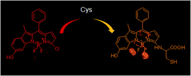
We and others have recently demonstrated that a change in electron-donating nature of the substituent on monochlorinated boron dipyrromethene (BODIPY) can result in a remarkable difference in reactivity toward nucleophilic reagents.8 By employing this promising feature, the selective detection of glutathione (GSH) over cysteine (Cys) and homocysteine (Hcy) has been obtained. However, few monochlorinated boron dipyrromethene-based probes have fulfilled the discrimination of Cys over Hcy and GSH. In the present study, we developed a 6-hydroxylindole-based monochlorinated BODIPY for highly discriminating detection of cysteine over Hcy and GSH based on the distinct electrophilic reactivity of the probe toward them. 6-Hydroxylindole was introduced to a monochlorinated BODIPY core, rendering the BODIPY core with electron-rich nature and enabling the difference in reactivity toward Cys, Hcy, and GSH, thus leading to the high selectivity of the probe toward nucleophilic analytes. Detailed studies demonstrated that replacement of chlorine with thiolate in Hcy and GSH through nucleophilic aromatic substitution (SNAr) reaction yields sulfenyl-substituted BODIPY with red-shifted absorption and emission features. The sulfenyl-substituted BODIPY derived from Cys is unstable and undetectable, which immediately undergoes intramolecular displacement with the amino group displacing of the sulfur function to form amino-substituted BODIPY, thus resulting in a significant hypsochromic shift in the optical spectra. These distinct fluorescence turn-on responses allow Cys to be distinguished from Hcy and GSH. We further testified the potential application of this probe for detection of Cys in living cells.
Cystathionine γ-lyase (CGL) is a pyridoxal 5′-phosphate (PLP)-dependent enzyme, which catalyzes the decomposition of cystathionine to generate cysteine and other transsulfurations.9 CGL plays a central role in amino acid catabolism and deregulation level of CGL is linked to cystathioninuria and cystinosis.10 Therefore, monitoring CGL activity could provide valuable insight into early detection of the related metabolic disorders. However, few fluorescent probes have been developed for evaluating CGL activity. Significantly, the fluorescent turn-on probe reported here is capable of assay of cystathionine γ-lyase under physiological conditions.
We assessed the reactivity of HO-BODIPY-Cl with Cys in buffer solutions (acetonitrile/HEPES buffer, 1:
1, v/v, 20 mM, pH 7.4) at 37 °C by monitoring the changes in UV-vis and fluorescence spectra. The reaction triggered a ratiometric absorption change over time and time-dependent fluorescence increase (Fig. 1). As can be seen in Fig. 1a, the absorption peaked at 557 nm and 600 nm gradually decreased, while a new absorption band at 520 nm was noted to increase simultaneously. This process was accompanied with an isosbestic point at 538 nm, suggesting the potential application of HO-BODIPY-Cl as a ratiometric and colorimetric probe for evaluation Cys under physiological condition. In the absence of Cys, HO-BODIPY-Cl displayed a weak emission centered at 645 nm. The addition of Cys (5 mM) to a solution of HO-BODIPY-Cl in buffer introduced a remarkable fluorescence increment, which was completed within 45 min. Accordingly, the fluorescence was changed from weak pink color to bight orange (Fig. S1†). The observed rate constant was then determined to be 0.085 min−1 by fitting the data with a pseudo-first-order model (Fig. S2†). The enhancement of fluorescence intensity after the reaction with Cys was as high as 10 fold, accompanied by a blue shift in the emission maxima from 645 nm to 586 nm, which enable HO-BODIPY-Cl to serve as a suitable fluorescence turn-on probe for Cys in aqueous solution.
The reaction between HO-BODIPY-Cl and Cys yields amino-substituted BODIPY (HO-BODIPY-N), which shows blue-shift in the maximum wavelengths of both the absorption and the fluorescence spectra compared to that of the original probe. The production of HO-BODIPY-N after the reaction was confirmed by HRMS analysis as well as high performance liquid chromatography (HPLC) (Fig. S3†). HPLC analysis of HO-BODIPY-Cl + Cys revealed that HO-BODIPY-Cl (retention time at 9.306 min) was converted efficiently into HO-BODIPY-N (retention time at 6.010 min). In HRMS, the mass peak at 466.1206 corresponding to [HO-BODIPY-N–H]− was noted. Though the photophysical properties of HO-BODIPY-N are stable in buffer, it was likely to undergo oxidation upon drying and purifying. Thus, we could not obtain pure product for NMR characterization and observe a triplet signal from the proton of SH. It is worth mentioning that signals of sulfenyl-substituted BODIPY (HO-BODIPY-S) in the optical response spectral were not observed on the experimental time scale, indicating that HO-BODIPY-S is unstable and undetectable, which immediately undergo intramolecular displacement to form amino-substituted BODIPY (Scheme 2). The formation of amino-substituted BODIPY was also demonstrated by the reaction between HO-BODIPY-Cl and N-acetyl-cysteine. N-Acetyl-cysteine is structurally similar to Cys but the amino is inactive. This structure feature makes only the product of sulfenyl-substituted BODIPY possible, thus resulting in the absorption and emission shifted to 581 nm and 608 nm (Fig. S4†).
The fluorescence change of HO-BODIPY-Cl treated with different concentrations of Cys was also recorded in Fig. 2. Upon addition of gradually increasing concentration of Cys, a progressive fluorescence intensity increase at 586 nm was observed, which is linearly dependent (R = 0.98) on the Cys concentration over a range of 0–100 μM, essentially falling within the physiological levels of Cys (< 360 μM). Therefore, probe HO-BODIPY-Cl can be employed as an ideal candidate for quantitative detection of Cys. The detection limit was then calculated to be 4.1 × 10⁻⁷ M, which is comparable to previously reported probes.12
We next evaluated the inference from Hcy and GSH (Fig. 3). As compared to Cys, both Hcy and GSH elicited inverse changes in the absorption spectra. A dramatic red-shift of the S0–S1 transition from 557 nm to 581 nm was introduced. Although Hcy and GSH also caused the blue-shift in the maximum wavelengths of fluorescence spectra, together with the fluoresce intensity increment after the reaction with HO-BODIPY-Cl, the fluorescence maximum located at 608 nm with red color, showing 22 nm red-shift relative to that of HO-BODIPY-Cl + Cys system. Both the UV-vis and emission spectra demonstrate the formation of single sulfenyl-substituted BODIPYs, HO-BODIPY-S-Hcy and HO-BODIPY-S-GSH after the reaction with Hcy and GSH, respectively. HPLC analysis and HRMS also confirmed the formation of HO-BODIPY-S-Hcy (retention time at 3.859 min, mass peak at 482.1516) and HO-BODIPY-S-GSH (retention time at 3.656 min, mass peak at 654.2011). As can be seen from Fig. 3f, the sulfenyl-substituted BODIPYs, HO-BODIPY-S-Hcy and HO-BODIPY-S-GSH, have minimal absorption at 500 nm compared to HO-BODIPY-N formed through the reaction of HO-BODIPY-Cl with Cys, therefore the selective excitation of HO-BODIPY-N with 500 nm can be performed. In this case, HO-BODIPY-Cl showed a very strong orange fluorescence in response to Cys upon excitation at 500 nm, while minimal fluorescence was observed with Hcy or GSH. Thus HO-BODIPY-Cl has a high selectivity for Cys, minimal disturbance by Hcy and GSH on the detection could be observed.
Based on the aforementioned experimental data and the reaction mechanism of two related thiol-response probes,8 we believe that the spectra change of HO-BODIPY-Cl induced by Cys was initiated by a two-step reaction: displacement of chlorine with thiolate to generate sulfenyl-substituted BODIPY and then intramolecular cyclization to yield amino-substituted BODIPY through a five-membered cyclic transition state. The amino-substituted BODIPY was not observed for Hcy and GSH. This difference between Cys and Hcy/GSH can be attributed to the kinetic rate of the intramolecular cyclization reactions. The intramolecular cyclization reaction to form the six-membered ring with Hcy or a macrocyclic ring with GSH may be kinetically unfavorable.
We then examined the reactivity of HO-BODIPY-Cl toward other related thiols and amino acids by means of fluorescence spectrometry (Fig. 4). HO-BODIPY-Cl showed negligible fluorescence ratio change (I586/I645) in response to these related species. Taken together, HO-BODIPY-Cl offers a good selectivity toward theses competing analytes, indicative of its capability for practical usefulness.
With these characterization studies in hand, the efficacy of HO-BODIPY-Cl for live-cell imaging of Cys was explored. As shown in Fig. 5, when cells successively pretreated with N-ethylmaleimide (a trapping reagent of thiols) were treated with HO-BODIPY-Cl for 20 min in the culture medium, only faint red fluorescence (606 nm–636 nm) signals was observed, which was ascribed to the unreacted probe. In contrast, the yellow channel (572–588 nm) is almost nonfluorescent. The ratio of the two emissions from yellow channel to red channel is about 0.1. More importantly, bright fluorescence in the yellow channel between 572–588 nm was observed in cells upon incubation with 100 μM Cys for 20 min and further incubation with HO-BODIPY-Cl for another 20 min in the same culture medium. The ratio was improved to be around 1.6, showing a ca. 16-fold enhancement. These results suggest that HO-BODIPY-Cl is a promising probe for living cells imaging of Cys.
This unique probe was also used to monitor the activities of cystathionine γ-lyase (CGL) in vitro. It is known that CGL catalyzes the decomposition of cystathionine to generate cysteine. Therefore, probe HO-BODIPY-Cl should give rise to a turn-on emission signal in the presence CGL and its substrate cystathionine. This was accomplished by mixing HO-BODIPY-Cl, CGL and cystathionine in buffer at 37 °C. The time dependent fluorescence changes were recorded in Fig. 6 and S5,† indicating a remarkable fluorescence increment accompanying by the emission maximum shifted from 645 nm to 586 nm. More importantly, minimal fluorescence enhancement was observed upon pretreatment with CGL inhibitor, propargylglycine (PAG), indicative of the blocking effect in formation of Cys. These results verify the capacity of HO-BODIPY-Cl for monitoring CGL activity.
Footnote |
† Electronic supplementary information (ESI) available: UV-vis absorption and emission spectra, pseudo first-order kinetic profile, 1H NMR, 13C NMR spectra of the new compounds. See DOI: 10.1039/c4ra11167b |
This journal is © The Royal Society of Chemistry 2014 |