Highly selective NO2 sensor at room temperature based on nanocomposites of hierarchical nanosphere-like α-Fe2O3 and reduced graphene oxide

Ying-li Dong, Xian-fa Zhang, Xiao-li Cheng, Ying-ming Xu*, Shan Gao, Hui Zhao and Li-hua Huo*
Key Laboratory of Functional Inorganic Material Chemistry, Ministry of Education, School of Chemistry and Materials Science, Heilongjiang University, Harbin 150080, People's Republic of China. E-mail: lhhuo68@yahoo.com; Fax: +86-0451-86608040; Tel: +86-0451-86608426

Received 10th September 2014 , Accepted 23rd October 2014

First published on 23rd October 2014


Abstract

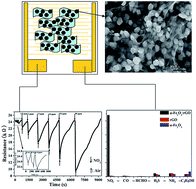
Nanosphere-like α-Fe2O3 modified reduced graphene oxide nanosheets were prepared by a simple hydrothermal method without any surfactant or template. The nanocomposites were characterized by X-ray diffraction (XRD), Raman spectra (RS), Fourier transform infrared (FT-IR) spectra, X-ray photoelectron spectroscopy (XPS), scanning electron microscopy (SEM), and transmission electron microscopy (TEM) techniques. The α-Fe2O3 nanospheres have a hierarchical structure, with diameter of about 40–50 nm, and grow uniformly on the surface of single graphene nanosheets. α-Fe2O3/rGO nanocomposites exhibit high response of 150.63% to 90 ppm NO2 at room temperature, 65.5 times higher than the response of pure graphene, and the detection limit for NO2 can be decreased down to 0.18 ppm. A mechanism is proposed for sensing of the nanocomposites: the high response of the nanocomposites to NO2 at room temperature is the synergistic effect of the two sensing materials and large specific surface area of the nanocomposites.


Introduction

There has always been great scientific interest in carbon materials because of their advantageous properties, such as cost-effectiveness, environmental friendliness, availability, and corrosion resistance.1 As the thinnest carbon material, graphene has been considered in the field of nanomaterials since 2008, and has outstanding physical and chemical properties. Graphene has been widely used in supercapacitors, Li-ion batteries, and gas sensors among other applications.1–4 In comparison with other gas sensing materials, graphene has the following advantages: excellent conductivity, very large specific surface area, exceptional low noise to signal ratios, and low working temperature. It has been investigated in detection of gases including NO2, NH3, H2O, and CO.5–7

However, graphene happens to reunite among sheets because of van der Waals interactions, resulting in low sensitivity and irreversibility of sensors.8,9 To overcome these problems, organic functional groups (sulfonated, ethylenediamine)10 and noble metals (Pd,11 Au,12 Ag12) have been investigated as modifying materials. Recently, semiconducting metal oxides, such as SnO2,13 NiO,14 WO3,15 ZnO,16 and Fe2O3,17 have been chosen as modifying materials because of their easy synthesis, low cost, and good stability. Their composites exhibit high sensitivity and reversibility at working temperature of 150–300 °C. Also, some metal oxide/graphene composites have been investigated under decreasing working temperature, and they show certain response to some test gases at room temperature. For example, Cu2O nanowire,18 Co3O4 nanoparticles,19 SnO2 nanoparticles,20 and indium-doped SnO2 nanoparticles21 have been studied in modification of reduced graphene oxide, exhibiting certain responses with long response-recovery characteristics to NO2. Radial flower-like SnO2,22 ZnO quantum dots,23 TiO2 nanoparticles,24 and Cu2O nanoparticles25 also have been considered as composites with graphene to detect NH3, HCHO, O2, and H2S, respectively, but with low responses at room temperature. However, the selectivity of most nanocomposite materials was not investigated in the cited reports. There is interest in investigation of new nanocomposites that show good selectivity to target gases at room temperature.

Ferric oxide nanomaterials are a kind of functional material whose composites with graphene have been applied mainly in Li-ion batteries,26 frictional materials,27 high-performance catalysts,28 supercapacitors,29 and biosensors.30 Two studies of Fe2O3–graphene nanocomposites used in gas sensors at high temperature were recently reported. Liang et al. prepared α-Fe2O3 nanoparticle modified graphene nanocomposites at 180 °C via an ethanol solvothermal route, which showed response of about 29 to 1000 ppm ethanol at 280 °C.17 In another paper, Fe2O3 nanoparticle-coated graphene nanosheets were obtained by a super critical CO2 assisted thermal method followed by vertical magnetic field assembly with directed flow. The material exhibited a CL emission of about 450 absorption units in response to 15 ppm H2S at 190 °C with good selectivity.31

In a previous investigation, we reported that the working temperature of the sensors could be decreased down to room temperature when the microstructure of the sensing materials was controlled through construction of the 3D hierarchical structure32 or adjustment of special morphology.33 Similarly, if hierarchical α-Fe2O3 nanomaterial with special morphology was used to modify graphene, it might be possible to decrease the working temperature of such nanocomposites.

There is a need for efficient sensors to selectively detect low concentrations of nitrogen dioxide (NO2) in a short time at low temperature. The rapid development of the automobile industry has caused nitrogen dioxide, produced mainly by automobiles and power plants, to become one of the main pollutants in the atmospheric environment. It is well known that NO2 can destroy the ozone layer, and can also do great harm to human health, e.g. respiratory system of human. According to reports, the Lethal Concentration 50 (LC50) of NO2 is 126 mg m−3, with an exposure time of no longer than 8 h to 3 ppm NO2.21

In this paper, we report a simple and low cost hydrothermal synthesis route to prepare α-Fe2O3/rGO nanocomposites at 120 °C, in which nanosphere-like α-Fe2O3 of 40–50 nm diameter are constructed by a few nanometer-sized nanoparticles and reduced graphene oxide (rGO) intercalated single sheets. These nanocomposites exhibit excellent response and selectivity to NO2 at room temperature.

Experimental

Preparation of α-Fe2O3/rGO nanocomposites

All the materials used were of analytical grade. 20 mg graphene oxide (GO) was prepared by natural flake graphite (325 mesh) according to the modified Hummers method,34 was dispersed in 20 mL deionized water and sonicated for 1 h. 10 mL 0.022 mol L−1 FeCl3 aqueous solution was added dropwise into the GO disperse solution with magnetic stirring for 30 min, then sonicated for 10 min. The solution was transferred into a Teflon-lined autoclave and maintained at 120 °C for 8 h. The final product of α-Fe2O3/rGO nanocomposite was obtained after vacuum filtration, washed with deionized water, and dried at 60 °C, as shown in Fig. 1a. Reduced graphene oxide (rGO) and pure α-Fe2O3 nanoparticles were obtained by a similar procedure only in the absence of FeCl3·6H2O and GO correspondingly.
image file: c4ra10136g-f1.tif
Fig. 1 The experimental reaction diagram of α-Fe2O3/rGO nanocomposites (a), schematic of α-Fe2O3/rGO nanocomposites on the sensor substrate (b) and schematic of sensing test (c).

Characterizations

The composition and phase purity of the as-synthesized samples were analyzed by powder X-ray diffraction (XRD) with monochromatized Cu Kα (λ = 0.15406 nm) by a Rigaku, D/MAX-3B instrument operating at 40 kV voltage and 50 mA current. The nanocomposites were analyzed using a Renishaw 1000 Micro-Raman spectrometer with a long-range 50× objective, 10S integration, and 10% laser power (457.9 nm excitation; 8 mW at 100%). The chemical compositions on the surface of the nanocomposites were detected by Fourier transform infrared (FT-IR) spectroscopy (Nexus, Thermo Nicolet). The sample was also analyzed by X-ray photoelectron spectroscopy (XPS, Kratos, ULTRA AXIS DLD) with monochromatized Al Kα ( = 1486.6 eV) radiation. All binding energies were calibrated by referencing to C1s (284.6 eV). The size and morphology of the samples were observed using a field emission scanning electron microscope (FESEM, FEI/Philips, XL-30). A JEM-2010 transmission electron microscope (TEM), operating at a 200 kV accelerating voltage, was used for TEM analysis. Specific surface area of the products was analyzed by nitrogen adsorption–desorption at 77 K using a Gas Sorption System (Micro-metrics Instruments, TriStar II 3020).

Gas sensor fabrication and sensing measurements

To prepare gas sensors composed of α-Fe2O3/rGO nanocomposites, 18 pairs of gold interdigitated electrodes were fabricated by an e-beam lithography process on an Al2O3 wafer. The size of wafer was 9.4 × 9.4 × 0.38 mm, on which the distance of electrodes was 50 μm. The α-Fe2O3/rGO nanocomposites were dispersed in ethanol to form a dispersion liquid, which was dropped (75 μL, 1.53 × 10⁻⁶ g μL−1) on the gold electrode of the sensor substrate uniformly by spin coating, as shown in Fig. 1b. To volatilize the solvent completely, the sensor devices were dried at 100 °C for 10 min before sensing measurements.

The gas-sensing properties of the nanocomposite sensors were tested in a closed container at room temperature (ca. 25 °C) by a dynamic gas test method with a gas inlet and a gas outlet using JF02E type gas sensor tester (Kunming, China). The different concentrations of test gas were obtained through mixing test gas and dry air, all from standard bottles and controlled by the mass flow controllers. The concentration of test gas was controlled at a constant rate of 200 standard cubic centimeter (sccm) per minute during the testing process as shown in Fig. 1c.

The sensitive degree of the sensors was detected by the change of the sensor resistance, and the data were collected using a computer. To begin the sensing measurement, the sensors were put into the closed container, and dry air flowed into the container to keep the container clean. Then the test gas flowed into the container, and changes of signals were collected by a computer during the gas passing. After 80 s, the test gas flow was stopped, but the dry air was kept circulating in the whole container. The response is defined as S = [(RaRg)/Rg] × 100%, in which Ra is the resistance of the sensors in the dry air flow and Rg is the resistance of the sensors in the test gas. Because of the long recovery time of graphene materials,6,35,36 the response time was controlled as 80 s. The recovery time is defined as the time needed to reach 63% of total signal change.

To investigate any influence of humidity on the gas sensing property of the nanocomposites, the responses of the nanocomposites to different relative humidities (11.3–75.3% RH) were also tested by a static gas test method. The test method used was as described in the literature.37 Table 1 shows standard equilibrium relative humidity in the confined space on the top of saturated salt solutions at room temperature (25 °C).37

Table 1 Standard equilibrium relative humidity in the confined space on the top of saturated salt solutions at room temperature (25 °C)
Salt LiCl MgCl2 Mg(NO3)2 NaCl
Humidity (%RH) 11.3 32.8 54.4 75.3


Results and discussion

Characterization of α-Fe2O3/rGO nanocomposites

Fig. 2 shows the XRD patterns of α-Fe2O3/rGO nanocomposites and graphene oxide. There is only one obvious peak centered at 2θ = 10.0° in Fig. 2b, corresponding to the (002) interplanar spacing of 0.9235 nm of graphene oxide.38 After α-Fe2O3 composited with GO, a few sharp diffraction peaks appeared at 2θ of 24.1°, 33.2°, 35.6°, 40.8°, 49.5°, 54.1°, 62.5°, and 64.1° corresponding to (012), (104), (110), (113), (024), (116), (214), and (300) crystal planes of hematite phase, respectively (Fig. 2a), which can be indexed to the rhombohedral structure of α-Fe2O3 (JCPDS no. 33-0664). No characteristic diffraction peak of graphite oxide can be seen, illustrating that the GO in the nanocomposites has been reduced completely. No peaks corresponding to any impurities are detected.
image file: c4ra10136g-f2.tif
Fig. 2 XRD patterns of α-Fe2O3/rGO nanocomposites (a) and graphene oxide (b).

Characteristics of carbon materials can be distinguished well by Raman spectra. In the carbon materials, the in-plane vibration of C sp2 atoms corresponds to G band, located at about 1587 cm−1; disorders and defects of the graphitic layer correspond to D band, located at about 1330 cm−1.39 The intensity ratio of D/G (ID/IG) indicates disorder and defect structures and defect density of carbon materials.39,40 Fig. 3 shows the Raman spectra of graphene oxide (a), rGO (b), and α-Fe2O3/rGO nanocomposites (c), with similar G bands and D bands, indicating the existence of carbon material in the nanocomposites. The ID/IG ratio increases from 0.7325 for GO to 0.8630 for rGO, suggesting higher defects and disorders of rGO. This is because more functional groups dropped out when graphene oxide was reduced to rGO. However, the ID/IG ratio of α-Fe2O3/rGO nanocomposites is the highest (0.9834) in the three materials, indicating the highest defects and disorders of carbon material in the nanocomposites, which might result from the α-Fe2O3 nanoparticles modified on the surface of rGO.


image file: c4ra10136g-f3.tif
Fig. 3 Raman spectra of graphene oxide (a), reduced graphene oxide (rGO) (b), and α-Fe2O3/rGO nanocomposites (c).

Fig. 4 shows the FT-IR spectra of the nanocomposites and related single materials. The FT-IR spectrum of graphene oxide (Fig. 4a) displays the characteristic absorption bands for the stretching vibration of hydroxyl groups (3376 cm−1), the stretching vibration of water molecules (3141 cm−1), the stretching vibration of carboxyl groups on the edges of the layer planes or conjugated carbonyl groups (1719 cm−1),41 the vibration of carboxyl C–O (1417 cm−1), epoxy C–O (1223 cm−1), and alkoxyl C–O (1053 cm−1) of graphene oxide.28 The band, located at 1621 cm−1 might be from skeletal vibration of unoxidized graphitic domains.42 In the FT-IR spectrum of rGO (Fig. 4b), there are three bands at 1719 (C[double bond, length as m-dash]O),41 1580 (C[double bond, length as m-dash]C),43 and 1223 cm−1 (epoxy C–O), but other functional groups from graphene oxide disappear. These changes suggest that graphene oxide was reduced completely by our synthetic method. In the case of the nanocomposites (Fig. 4c), absorption bands of 1719 cm−1 (C[double bond, length as m-dash]O), 1580 cm−1 (C[double bond, length as m-dash]C), and 1223 cm−1 (epoxy C–O) are also found, suggesting that rGO indeed existed in the nanocomposites. In addition, there are two strong absorption bands located at 550 and 470 cm−1 (Fig. 4c), which are the characteristic Fe–O vibration in α-Fe2O3 nanomaterial.44 In conclusion, FT-IR spectra analysis confirms that the product is an α-Fe2O3/rGO nanocomposite.


image file: c4ra10136g-f4.tif
Fig. 4 FT-IR spectra of graphene oxide (a), rGO (b), and α-Fe2O3/rGO nanocomposites (c).

To research the surface compositions and chemical states of α-Fe2O3/rGO nanocomposites, XPS analysis was carried out (Fig. 5). The XPS full survey spectrum (Fig. 5a) indicates that the nanocomposites contain O, Fe, and C elements with sharp peaks located at binding energies of 973.5 (auger electron peak of O), 898.3, 884.0, 786.2 (auger electron peak of Fe), 847.1 (Fe2s), 724.5 (Fe2p), 531.6 (O1s), 284.6 (C1s), 98.5 (Fe3s), and 55.6 eV (Fe3p), respectively.


image file: c4ra10136g-f5.tif
Fig. 5 XPS spectra of full survey (a), the fine spectrum of Fe2p (b), the fine spectrum of C1s of graphene oxide (c), reduced graphene oxide (d), and α-Fe2O3/rGO nanocomposites (e).

The fine spectrum of Fe2p (Fig. 5b) shows the chemical state of Fe. Two distinct wide peaks, located at binding energies of 713.3 and 726.2 eV for Fe2p3/2 and Fe2p1/2, respectively, are characteristic of Fe3+ species in Fe2O3, in good agreement with previous reports.45,46

To gain further insights into chemical states and changes of C elements in α-Fe2O3/rGO nanocomposites, the fine spectra of C1s of graphene oxide, rGO, and the nanocomposites were compared. As shown in Fig. 5c, four wide peaks can be observed in the fine spectrum of C1s of graphene oxide, located at 284.3, 286.4, 287.6, and 288.6 eV, corresponding to C–C, C–O (epoxy and alkoxy), C[double bond, length as m-dash]O (carbonyl), and O–C[double bond, length as m-dash]O (carboxyl) of GO, respectively.47 Table 2 shows the percent content of chemical states of C1s in three materials. As can be seen, the percent content of C–O is the highest in graphene oxide, this is because many epoxy, alkoxy, carbonyl, and carboxyl groups exist on the surface of graphene oxide.

Table 2 The percent contents of chemical states of C1s (%) in graphene oxide, rGO, and α-Fe2O3/rGO nanocomposites
Materials The chemical states of C1s (%)
C–C C–O C[double bond, length as m-dash]O O–C[double bond, length as m-dash]O
Graphene oxide 39.86 50.4 5.64 4.09
RGO 88.5 7.64 1.87 1.99
α-Fe2O3/rGO nanocomposites 50.12 25.59 13.91 10.37


After graphene oxide was reduced, four wide peaks were observed in the fine spectrum of C1s of rGO, located at 284.6, 286.0, 287.3, and 288.6 eV (Fig. 5d). The binding energy positions are similar to those of GO. The percent content of C–C is the highest in rGO, however, with the percent contents of the rest of the valence bonds decreasing (see Table 2) in comparison with those of GO. That is, the numbers of epoxy, alkoxy, and carboxyl groups on the edges of rGO decrease, resulting from deoxygenation and reduction of graphene oxide, which confirms the consequence of FT-IR analysis.

From the fine spectrum of C1s in the nanocomposites (Fig. 5e), four wide peaks are also seen at 284.6, 286.0, 287.5, and 288.9 eV, which are consistent with those of rGO. The percent contents of all oxygen-containing functional groups in the nanocomposites increase, but the percent content of C–C is still the highest. This indicates that there is obvious bonding interaction between the modified α-Fe2O3 particles and carbon material, with some oxygen-containing functional groups preserved in the composites, although GO is reduced during the nanocomposite synthesis. This is consistent with the above analysis results.

The morphology of the α-Fe2O3/rGO nanocomposites and the distribution of the oxide on the rGO layer can be observed from SEM (Fig. 6b) and TEM (Fig. 6c and d) images. The oxide particles are uniformly distributed on the surface of the rGO layer (Fig. 6b). They have an irregular hierarchical sphere-like assembly with a size of about 40–50 nm, constructed by a few nanometer-sized smaller particles (Fig. 6d). In comparison with the pure rGO (Fig. 6a), the monolayer of rGO can be clearly seen in the nanocomposites (Fig. 6b and d). This indicates that the existence of α-Fe2O3 nanospheres prevents the reunion of rGO layers. This will be of benefit for the nanocomposites in adsorption and reaction with test gas.


image file: c4ra10136g-f6.tif
Fig. 6 SEM images of reduced graphene oxide (a) and α-Fe2O3/rGO nanocomposites (b). TEM images of α-Fe2O3/rGO nanocomposites (c and d).

Surface information of the α-Fe2O3/rGO nanocomposites was further obtained by nitrogen adsorption and desorption measurements. Fig. 7 shows representative N2 adsorption and desorption isotherms of the nanocomposites. The specific surface area of the nanocomposites was calculated to be 193.15 m2 g−1 using the Brunauer–Emmett–Teller (BET) method. The measured value is larger than that of rGO (120.20 m2 g−1), because the existence of α-Fe2O3 nanospheres prevents reunion of the rGO layers.


image file: c4ra10136g-f7.tif
Fig. 7 Nitrogen adsorption–desorption isotherm of α-Fe2O3/rGO nanocomposites.

Gas sensing property of the nanocomposites

Fig. 8a shows the responses of α-Fe2O3/rGO nanocomposites to a series of concentrations of NO2 at room temperature. Increasing the concentration of NO2 causes the responses of the nanocomposites to increase, and there are almost linear relationships in the concentration ranges 0.18–9 ppm (R2 = 0.99017) and 9–90 ppm (R2 = 0.99151). After flowing into 54 ppm NO2 for 80s, an 88.27% response increment can be observed, which is much larger than that of SnO2/rGO composites (6.5%, 50 ppm),20 and a little larger than Co3O4/rGO composites (80%, 60 ppm)19 to NO2. The nanocomposites can detect concentration of NO2 as low as 0.18 ppm.
image file: c4ra10136g-f8.tif
Fig. 8 (a) Exponential curve of the nanocomposites' response as a function of NO2 concentration. (b) Dynamic responses of α-Fe2O3/rGO nanocomposites to different concentrations of NO2. (c) Response comparison of α-Fe2O3/rGO nanocomposites, reduced graphene oxide, and α-Fe2O3 to 18–90 ppm NO2. (d) Response comparison of α-Fe2O3/rGO nanocomposites, reduced graphene oxide, and α-Fe2O3 to different gases at room temperature.

Fig. 8b shows dynamic responses of α-Fe2O3/rGO nanocomposites to 0.18–90 ppm NO2. The recovery time of α-Fe2O3/rGO nanocomposites was in the range of 44–1648 s. The recovery time is 44 s when the concentration of NO2 was 0.18 ppm.

To compare gas responses of the α-Fe2O3/rGO nanocomposites, reduced graphene oxide and pure α-Fe2O3 sensors were also fabricated using the same conditions and the sensing properties were tested to a series of gases under the same conditions at room temperature (Fig. 8c and d). As shown in Fig. 8c, rGO and α-Fe2O3 sensors are almost insensitive to NO2 at 54 ppm concentration. Increasing the NO2 concentration to 90 ppm, the rGO sensor exhibits 2.29% response to NO2, which is still much lower than that of the nanocomposite sensor (150.63%). This indicates better sensing property of nanocomposites to NO2 than that of single sensing materials.

Fig. 8d shows the responses of the three materials to NO2 (54 ppm), CO (54 ppm), HCHO (54 ppm), H2S (0.1%), NH3 (0.1%), and C2H5OH (54 ppm). The three sensors are almost all insensitive to CO and HCHO, but show low and similar responses to H2S, NH3, and C2H5OH, with the nanocomposites being a little more sensitive to H2S (4.56%) than the others. The nanocomposites and rGO show similar and a little larger responses (4.4%) to NH3 than α-Fe2O3, and α-Fe2O3 exhibits twice the response (6.96%) of rGO and nanocomposite sensors to C2H5OH. However, all the responses of the three sensors to these three gases were negligible in comparison with that of nanocomposites to NO2. The selectivity coefficients of the nanocomposites to NO2 and other gases are in the range of 19.34–275.8. This indicates that the nanocomposites show excellent selectivity to NO2 at room temperature.

Fig. 9 shows the responses of α-Fe2O3/rGO nanocomposites to the humidity from 11.3 to 75.3% RH at room temperature. It can be seen that the relative responses of the nanocomposites to the humidity in the whole measured range are 0–86.48%. This means that the low humidity (<54.4% RH) has no obvious effect on the NO2 gas (90 ppm) sensing property of the nanocomposites. Even at high humidity (75.3% RH), the nanocomposites still exhibit almost twice the response to NO2 (90 ppm) than to humidity (75.3% RH). So any influence of water vapors on the NO2 gas sensing property of the nanocomposites can be disregarded in this study.


image file: c4ra10136g-f9.tif
Fig. 9 Responses of α-Fe2O3/rGO nanocomposites to humidity from 11.3 to 75.3% RH at room temperature.

NO2 sensing mechanism of the nanocomposites

rGO possesses p-type semiconductor characteristics.6 The sensing mechanism of rGO to NO2 (oxidizing gas) can be described as follows: NO2 captures an electron from rGO, which leads to an increase in hole density, resulting in the resistance of rGO decrease.48 The reaction can be illustrated as follows:
 
NO2 (gas) + e ↔ NO2 (1)

α-Fe2O3 is well known as an n-type semiconductor with oxygen vacancies or metal ions as electron donors. The oxygen molecules in air act as acceptors by trapping electrons from the α-Fe2O3 conduction band, and become chemisorbed oxygen O2 (<100 °C) on the surface of sensing material,49 illustrated as follows:

 
O2 (gas) → O2 (ads) (2)
 
O2 (ads) + e → O2 (ads) (<100 °C) (3)

However, pure α-Fe2O3 is almost insensitive to NO2 at room temperature. After α-Fe2O3 composited with rGO, α-Fe2O3/rGO nanocomposites exhibited good response to NO2, which might be explained by the following sensing mechanism:

When α-Fe2O3/rGO nanocomposites are exposed to NO2 (shown in Fig. 10), an electron of rGO is captured by NO2, which leads to decreased resistance. At the same time, NO2 reacts with O2 (ads) on the surface of α-Fe2O3 of the nanocomposites, forming an intermediate complex NO3.50 The reaction between O2 (ads) on the surface of α-Fe2O3 and NO2 molecules can be described as follows:

 
2NO2 (gas) + O2 (ads) → 2 NO3 (ads). (4)


image file: c4ra10136g-f10.tif
Fig. 10 Proposed sensing mechanism of α-Fe2O3/rGO nanocomposites to NO2.

The reaction of NO2 and O2 (ads) leads to unbalance of charge on the surface of α-Fe2O3. rGO provides more electrons to α-Fe2O3 to form O2 (<100 °C) on the surface of α-Fe2O3; in consequence more holes are produced in rGO, resulting in decrease of nanocomposite resistance.

When the nanocomposites are exposed to air again, NO2(ads) species desorb, leaving the electrons to the nanocomposites. The electrons combine with the holes again, making the resistance of the nanocomposites increase to the starting value.

In addition, there is another possible reason to explain such excellent sensing property of the nanocomposites to NO2 at room temperature: uniformly distributed α-Fe2O3 nanospheres can separate rGO layers perfectly, especially these nanospheres as they have a hierarchical nanostructure further assembled by a few nanometer-sized particles. The specific surface area of the composites increases greatly compared with that of rGO, which is of benefit for more NO2 molecules to adsorb and react on the surface of the nanocomposites. As a consequence, the nanocomposites exhibit high response to NO2.

Conclusions

Hierarchical nanosphere-like α-Fe2O3 was used to modify reduced graphene oxide nanosheets by a simple hydrothermal method without any surfactant or template. α-Fe2O3 nanospheres distribute uniformly on the surface of rGO single sheets. As a result of modification of the α-Fe2O3 nanospheres, the response of the rGO was improved greatly. The gas sensing responses of the resulting nanocomposites were enhanced significantly for NO2 compared with pure graphene and α-Fe2O3 at room temperature. The synergistic effect of these two single sensing materials and large specific surface area of the nanocomposites results in a high response of the nanocomposites to NO2 at room temperature. As these nanocomposites can be prepared simply, using inexpensive experiments, and because of their high response and selectivity, they have potential for use in NO2 sensors.

Acknowledgements

This work is supported by the National Natural Science Foundation of China (61271126, 21201060 and 21305033), Program for Innovative Research Team in University (IRT-1237), Heilong-jiang Educational Department (2013TD002, 2011CJHB006, 12541613), and Youth Foundation of Harbin (2013RFQXJ142).

Notes and references

  1. S. Basu and P. Bhattacharyya, Sens. Actuators, B, 2012, 173, 1 CrossRef CAS.
  2. K. Zhang, L. L. Zhang, X. S. Zhao and J. S. Wu, Chem. Mater., 2010, 22, 1392 CrossRef CAS.
  3. E. J. Yoo, J. Kim, E. Hosono, H. S. Zhou, T. Kudo and I. Honma, Nano Lett., 2008, 8, 2277 CrossRef CAS PubMed.
  4. K. R. Ratinac, W. R. Yang, S. P. Ringer and F. Braet, Environ. Sci. Technol., 2010, 44, 1167 CrossRef CAS PubMed.
  5. R. Arsat, M. Breedon, M. Shafiei, P. G. Spizziri, S. Gilje, R. B. Kaner, K. Kalantar-zadeh and W. Wlodarski, Chem. Phys. Lett., 2009, 467, 344 CrossRef CAS.
  6. J. D. Fowler, M. J. Allen, V. C. Tung, Y. Yang, R. B. Kaner and B. H. Weiller, ACS Nano, 2009, 3, 301 CrossRef CAS PubMed.
  7. I. Jung, D. Dikin, S. Park, W. W. Cai, S. L. Mielke and R. S. Ruoff, J. Phys. Chem. C, 2008, 112, 20264 CAS.
  8. M. W. K. Nomani, R. Shishir, M. Qazi, D. Diwan, V. B. Shields, M. G. Spencer, G. S. Tompa, N. M. Sbrockey and G. Koley, Sens. Actuators, B, 2010, 150, 301 CrossRef CAS.
  9. Y. P. Dan, Y. Lu, N. J. Kybert, Z. T. Luo and A. T. C. Johnson, Nano Lett., 2009, 9, 1472 CrossRef CAS PubMed.
  10. W. J. Yuan, A. R. Liu, L. Huang, C. Li and G. Q. Shi, Adv. Mater., 2013, 25, 766 CrossRef CAS PubMed.
  11. W. W. Li, X. M. Geng, Y. F. Guo, J. Z. Rong, Y. P. Gong, L. Q. Wu, X. M. Zhang, P. Li, J. B. Xu, G. S. Cheng, M. T. Sun and L. W. Liu, ACS Nano, 2011, 5, 6955 CrossRef CAS PubMed.
  12. V. Tjoa, W. Jun, V. Dravid, S. Mhaisalkar and N. Mathews, J. Mater. Chem., 2011, 21, 15593 RSC.
  13. G. Neria, S. G. Leonardi, M. Latino, N. Donatoc, S. Baek, D. E. Conte, P. A. Russo and N. Pinna, Sens. Actuators, B, 2013, 179, 61 CrossRef.
  14. L. T. Hoa, H. N. Tien, V. H. Luan, J. S. Chung and S. H. Hur, Sens. Actuators, B, 2013, 185, 701 CrossRef CAS.
  15. X. Q. An, J. C. Yu, Y. Wang, Y. M. Hu, X. L. Yu and G. J. Zhang, J. Mater. Chem., 2012, 22, 8525 RSC.
  16. J. Yi, J. M. Lee and W. I. Park, Sens. Actuators, B, 2011, 155, 264 CrossRef CAS.
  17. S. M. Liang, J. W. Zhu, C. Wang, S. T. Yu, H. P. Bi, X. H. Liu and X. Wang, Appl. Surf. Sci., 2014, 292, 278 CrossRef CAS.
  18. S. Z. Deng, V. Tjoa, H. M. Fan, H. R. Tan, D. C. Sayle, M. Olivo, S. Mhaisalkar, J. Wei and C. H. Sow, J. Am. Chem. Soc., 2012, 134, 4905 CrossRef CAS PubMed.
  19. N. Chen, X. G. Li, X. Y. Wang, J. Yu, J. Wang, Z. N. Tang and S. A. Akbar, Sens. Actuators, B, 2013, 188, 902 CrossRef CAS.
  20. X. Liu, J. S. Cui, J. B. Sun and X. T. Zhang, RSC Adv., 2014, 4, 22601 RSC.
  21. S. M. Cui, Z. H. Wen, E. C. Mattson, S. Mao, J. B. Chang, M. Weinert, C. J. Hirschmugl, M. Gajdardziska-Josifovsk and J. H. Chen, J. Mater. Chem. A, 2013, 1, 4462 CAS.
  22. Q. Q. Lin, Y. Li and M. J. Yang, Sens. Actuators, B, 2012, 173, 139 CrossRef CAS.
  23. Q. W. Huang, D. W. Zeng, H. Y. Li and C. S. Xie, Nanoscale, 2012, 4, 5651 RSC.
  24. J. Zhang, C. Zhao, P. A. Hu, Y. Q. Fu, Z. l. Wang, W. W. Cao, B. Yang and F. Placido, RSC Adv., 2013, 3, 22185 RSC.
  25. L. S. Zhou, F. P. Shen, X. K. Tian, D. H. Wang, T. Zhang and W. Chen, Nanoscale, 2013, 5, 1564 RSC.
  26. G. W. Zhou, J. L. Wang, P. F. Gao, X. W. Yang, Y. S. He, X. Z. Liao, J. Yang and Z. F. Ma, Ind. Eng. Chem. Res., 2013, 52, 1197 CrossRef CAS.
  27. H. J. Song, X. H. Jia, N. Li, X. F. Yang and H. Tang, J. Mater. Chem., 2012, 22, 895 RSC.
  28. S. Guo, G. K. Zhang, Y. D. Guo and J. C. Yu, Carbon, 2013, 60, 437 CrossRef CAS.
  29. X. F. Xia, Q. L. Hao, W. Lei, W. J. Wang, D. P. Sun and X. Wang, J. Mater. Chem., 2012, 22, 16844 RSC.
  30. M. Y. Wang, T. Shen, M. Wang, D. E. Zhang, Z. W. Tong and J. Chen, Sens. Actuators, B, 2014, 190, 645 CrossRef CAS.
  31. Z. X. Jiang, J. Li, H. Aslan, Q. Li, Y. Li, M. L. Chen, Y. D. Huang, J. P. Froning, M. Otyepka, R. Zboril, F. Besenbacher and M. D. Dong, J. Mater. Chem. A, 2014, 2, 6714 CAS.
  32. X. L. Cheng, Z. M. Rong, X. F. Zhang, Y. M. Xu, S. Gao, H. Zhao and L. H. Huo, Sens. Actuators, B, 2013, 188, 425 CrossRef CAS.
  33. L. H. Huo, Q. Li, H. Zhao, L. J. Yu, S. Gao and J. G. Zhao, Sens. Actuators, B, 2005, 107, 915 CrossRef CAS.
  34. W. S. Hummers and R. E. Offeman, J. Am. Chem. Soc., 1958, 80, 1339 CrossRef CAS.
  35. M. G. Chung, D. H. Kim, H. M. Lee, T. Kim, J. H. Choi, D. K. Seo, J. B. Yoo, S. H. Hong, T. J. Kang and Y. H. Kim, Sens. Actuators, B, 2012, 166–167, 172 CrossRef CAS.
  36. G. H. Lu, S. J. Park, K. H. Yu, R. S. Ruoff, L. E. Ocola, D. Rosenmann and J. H. Chen, ACS Nano, 2011, 5, 1154 CrossRef CAS PubMed.
  37. Y. Yao, X. D. Chen, H. H. Guo and Z. Q. Wu, Appl. Surf. Sci., 2011, 257, 7778 CrossRef CAS.
  38. S. Mao, H. H. Pu and J. H. Chen, RSC Adv., 2012, 2, 2643 RSC.
  39. K. N. Kudin, B. Ozbas, H. C. Schniepp, R. K. Prud'homme, I. A. Aksay and R. Car, Nano Lett., 2008, 8, 36 CrossRef CAS PubMed.
  40. H. L. Wang, J. T. Robinson, X. L. Li and H. J. Dai, J. Am. Chem. Soc., 2009, 131, 9910 CrossRef CAS PubMed.
  41. G. I. Titelman, V. Gelman, S. Bron, R. L. Khalfin, Y. Cohen and H. Bianco-Peled, Carbon, 2005, 43, 641 CrossRef CAS.
  42. S. Stankovich, R. D. Piner, S. T. Nguyen and R. S. Ruoff, Carbon, 2006, 44, 3342 CrossRef CAS.
  43. M. B. Avinash, K. S. Subrahmanyam, Y. Sundarayya and T. Govindaraju, Nanoscale, 2010, 2, 1762 RSC.
  44. G. K. Pradhan and K. M. Parida, ACS Appl. Mater. Interfaces, 2011, 3, 317 CAS.
  45. N. S. McIntyre and D. G. Zetaruk, Anal.Chem., 1977, 49, 1521 CrossRef CAS.
  46. X. L. Hu, J. C. Yu, J. M. Gong, Q. Li and G. S. Li, Adv. Mater., 2007, 19, 2324 CrossRef CAS.
  47. H. Feng, X. D. Wang and D. Z. Wu, Ind. Eng. Chem. Res., 2013, 52, 10160 CrossRef CAS.
  48. R. K. Joshi, H. Gomez, F. Alvi and A. Kumar, J. Phys. Chem. C, 2010, 114, 6610 CAS.
  49. D. H. Yoon and G. M. Choi, Sens. Actuators, B, 1997, 45, 251 CrossRef CAS.
  50. S. M. Cui, H. H. Pu, E. C. Mattson, G. H. Lu, S. Mao, M. Weinert, C. J. Hirschmugl, M. Gajdardziska-Josifovskab and J. H. Chen, Nanoscale, 2012, 4, 5887 RSC.

This journal is © The Royal Society of Chemistry 2014
Click here to see how this site uses Cookies. View our privacy policy here.