
Refining nanotopographical features on bone implant surfaces by altering surface chemical compositions
Xiaobing Zhaoab,
Guocheng Wang†
*b,
Hai Zhenga,
Zufu Lub,
Xingbao Chenga and
Hala Zreiqat*b
aSchool of Materials Science and Engineering, Changzhou University, Changzhou 213164, China
bBiomaterials and Tissue Engineering Research Unit, School of AMME, The University of Sydney, Sydney 2006, Australia. E-mail: gwang@cicbiomagune.es; hala.zreiqat@sydney.edu.au; Tel: +61 2 9351 2392
First published on 24th September 2014
Abstract
Surface nanotopographic and chemical modification are the most often used strategies to improve the performance of the currently used implants due to the well-documented effects of nanotopographic features and surface chemistry on the osseointegration of dental and orthopaedic implants. In this study, a dual-modification of surface chemistry and nanotopography was realized in one single process and a correlation between the surface chemistry and nanotopography was found. We used Nb2O5 to change the surface chemical composition of the plasma sprayed TiO2 coatings, which in turn refined the shapes and sizes of nanotopographic features due to the modulation of the rapid solidification process of plasma spraying. The refinement of nanotopographic features is dose-dependent, and the most prominent effect is found at the 50 wt% Nb2O5 doping where a solid solution with a formula of Ti0.95Nb0.95O4 was formed. The introduction of Nb2O5 into the TiO2 matrix also enhanced the corrosion resistance of TiO2 coatings and improved the bonding strength between the coating and the substrate, which are also dose-dependent. Moreover, it was found that cells on the 50 wt% Nb2O5/TiO2 coating with a nanoplate structure showed the highest viability over the culture period. This study demonstrates the potential use of TiO2 coatings doped with Nb2O5 for enhancing the bioactivity of the currently used metallic dental and orthopaedic implants.
1. Introduction
Demand for orthopaedic implants is increasing dramatically, largely due to the growing prevalence of osteoarthritis as the population ages and road traffic accidents causing a large number of trauma injuries.1 Titanium (Ti) and its alloys are extensively used in orthopaedics due to their excellent properties such as low specific weight, high corrosion resistance and good biocompatibility.2,3 The main problem of Ti implants is their suboptimal surface properties including insufficient bioactivity and corrosion resistance properties.4,5 Titanium dioxide (TiO2) has been used as a biomedical coating on Ti and its alloy implants due to its chemical stability, low cost, non-toxicity and biocompatibility.6 It has been clinically used in applications such as artificial heart valves and vascular stents,1,7 however, its use in orthopaedic applications is limited due to its poor bioactivity that results in the poor interfacial bonding onto the host bone. To enhance the bioactivity of the TiO2 coated implants, approaches include changing the surface chemistry and adjusting the topography of the TiO2 coating have been utilized.Niobium oxide (Nb2O5) as a biomedical material has good corrosion resistance and outstanding biocompatibility.8 Nb has recently received great attention in the biomedical field either as alloy elements to improve the corrosion resistance of some metal implants or as a second phase for enhancing the biocompatibility of some biomaterials.9–11 Wang et al.12 and Zhao et al.13 demonstrated that Nb modified biomedical metal materials had improved corrosion resistance and enhanced biocompatibility due to the formation of a Nb2O5 layer on the metal surface which prevents metal ion release. We recently incorporated SiO2 and Nb2O5 into plasma sprayed TiO2 coatings, and compared their individual effects on the bioactivity and electrochemistry properties of the TiO2 coatings. It was demonstrated that plasma sprayed TiO2 coatings doped with Nb2O5 on the surface of commercially pure Ti implants, had enhanced chemical stability and bioactivity, compared to the pure TiO2 and the SiO2 doped TiO2 coatings, which was ascribed to the changes in the surface chemistry and the generation of special nanoplate network structure.14 This result implied the potential use of Nb2O5 to enhance the bioactivity of the TiO2 coated implants and also pointed out possibility of refining surface nanotopography by changing the surface chemistry, thus realizing a dual-modification of the implant surface in one single physical process of plasma spraying.
In this study, we try to control the nanotopographical features by adjusting the chemical composition of plasma sprayed TiO2 coatings and undertake an in-depth investigation of the effects of the Nb2O5 dopant on the surface nanotopographies of the TiO2 coating. To validate the potential application of the Nb2O5 doped TiO2 as a biomedical coating to improve the overall performance of the Ti and its alloy implants, those properties of critical importance for the biomedical application were evaluated including corrosion resistance, bonding strength and bioactivity.
2. Experimental
2.1. Coating fabrication
Commercial TiO2 (P25, Deggusa) and Nb2O5 (Shanghai CNPC powder material Co., Ltd, China) powders were used as feedstocks, with a particle size around 30 nm and 10 μm, respectively. To improve the flowability of the composite powders and to prevent them from agglomeration in the powder feeding process, TiO2 and Nb2O5 powders with weight ratios of 10![[thin space (1/6-em)]](https://www.rsc.org/images/entities/char_2009.gif) :0, 9:1, 7:3, 5:5 and 0:10 were thoroughly mixed by ball-milling and reconstituted using the following way: 100 g composite powders, 50 mL deionized water and 10 mL poly(vinylalcohol) (PVA) solution (5 wt%) were mixed in the ball milling tank for 2 hours, dried at 80 °C for 24 hours and then sieved by 80 mesh sieves. Only powders below 80 meshes were used to deposit the coatings.
:0, 9:1, 7:3, 5:5 and 0:10 were thoroughly mixed by ball-milling and reconstituted using the following way: 100 g composite powders, 50 mL deionized water and 10 mL poly(vinylalcohol) (PVA) solution (5 wt%) were mixed in the ball milling tank for 2 hours, dried at 80 °C for 24 hours and then sieved by 80 mesh sieves. Only powders below 80 meshes were used to deposit the coatings.
An atmospheric plasma spraying (APS) system (9M, Sulzer Metco, USA) was used to produce the coatings on commercially pure titanium (cp-Ti) discs with a dimension of 10 mm × 10 mm × 1 mm. Prior to plasma spraying, the cp-Ti discs were ultrasonically cleaned in absolute ethanol and grit-blasted with brown corundum. After plasma spraying, the coatings were ultrasonically cleaned in acetone and deionized water. The spraying parameters are listed in Table 1.
| Power (kW) | 42 |
| Primary gas pressure (psi) | 75 |
| Secondary gas pressure (psi) | 50 |
| Primary gas (Ar) flow rate (slpm) | 40 |
| Secondary gas (H2) flow rate (slpm) | 12 |
| Spraying distance (mm) | 100 |
| Powder feed rate (g min−1) | 20 |
2.2. Coating characterization
The phase structures of the plasma sprayed TiO2, 10%Nb2O5/TiO2, 30%Nb2O5/TiO2, 50%Nb2O5/TiO2 and Nb2O5 coatings were examined by X-ray diffraction (XRD) employing a diffractometer of Shimadzu 6000 with Cu Kα radiation (λ = 1.5418 Å). Data was obtained from 20–80° (2θ) with a step size of 0.02° at a scanning rate of 4° min−1. The surface and cross-sectional morphologies of the coatings were observed by field emission scanning electron microscopy (FESEM, Zeiss Ultra Plus) and scanning electron microscopy (SEM) (Hitachi S3800), respectively. The bonding strength between the coating and the Ti substrate was measured in accordance with ASTM C-633-79 procedure.15 Four samples were tested independently for statistical analysis.The surface roughness (Ra) of the coating surfaces was determined by measuring four randomly chosen tracks on each type of coating samples using a surface profilometer (Hommelwerke T8000-C, Germany). Data are presented as mean ± S.D. The corrosion behaviour of the coatings was evaluated by electrochemical workstation (CS400, China) in simulated body fluid (SBF) at 37 °C. A standard three-electrode system was utilized for the electrochemical measurements. The auxiliary and reference electrodes were copper and saturated calomel electrode (SCE), respectively. When the open-circuit potential became almost steady, the coatings with approximate 1 cm2 exposed to the SBF solution were scanned at a potential sweep rate of 5 mV s−1.
2.3. Cell culture, adhesion and proliferation
Cell culture and cell seeding on the TiO2, 10%Nb2O5/TiO2, 30%Nb2O5/TiO2, 50%Nb2O5/TiO2 and Nb2O5 coatings were performed as previously described.15–18 Briefly, primary human osteoblasts (HOBs) were first isolated from normal human trabecular bone, with a permission to use discarded human tissue granted by the Human Ethics Committee of the University of Sydney and informed consent was obtained. HOBs were cultured at 37 °C in an atmosphere of 5% CO2. The medium was refreshed every three days and HOBs at passage 3 were used for the compatibility tests of the developed coatings. 0.5 mL cell suspension with a density of 8.0 × 10⁴ cells per mL was seeded on the coating surface. After culturing for 2 hours, HOBs were fixed in a 4% paraformaldehyde solution. For fluorescence microscopy observation, HOBs cultured on the TiO2, 10%Nb2O5/TiO2, 30%Nb2O5/TiO2, 50%Nb2O5/TiO2 and Nb2O5 coatings were first permeabilized with Triton-X100 (Amresco, USA) for 4 min at room temperature and then stained with rhodamine phalloidin (Invitrogen Detection Technologies, USA) at room temperature for 40 min, followed by rinses with PBS for three times. The cytoskeleton of HOBs was visualized using a Leica TCS SPII Multi-photon Microscope. For SEM observation, HOBs were dehydrated in a series of graded ethanol solution (30, 50, 70, 90, 95, and 100%), and dried in hexamethyldisilizane for 3 min, followed by gold-sputtering prior to SEM observation. A Zeiss Ultra Plus Field Emission scanning electron microscope was used to observe the cell morphology.AlarmarBlue™ assay (Abd Serotec Ltd., UK) was used to determine HOBs proliferation. Four coating samples were tested for statistical analysis. After 3, 7 and 14 days of culturing, cultured medium was replaced by 1 mL fresh medium with 10% alamarBlue™. After incubation for 5 h, 100 μL culture medium was transferred to a 96-well plate to measure its optical density using an enzyme labeling instrument (MULTISKAN, Thermo Electron Corporation, China) at extinction wavelengths of 570 and 600 nm. The operation of alamarBlue™ assay and calculation of HOBs proliferation was performed strictly according to the user instruction from the company. More details about the proliferation test are detailedly described in our previous work.19
2.4. Statistical analysis
For statistical analysis, SPSS 17.0 program was used and the data were expressed as mean ± S.D. Levene's test was performed to determine the homogeneity of variance for all the data. Tukey HSD post hoc tests were used for the data with homogeneous variance. Tamhane's T2 post hoc was employed in the case that the tested group did not have a homogeneous variance. A p-value of less than 0.05 was considered significant.3. Results
3.1. Phase composition
Fig. 1 shows the XRD patterns of the as-sprayed TiO2, 10%Nb2O5/TiO2, 30%Nb2O5/TiO2, 50%Nb2O5/TiO2 and Nb2O5 coatings. The TiO2 coating is mainly composed of rutile phase (JCPDS no. 21-1276), while the Nb2O5 coating consisted of pseudo-hexagonal phase (JCPDS no. 28-0317). The 10%Nb2O5/TiO2 and 30%Nb2O5/TiO2 were composed of rutile phase, indicating that addition of a small amount of Nb2O5 into TiO2 coating (below 30%) did not change the rutile phase structure, despite of the shift of the XRD peaks to low angle due to the intercalation of Nb into the lattice of TiO2. When the Nb2O5 doped amount was up to 50 wt%, tetragonal phase Ti0.95Nb0.95O4 (JCPDS 47-0024) solid solution was formed, accompanied by formation of a small amount of anatase phase. | ||
| Fig. 1 XRD patterns of the (a) TiO2, (b) 10%Nb2O5/TiO2, (c) 30%Nb2O5/TiO2, (d) 50%Nb2O5/TiO2 and (e) Nb2O5 coatings. | ||
3.2. Surface and cross-sectional morphology and interfacial bonding
Fig. 2 displays the surface morphologies of the TiO2, 10%Nb2O5/TiO2, 30%Nb2O5/TiO2, 50%Nb2O5/TiO2 and Nb2O5 coatings. All coatings exhibit a surface with micro-roughness (Fig. 2a), which is one of the typical characteristics of plasma sprayed coatings.20 Higher magnification images (Fig. 2b–f) clearly show that the nanostructures of the TiO2, 10%Nb2O5/TiO2, 30%Nb2O5/TiO2, 50%Nb2O5/TiO2. The nanostructure of the pure TiO2 coating was composed of well-defined nanograins with a size around 25 nm (Fig. 2b). The TiO2 coating doped with 10%Nb2O5 shared a similar nanostructure to that of the pure TiO2 coating, however, the nanograins on the surface were stacked forming small aggregates (Fig. 2c). For the TiO2 coating with 30%Nb2O5, the grain size in its nanostructure is less-defined and obviously smaller (Fig. 2d). The nanostructure of the pure Nb2O5 coating resembles that of the pure TiO2 coating in terms of both the shape and the size of the nanograins (Fig. 2f). It is remarkable and interesting that nanoplate network structures differing from those seen on the other coatings was formed on 50%Nb2O5/TiO2 coating, as shown in (Fig. 2e), indicating the great influence imposed by the amount of Nb2O5 dopant on the topography of TiO2 coatings. Fig. 3 presents the surface roughness of these five types of coatings. The TiO2 coating and the Nb2O5/TiO2 composite coatings have similar roughnesses with Ra values between 7 and 10 μm, while the Nb2O5 coatings have a much lower surface roughness of only 2 μm.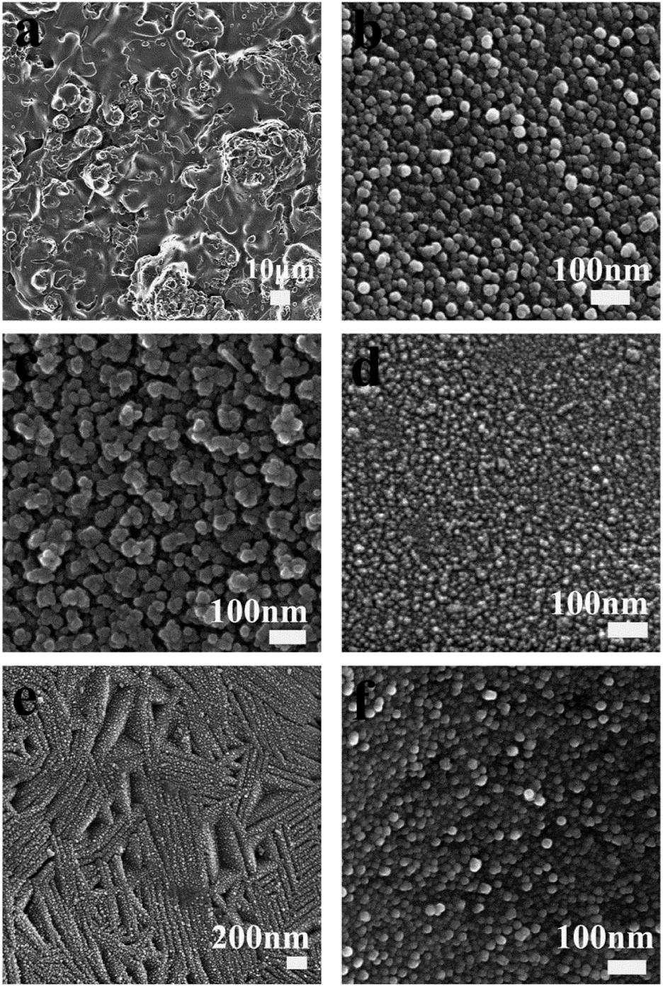 | ||
| Fig. 2 SEM images of the surface of the TiO2 coatings doped with different percentage of Nb2O5: (a) Low magnification image of the pure TiO2 coating showing the typical rough surface of plasma sprayed coatings; High magnification views of: (b) the pure TiO2 coating, (c) 10%Nb2O5/TiO2; (d) 30%Nb2O5/TiO2; (e) 50%Nb2O5/TiO2 and (f) Nb2O5 coatings. | ||
 | ||
| Fig. 3 Surface roughness of the (a) TiO2, (b) 10%Nb2O5/TiO2, (c) 30%Nb2O5/TiO2, (d) 50%Nb2O5/TiO2 and (e) Nb2O5 coatings, *p < 0.05. | ||
Fig. 4a–e depict the cross-sectional morphologies of the TiO2, Nb2O5 doped TiO2 and Nb2O5 coatings. The TiO2 coating and the doped TiO2 coatings have a similar thickness of about 40 μm, but the thickness of Nb2O5 coating is only 20 μm, which may be due to the low deposition efficiency of Nb2O5 powders driven by its lower density during the reconstituted process. The bonding strength of all types of coatings is presented in Fig. 4f. The TiO2, 10%Nb2O5/TiO2, 30%Nb2O5/TiO2 and the Nb2O5 coatings have similar bonding strength of around 20 MPa, while the 50%Nb2O5/TiO2 coating displayed the highest bonding strength (31.0 ± 2.9 MPa) among all the types of coatings tested.
 | ||
| Fig. 4 Cross-sectional images of the (a) TiO2, (b) 10%Nb2O5/TiO2, (c) 30%Nb2O5/TiO2, (d) 50%Nb2O5/TiO2, (e) Nb2O5 coatings and (f) their bonding strength, *p < 0.05. | ||
3.3. Corrosion resistance
Fig. 5 presents the potentiodynamic polarization curves of the TiO2, 10%Nb2O5/TiO2, 30%Nb2O5/TiO2, 50%Nb2O5/TiO2 and Nb2O5 coatings tested in SBF solution at 37 °C. The corrosion potential (Ecorr) and corrosion current density (Icorr) values of all the coatings are listed in Table 2. The electrode potential is an indicator of corrosion activity; the more negative the potential is, the worse the corrosion resistance will be. The Ecorr of the Nb2O5 coating is −200 mV, much higher than that of the TiO2 coating (−347 mV), indicating that the Nb2O5 coating exhibits better corrosion resistance than that of the TiO2 coating. The addition of 10% and 50%Nb2O5 improved the corrosion resistance of the TiO2 coating, reflecting in the increase in the Ecorr of the 10% and 50%Nb2O5 doped coatings, compared to that of the TiO2 coating (Table 2). It is worth noting that the Ecorr of the 30%Nb2O5/TiO2 coating (−175 mV) is the highest among the five types of coatings tested, suggesting that the addition of 30%Nb2O5 in the TiO2 coating results in a coated surface with the best corrosion resistance. The Nb2O5 coating has the lowest Icorr value while the TiO2 coating has the highest, indicating that the TiO2 coating has the fastest corrosion rate while the Nb2O5 coating displaying the slowest corrosion rate. The corrosion rate for the Nb2O5 doped TiO2 coatings falls in between those of TiO2 and Nb2O5 coatings. The 30%Nb2O5/TiO2 coating exhibits the lowest corrosion rate and no significant difference can be found between the other two Nb2O5 doped TiO2 coatings. These results suggest that Nb2O5 has a dose-dependent effect on the improvement of the corrosion resistance of the TiO2 coating. | ||
| Fig. 5 Potentiodynamic polarization curves of the TiO2, 10%Nb2O5/TiO2, 30%Nb2O5/TiO2, 50%Nb2O5/TiO2 and Nb2O5 coatings. | ||
| Coating | Ecorr (mV) | Icorr (μA cm−2) |
|---|---|---|
| TiO2 | −347 | 2.25 |
| 10%Nb2O5/TiO2 | −326 | 1.57 |
| 30%Nb2O5/TiO2 | −175 | 1.05 |
| 50%Nb2O5/TiO2 | −312 | 1.56 |
| Nb2O5 | −200 | 0.74 |
3.4. Osteoblast adhesion and proliferation
Fig. 6 depicts representative confocal images of HOBs seeded on the surface of the TiO2, 10%Nb2O5/TiO2, 30%Nb2O5/TiO2, 50%Nb2O5/TiO2 and Nb2O5 coatings for 2 hours. After culturing for 2 hours, HOBs flattens on the surfaces of all the coatings. However, fewer HOBs attached to the surface of the 10%Nb2O5/TiO2 coating (Fig. 6b), compared to those on the surface of the TiO2, 30%Nb2O5/TiO2, 50%Nb2O5/TiO2 and Nb2O5 coatings (Fig. 6a and c–e).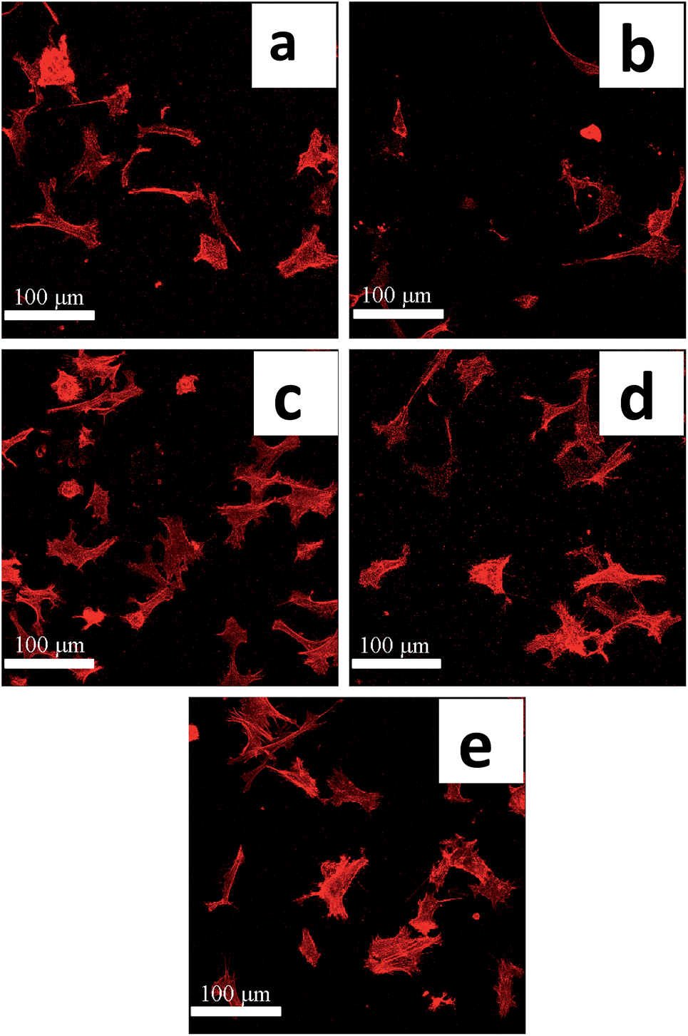 | ||
| Fig. 6 Confocal images of HOBs seeded for 2 hours on the surface of the (a) TiO2, (b) 10%Nb2O5/TiO2, (c) 30%Nb2O5/TiO2, (d) 50%Nb2O5/TiO2 and (e) Nb2O5 coatings. | ||
Fig. 7 depicts SEM images of HOBs cultured for 2 hours on the surface of the TiO2, 10%Nb2O5/TiO2, 30%Nb2O5/TiO2, 50%Nb2O5/TiO2 and Nb2O5 coatings. HOBs cultured on all the types of coatings attach well to the coating surface. Cells cultured on the TiO2 and the Nb2O5 doped TiO2 coatings (Fig. 7a–h) spread out and interact with the coated surfaces, while those on the Nb2O5 coatings were less spread.
 | ||
| Fig. 7 SEM micrographs of HOBs cultured on the (a and b) TiO2, (c and d) 10%Nb2O5/TiO2, (e and f) 30%Nb2O5/TiO2, (g and h) 50%Nb2O5/TiO2 and (i and j) Nb2O5 coatings (images (b) (d) (f) (h) and (j) are under higher magnifications). | ||
Fig. 8 depicts the proliferation of HOBs on the TiO2, 10%Nb2O5/TiO2, 30%Nb2O5/TiO2, 50%Nb2O5/TiO2 and Nb2O5 coatings. The proliferation rates of HOBs on all coatings increase with the increase in the culture time. In the initial 3 days of culture, the HOB proliferation rate on 50%Nb2O5/TiO2 coating is higher than those on the other coatings. After culturing for 7 days, similar proliferation rates was seen for TiO2, 30%Nb2O5/TiO2, 50%Nb2O5/TiO2 and Nb2O5, which are significantly higher than that of HOBs cultured on the 10%Nb2O5/TiO2. At day 14, significantly higher proliferation rate was observed for the HOBs cultured on the 50%Nb2O5/TiO2 coating, compared to those cultured on the other coatings.
 | ||
| Fig. 8 HOB proliferation on the TiO2, 10%Nb2O5/TiO2, 30%Nb2O5/TiO2, 50%Nb2O5/TiO2 and Nb2O5 coatings after culturing for 3, 7 and 14 days, *p < 0.05. | ||
The in vitro bioactivity studies suggest that the addition of 30% and in particular 50%Nb2O5 enhances the bioactivity of the TiO2 coating, whereas the addition of 10%Nb2O5 slightly compromises the bioactivity of the TiO2 coating, indicating that the impact of Nb2O5 on the bioactivity of TiO2 coatings is dependent on its doped amount.
4. Discussion
The surface characteristics of artificial implant materials, such as chemical composition, topography, roughness and hydrophilicity, are critical for the success of the implant.21–23 In this study, to improve the surface properties of TiO2 coatings for their better application as biomedical coatings, a chemically stable and biocompatible oxide (Nb2O5)10,13,24 was incorporated into TiO2 coatings using plasma spraying, which not only improved the corrosion resistance properties, but also enhanced the bioactivity and biocompatibility of the TiO2 coatings.14 Importantly, we found that the addition of Nb2O5 can refine the nanotopographic features of the TiO2 coating, thus providing an approach capable of changing surface chemistry and refining surface nanotopography in one simple physical process. As the importance of both surface chemistry and nanotopography on the biological performance of biomedical implants, the findings in our study may provide new insight of innovative application of traditional plasma spaying in future biomedical implant coating design.Our results demonstrated that small amounts of Nb2O5 does not affect the crystalline structures of the TiO2 coatings as Nb is intercalated into the spaces between TiO2 octahedron sheets during the coating process (solidification).25–27 This intercalation resulted in shifting of the TiO2 peaks to lower angles in the XRD pattern for the Nb2O5 doped TiO2 coatings (Fig. 1b and c). In contrast, increase in the doped amount of Nb2O5 to 50 wt% resulted in the formation of a new solid solution phase (Ti0.95Nb0.95O4) and a small amount of anatase phase (Fig. 1d) on the TiO2 coatings (Fig. 1d). Since TiO2 coating exists in the form of rutile phase in the TiO2 coatings (Fig. 1a), the formation of the small amount of anatase phase is possible because Nb doping inhibits phase transformation of TiO2 from anatase to rutile in the solidification process. This is in agreement with others who demonstrated the inhibition effect of Nb on the anatase-rutile transformation.27,28 Interestingly, accompanying the changes in the crystalline phase, the nanotopography of the coatings evolved from nanostructures composed of nanosized spherical grains (the TiO2, Nb2O5, 10 wt% and 30 wt% Nb2O5/TiO2 coatings) into nanoplate network structures (the 50%Nb2O5/TiO2 coatings) (Fig. 2a–f). The formation of this special nanostructure was supposed to be ascribed to the recrystallization of the molten particles during the rapid solidification process of plasma spraying.21 Under the unique thermal conditions of plasma spraying (high flame temperature and ultra-rapid cooling rate), the growth of crystallites in the process of recrystallization is strongly suppressed, leading to the formation of the nano-sized grains.21,29 The formation of this nanostructure, which is based on the rapid solidification under the high cooling rate, is influenced by many factors including the temperature of the substrate, spraying processing parameters (e.g. powers, ratio of H2/Ar) and the auxiliary cooling conditions.30 In addition, the thermo-physical property of the spraying material itself is also of great importance to recrystallization process, thus influencing the final size and shape of the nanotopographic features on the surface. Among all the Nb2O5/TiO2 coatings, the most prominent changes in the nanotopography occurred in the TiO2 coating incorporated by 50%Nb2O5, was thought to be ascribed to the formation of the new solid solution phase which is highly possible to cause the changes in the thermodynamics of the recrystallization process.
The interfacial bonding between the coating and the underlying substrate plays an important role in the long-term stability of the implants and a strong bonding between the implant and the coating is a critical in avoiding coating delamination.31,32 Among all the five types of coatings tested in this study, the 50%Nb2O5/TiO2 coating exhibits the highest bonding strength to the underlying substrate (Fig. 4f), whereas addition of 10 wt% and 30 wt% Nb2O5 has no prominent effect on the bonding strength of the TiO2 coating. Coefficient of thermal expansion (CET) is a critical factor in influencing the quality of the coatings and their interface with the underlying substrates. The CETs of TiO2, Nb2O5 and cp-Ti were reported to be 8.0–10.0 × 10⁻⁶ K−1,33 5.3–5.9 × 10⁻⁶ K−1,34 and 9.7–9.9 × 10⁻⁶ K−1,35 respectively. Due to the mismatch of the CETs between the cp-Ti and Nb2O5, the addition of 10% and 30% into the TiO2 matrix does not significantly improve the bonding strength of the TiO2 coating (Fig. 4f). However, prominent improvement in the interfacial bonding strength is observed when the amount of Nb2O5 increases to 50%, which is highly possible because of the formation of the new Ti0.95Nb0.95O4 phase. We speculate that Ti0.95Nb0.95O4 phase might have a CET much closer to that of cp-Ti substrate. In-depth investigation into the effect of Nb2O5 amount on the CETs of Nb2O5/TiO2 composite coatings will be carried out in our future study to validate this hypothesis.
Corrosion resistance is one of the most important surface properties determining the overall success of an orthopaedic implant; it influences the stability and biosafety of the implants by inhibiting the release of non-compatible metal ions which can cause allergic and toxic reactions.5,36 This study demonstrated that plasma sprayed Nb2O5 coatings have superior corrosion resistance compared to the TiO2 coatings, reflected in both Ecorr and Icorr values (Fig. 5 and Table 2). We further confirmed that the addition of Nb2O5 into TiO2 matrix improves the corrosion resistance of the TiO2 coating in a dose-dependent manner. Compared to the TiO2 coatings doped with 10% and 50%Nb2O5, 30%Nb2O5/TiO2 coatings display better corrosion resistance and a lower corrosion rate (Table 2). The corrosion resistance ability of a material is influenced by its physical chemistry properties. It was reported that there is a negative correlation between the pitting potential of the oxide coated metal and the point of zero charge (PZC) of the oxide.13 A lower PZC indicates a higher pitting potential and better corrosion resistance.37 The PZC of Nb2O5 was reported to be 2.8, much lower than that of TiO2 (5.2), resulting in a better corrosion resistance of Nb2O5 than that of TiO2.13,37,38 As the 10%Nb2O5/TiO2 and 30%Nb2O5/TiO2 coatings are composed of TiO2 and Nb2O5 phases, the PZCs of these composite coatings should fall between the PZCs of the TiO2 and Nb2O5. Due to the increased amount of Nb2O5, the PZC of the 30%Nb2O5/TiO2 coating is expected to be lower compared to that of the 10%Nb2O5/TiO2 coating, which accounts for the better corrosion resistance of the 30%Nb2O5/TiO2 coating. However, as the doped amount of Nb2O5 escalated up to 50%, the corrosion resistance of the TiO2 coating is not further improved. To explain this, we hypothesized that the PZC of the 50%Nb2O5/TiO2 coating is possibly higher than that of the 30%Nb2O5/TiO2 coating due to the formation of the new solid solution of Ti0.95Nb0.95O4.
Cell adhesion and proliferation are two important biological reactions of the osteoblast to the underlying substrate surface, which can be greatly influenced by the implant surface properties. The TiO2 and Nb2O5 coatings have comparable biocompatibility (here specific to cellular adhesion and proliferation). In agreement with our previous finding14 and that of others,12,13 we demonstrated in this study that the incorporation of Nb2O5 into TiO2 matrix improves the biocompatibility of the TiO2 coatings, however obvious enhancement effect is only observed when the amount of Nb2O5 increases up to 50% (Fig. 6–8). Although the exact mechanism(s) behind this dose-dependent effect of Nb2O5 on the biocompatibility is still unclear, we speculate that the differences in the surface chemistry and topography among these different types of coatings resulted in this dose-dependent effect on the coatings biocompatibility.
Compared to the TiO2 and the other Nb2O5 doped TiO2 coatings, 50%Nb2O5/TiO2 coatings not only have modified surface chemistry but also possess special surface topography. The addition of 50%Nb2O5 into TiO2 matrix changes the surface chemical compositions such that a larger amount of Nb element is exposed on the surface to interact with the seeded osteoblasts. Nb is a biocompatible element and has been recently used to enhance the biocompatibility of biomaterials.9–13 For example, Ding et al.39 reported that Nb doped TiO2 nanotubes displayed an enhanced in vitro bioactivity and promoted mesenchymal stem cell adhesion and the formation of extracellular matrix, compared to the undoped TiO2 nanotubes. Topographically, 50%Nb2O5/TiO2 coating not only possesses increased surface micro-roughness but also have unique surface nanostructure, contributing to its enhanced biocompatibility. Micro-roughness has been reported to increase bone anchorage and reinforce the biomechanical interlocking between the bone and the implant.40 The effect of nanostructure on cell behaviors has been widely studied and it was revealed that the influence of the nanotopographical features is dependent on their physical status, e.g. shapes and sizes.41–43 We previously demonstrated that the nanosized rod-shaped hydroxyapatite more efficiently enhanced the cellular activity compared to nanosized spherical hydroxyapatite.44 In this study, the nanoplate formed on the surface of the 50%Nb2O5/TiO2 coating exhibits a rod-liked shape with a high aspect ratio, which is the predominant topographical difference of the 50%Nb2O5 doped coating from the others. Therefore, it is plausible to suggest that the unique nanostructure formed on the 50%Nb2O5/TiO2 coating also accounts largely for the better biocompatibility of the coating.
In summary, in this study, we establish that incorporation of Nb2O5 can improve the corrosion resistance, bonding strength and biocompatibility of TiO2 coatings in a dose-dependent manner. This study indicates the potential application of Nb2O5 in improving the long-term performance of Ti-based implants through changes in surface chemistry and surface nanotopography.
5. Conclusions
To improve the biological performance of currently used Ti-based implants and shed light on the potential of Nb2O5 for biomedical application, TiO2 coatings doped with different percents of Nb2O5 were deposited on cp-Ti substrates by APS. Coating properties relating to the long-term performance, e.g. corrosion resistance, interfacial bonding strength and biocompatibility were studied in correlation with the Nb2O5 dopant amount. The addition of Nb2O5 does not change the crystalline structure of the TiO2 at low amounts (10% and 30%), however, a new solid solution phase is formed when the doped amount of Nb2O5 increases to 50%, which is highly possible reason for the formation of the unique nanotopography composed of nanoplate network. Results demonstrated a dose-dependent effect of Nb2O5 on the corrosion resistance, biocompatibility and interfacial bonding strength of the TiO2 coating. The 50%Nb2O5 doped TiO2 coating exhibited the best biocompatibility and the highest bonding strength. However, improvement in the corrosion resistance of the 50%Nb2O5 coatings was relatively less predominant than the 30%Nb2O5 doped TiO2 coating. Further study will be carried out to find the best composition ratio of Nb2O5 and TiO2 between 30% and 50%, with which the composite coating possesses the best balanced overall properties.Acknowledgements
The authors give thanks to Jiangsu Overseas Research & Training Program for University Prominent Young & Middle-aged Teachers and Presidents and Australia National Health and Medical Research Council (NHMRC), Australian Research Council (ARC) and the Rebecca Cooper Foundation. The authors thank Australian Centre for Microscopy & Microanalysis for their assistance in Micro/Nano analysis.Notes and references
- M. Geetha, A. K. Singh, R. Asokamani and A. K. Gogia, Prog. Mater. Sci., 2009, 54, 397 CrossRef CAS PubMed.
- X. Zhu, J. Chen, L. Scheideler, R. Reichl and J. Geis-Gerstorfer, Biomaterials, 2004, 25, 4087 CrossRef CAS PubMed.
- M. Long and H. J. Rack, Biomaterials, 1998, 19, 162 CrossRef.
- J. M. Gomez-Vega, E. Saiz and A. P. Tomsia, J. Biomed. Mater. Res., 1999, 46, 549 CrossRef CAS.
- N. Zaveri, M. Mahapatra, A. Deceuster, Y. Peng, L. Li and A. Zhou, Electrochim. Acta, 2008, 53, 5022 CrossRef CAS PubMed.
- A. Ochsenbein, F. Chai, S. Winter, M. Traisne, J. Breme and H. F. Hildebrand, Acta Biomater., 2008, 4, 1506 CrossRef CAS PubMed.
- H. Shao, C. Yu, X. Xu, J. Wang, R. Zhai and X. Wang, Appl. Surf. Sci., 2010, 257, 1649 CrossRef CAS PubMed.
- H. Matsuno, A. Yokoyama, F. Watari, M. Uo and T. Kawasaki, Biomaterials, 2001, 22, 1253 CrossRef CAS.
- R. L. Karlinsey, A. T. Hara, K. Yi and C. W. Duhn, Biomed. Mater., 2006, 1, 16 CrossRef CAS PubMed.
- E. Eisenbarth, D. Velten and J. Breme, Biomol. Eng., 2007, 24, 27 CrossRef CAS PubMed.
- F. Y. Zhou, B. L. Wang, K. J. Qiu, W. J. Lin, L. Li, Y. B. Wang, F. L. Nie and Y. F. Zheng, Mater. Sci. Eng., C, 2012, 32, 851 CrossRef CAS PubMed.
- Y. B. Wang and Y. F. Zheng, Mater. Lett., 2009, 63, 1293 CrossRef CAS PubMed.
- T. Zhao, Y. Li, Y. Xiang, X. Zhao and T. Zhang, Surf. Coat. Technol., 2011, 205, 4404 CrossRef CAS PubMed.
- X. Zhao, G. Wang, H. Zheng, Z. Lu, X. Zhong, X. Cheng and H. Zreiqat, ACS Appl. Mater. Interfaces, 2013, 5, 8203 CAS.
- G. Wang, F. Meng, C. Ding, P. K. Chu and X. Liu, Acta Biomater., 2010, 6, 990 CrossRef CAS PubMed.
- G. Wang, Z. Lu, X. Liu, X. Zhou, C. Ding and H. Zreiqat, J. R. Soc., Interface, 2011, 8, 1192 CrossRef CAS PubMed.
- G. Wang, Z. Lu, D. Dwarte and H. Zreiqat, Mater. Sci. Eng., C, 2012, 32, 1818 CrossRef CAS PubMed.
- G. Wang, Z. Lu, K. Y. Xie, W. Y. Lu, S. I. Roohani-Esfahani, A. Kondyurin and H. Zreiqat, J. Mater. Chem., 2012, 22, 19081 RSC.
- G. Wang, X. Liu, J. Gao and C. Ding, Acta Biomater., 2009, 5, 2270 CrossRef CAS PubMed.
- C. W. Kang and H. W. Ng, Surf. Coat. Technol., 2006, 200, 5462 CrossRef CAS PubMed.
- G. Wang, X. Liu, H. Zreiqat and C. Ding, Colloids Surf., B, 2011, 86, 267 CrossRef CAS PubMed.
- R. G. Flemming, C. J. Murphy, G. A. Abrams, S. L. Goodman and P. F. Nealey, Biomaterials, 1999, 20, 573 CrossRef CAS.
- M. Lampin, R. Warocquier-Clerout, C. Legris, M. Degrange and M. F. Sigot-Luizard, J. Biomed. Mater. Res., 1997, 36, 99 CrossRef CAS.
- E. Eisenbarth, D. Velten, M. Müller, R. Thull and J. Breme, J. Biomed. Mater. Res., Part A, 2006, 79, 166 CrossRef CAS PubMed.
- K. A. Michalow, D. Flak, A. Heel, M. Parlinska-Wojtan, M. Rekas and T. Graule, Environ. Sci. Pollut. Res., 2012, 19, 3696 CrossRef CAS PubMed.
- C. Liu, L. Miao, J. Zhou, R. Huang and S. Tanemura, J. Mater. Chem., 2012, 22, 14180 RSC.
- M. Hirano and Y. Ichihashi, J. Mater. Sci., 2009, 44, 6135 CrossRef CAS.
- J. Arbiol, J. Cerdà, G. Dezanneau, A. Cirera, F. Peiró, A. Cornet and J. R. Morante, J. Appl. Phys., 2002, 92, 853 CrossRef CAS PubMed.
- F. I. Trifa, G. Montavon, C. Coddet, P. Nardin and M. Abrudeanu, Mater. Charact., 2005, 54, 157 CrossRef CAS PubMed.
- S. Sampath and H. Herman, J. Therm. Spray Technol., 1996, 5, 445 CrossRef CAS.
- Y. C. Yang and B. Y. Chou, Mater. Chem. Phys., 2007, 104, 312 CrossRef CAS PubMed.
- W. Q. Yan, T. Nakamura, K. Kawanabe, S. Nishigochi, M. Oka and T. Kokubo, Biomaterials, 1997, 18, 1185 CrossRef CAS.
- S. P. Singh, K. Pal, A. Tarafder, M. Das, K. Annapurna and B. Karmakar, Bull. Mater. Sci., 2010, 33, 33 CrossRef CAS PubMed.
- W. R. Manning, O. Hunter Jr, F. W. Calderwood and D. W. Stacy, J. Am. Ceram. Soc., 1972, 55, 342 CrossRef CAS PubMed.
- S. Zinelis, A. Tsetsekou and T. Papadopoulos, J. Prosthet. Dent., 2003, 90, 332 CrossRef CAS.
- R. Karpagavalli, A. Zhou, P. Chellamuthu and K. Nguyen, J. Biomed. Mater. Res., Part A, 2007, 83, 1087 CrossRef PubMed.
- P. M. Natishan, E. McCafferty and G. K. Hubler, J. Electrochem. Soc., 1988, 135, 321 CrossRef CAS PubMed.
- M. Kosmulski, J. Colloid Interface Sci., 2006, 298, 730 CrossRef CAS PubMed.
- D. Ding, C. Ning, L. Huang, F. Jin, Y. Hao, S. Bai, Y. Li, M. Li and D. Mao, Nanotechnology, 2009, 20, 305103 CrossRef PubMed.
- G. Mendonca, D. B. S. Mendonca, F. J. L. Aragão and L. F. Cooper, Biomaterials, 2008, 29, 3822 CrossRef CAS PubMed.
- T. Amna, M. S. Hassan, W. S. Shin, H. V. Ba, H. K. Lee, M. S. Khil and I. H. Hwang, Colloids Surf., B, 2013, 101, 424 CrossRef CAS PubMed.
- L. Zhao, L. Liu, Z. Wu, Y. Zhang and P. K. Chu, Biomaterials, 2012, 33, 2629 CrossRef CAS PubMed.
- C. Y. Chiang, S. H. Chiou, W. E. Yang, M. L. Hsu, M. C. Yung, M. L. Tsai, L. K. Chen and H. H. Huang, Dent. Mater., 2009, 25, 1022 CrossRef CAS PubMed.
- S. I. Roohani-Esfahani, S. Nouri-Khorasani, Z. Lu, R. Appleyard and H. Zreiqat, Biomaterials, 2010, 31, 5498 CrossRef CAS PubMed.
Footnote |
| † Present addresses, PP Miramon (CIC BIOMAGUNE) No. 182, PTA C, 20009, Donostia/S Sebstian, Gipuzkoa, Spain; Tel: +34 943005321. |
| This journal is © The Royal Society of Chemistry 2014 |