Haitao Wangab,
Fu-Quan Baib,
Xiaoshi Jiaa,
Di Caoa,
Ravva Mahesh Kumarc,
Jean-Luc Brédasc,
Songnan Qud,
Binglian Baie,
Hong-Xing Zhang*b and
Min Li*a
aKey Laboratory of Automobile Materials (MOE), College of Materials Science and Engineering, Jilin University, Changchun 130012, China. E-mail: minli@mail.jlu.edu.cn
bKey Laboratory of Theoretical and Computational Chemistry, Institute of Theoretical Chemistry, Jilin University, Changchun 130023, China. E-mail: zhanghx@mail.jlu.edu.cn
cDivision of Physical Sciences and Engineering, King Abdullah University of Science and Technology–KAUST, Thuwal 23955-6900, Kingdom of Saudi Arabia
dKey Laboratory of Excited State Processes, Changchun Institute of Optics, Fine Mechanics and Physics, Chinese Academy of Sciences, Changchun 130033, China
eCollege of Physics, Jilin University, Changchun 130012, China
First published on 9th October 2014
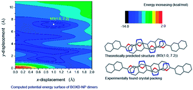
The molecular aggregation structure of 5,5′-bis(naphthalen-2-yl)-2,2′-bi(1,3,4-oxadiazole) (BOXD-NP) was studied by computing the intermolecular interaction potential energy surface (PES) at density functional theory level based on a dimer model. All B3LYP, CAM-B3LYP and M062x functionals can yield a reliable isolated molecular geometry. The conformation of BOXD-NP obtained with all methods is perfectly planar, indicating good conjugation ability between oxadiazole and naphthalene rings. The vibrational frequencies of BOXD-NP were also calculated using the B3LYP/6-311+G** method, which showed great consistency with the experimental observations and makes the assignments of the IR spectra more solid. It was revealed that the lowest excited state of BOXD-NP should be assigned as a highly allowed π–π* state by TD-DFT calculation. Considering the non-covalent interactions in molecular aggregates, the M062x functional was applied in the construction of the PES. Besides the packing structure found in the crystals, PES also predicted several stable structures, indicating that PES has great ability in guiding molecular self-assembly. Symmetry Adapted Perturbation Theory (SAPT) analysis on these energy-minimum molecular stacking structures revealed that London dispersion forces are the strongest attractive component in the binding.
The direct reason for this situation might be ascribed to the fact that there is no efficient strategy to obtain the detailed structural information of the active thin-film components. Exploring the molecular aggregation structures is still challenging for either theoretical or experimental investigations.21–23 As for the theoretical work, it is always hard to involve Van der Waals interactions into large systems. Until recently, great progress has been made by empirical correction for long-range dispersion effect (DFT-D)24 or doubling the amount of non-local exchange (M062x),25 which are based on different idea, but both yield quite well results. And for the experimental case, it is usually hard to get the detailed structural information, except some valuable results obtained from single crystals26–32 and Langmuir–Blodgett films.33,34
Even so, these static structures observed in experiment, for example in single crystal X-ray diffraction, are generally obtained by chance. It is desirable if we can predict or control the molecular packing order. The molecular self-assembled structures are usually precisely controlled by many types of weak intermolecular interactions, for example dipole–dipole interactions, hydrogen bonding, and Van der Waals forces. It could be expected that the intermolecular interaction potential energy surface (PES) and further energy decomposition analysis would have great ability in guiding the molecular self-assembly. Here we report the computed intermolecular interaction potential energy surface of 5,5′-bis(naphthalen-2-yl)-2,2′-bi(1,3,4-oxadiazole) (BOXD-NP, see Fig. 1) at M062x/6-31G** theoretical level based on a dimer model. The stable dimer structures predicted by the PES are further compared with the structure that was found in the crystalline state. Symmetry Adapted Perturbation Theory (SAPT) analysis on these theoretically predicted stable molecular stacking structures and crystal packing was carried out to get deep understanding of the molecular self-assembly.
The monomer structure of BOXD-NP in the ground state was optimized at density functional theory (DFT) level (B3LYP, CAM-B3LYP and M062x). In all calculations for the monomer, the standard 6-311+G** basis set was employed. The intermolecular interaction potential energy surface (PES) was constructed by calculating the single point energies with a face-to-face dimer model, where the non-covalent interactions were considered by applying M062x functional, which has been proved having well performance in investigations of main-group thermo-chemistry, kinetics, non-covalent interactions, and electronic structures.25 The monomer geometry obtained at M062x/6-311+G** level was used for the further PES scan of BOXD-NP dimers. The energy scan starts from the ideal face-to-face π-stacked structure with a intermolecular separation of 3.34 Å (taken from the crystal data),35 and then scan over the displacement along both molecular long axis (y-displacement, y for short, 0 ≤ y ≤ 11.0 Å) and short axis (x-displacement, x for short, 0 ≤ x ≤ 2.0 Å) with the steps of 0.2 Å. All these calculations of single point energy for molecular dimers were done at M062x/6-31G** level, and the function counterpoise procedure was utilized to correct the basis set superposition error (BSSE). The excitation energy for monomer was computed by TD-DFT approach (TD-B3LYP, TD-CAM-B3LYP and TD-M062x). Considering the non-covalent interactions would play an important role in determining the electronic and optical properties of molecular aggregates, TD-M062x functional only was employed to predict the electronic excitation energy for the BOXD-NP dimers. Similarly, standard 6-311+G** and 6-31G** basis sets were used for monomer and dimer calculations, respectively. All of the calculations were carried out with Gaussian 09 software package (version A.02).36
The energy decomposition analysis was carried out with an Open-Source ab initio Electronic Structure Package PSI 4.0.0-beta4 driver (SAPT0).37
Crystal dataa | B3LYP | CAM-B3LYP | M062x | |
---|---|---|---|---|
a Crystal data was taken from ref. 35. | ||||
Bond lengths (Å) | ||||
O1–C1 | 1.357 | 1.357 | 1.348 | 1.345 |
O1–C2 | 1.364 | 1.365 | 1.356 | 1.354 |
N1–C1 | 1.289 | 1.297 | 1.286 | 1.288 |
N1–N2 | 1.403 | 1.383 | 1.379 | 1.378 |
N2–C2 | 1.299 | 1.303 | 1.293 | 1.295 |
C1–C1′ | 1.443 | 1.442 | 1.445 | 1.448 |
C2–C3 | 1.459 | 1.455 | 1.456 | 1.458 |
C3–C12 | 1.371 | 1.383 | 1.373 | 1.375 |
C3–C4 | 1.418 | 1.423 | 1.418 | 1.420 |
C6–C7 | 1.419 | 1.418 | 1.416 | 1.418 |
C6–C11 | 1.423 | 1.432 | 1.420 | 1.422 |
C7–C8 | 1.360 | 1.375 | 1.367 | 1.370 |
C8–C9 | 1.404 | 1.416 | 1.413 | 1.416 |
C9–C10 | 1.360 | 1.373 | 1.366 | 1.369 |
C10–C11 | 1.413 | 1.421 | 1.418 | 1.420 |
C4–C5 | 1.363 | 1.371 | 1.363 | 1.367 |
C5–C6 | 1.417 | 1.420 | 1.417 | 1.420 |
C11–C12 | 1.410 | 1.413 | 1.411 | 1.413 |
![]() |
||||
Angles (°) | ||||
C1–O1–C2 | 101.84 | 102.47 | 102.51 | 102.37 |
C1–N1–N2 | 105.54 | 106.14 | 106.07 | 105.91 |
C2–N2–N1 | 106.25 | 106.92 | 106.83 | 106.71 |
N1–C1–O1 | 113.78 | 112.78 | 112.85 | 113.10 |
N2–C2–O1 | 112.58 | 111.70 | 111.74 | 111.92 |
O1–C1–C1′ | 118.28 | 118.84 | 118.85 | 118.78 |
O1–C2–C3 | 119.18 | 119.31 | 119.24 | 119.31 |
The main geometry parameters are collected in Table 1. For comparison, the crystal data is also included in Table 1. The values of bond lengths and angles obtained in calculation are very close to that from crystal data; it could be noticed that the biggest difference between the calculation and experimental found was the bond length of N1–N2 (0.025 Å), and the angle of N1–C1–O1 (1°). The root mean square (rms) deviation between the calculated and experimental results was found to be less than 0.01 Å for bond lengths and 0.7° for angles for all these three methods. This comparison indicates that all the three methods can get reliable ground state molecular geometry.
Calculated | Observed | Assignmentc (main contributions) | ||
---|---|---|---|---|
Freq.a (cm−1) | Int. | Freq. (cm−1) | Int.b | |
a The frequency data are scaled from the calculated B3LYP/6-311+G** data by a factor of 0.9688.42b vs, very strong; s, strong; m, medium; sh, shoulder; w, weak.c NP, naphthalene; OXD, oxadiazole; def., deformation; υ, stretching vibration; δ(Ar–H), Ar–H in plane bending; α, in plane bending; ε(Ar–H), Ar–H out of plane bending; τ, out of plane bending. | ||||
3102 | 4.70 | υs, υas(Ar–H) | ||
3092 | 49.35 | 3060 | m | |
3089 | 1.60 | |||
3080 | 41.65 | 3052 | m | |
3072 | 11.62 | |||
3068 | 0.44 | |||
3064 | 2.91 | |||
1614 | 7.62 | 1628 | m | NP ring def., υ(C![]() |
1590 | 21.86 | 1600 | m | |
1557 | 3.36 | 1577 | m | |
1528 | 395.92 | 1549 | vs | NP, OXD ring def., υ(C2–N2, C2′–N2′) (−) |
1485 | 232.57 | 1498 | vs | υ(C![]() |
1446 | 68.25 | 1464 | s | |
1440 | 50.83 | 1453 | sh | NP, OXD ring def., υ(C1–N1, C1′–N1′) (−) |
1422 | 44.07 | 1437 | s | δ(Ar–H) |
1365 | 0.35 | 1400 | w | NP ring def., υ(C![]() |
1354 | 6.77 | 1387 | w | |
1349 | 33.38 | 1361 | s | δ(Ar–H) |
1274 | 12.42 | 1298 | m | α(N2–C2–C3–C12), δ(Ar–H) |
1250 | 20.93 | 1272 | m | α(C5–C6–C7) (C10–C11–C12), δ(Ar–H) |
1226 | 12.58 | 1240 | m | α(C5–C6–C11) (C6–C11–C12), δ(Ar–H) |
1189 | 16.53 | 1201 | s | To and fro (C5–C6), δ(Ar–H) |
1146 | 14.93 | 1153 | s | δ(Ar–H) |
1140 | 0.32 | |||
1131 | 3.99 | α(C1–N1–N2) (O1–C2–N2), δ(Ar–H) | ||
1114 | 52.20 | 1129 | s | |
1073 | 97.25 | 1084 | vs | α(C3–C2–N2) (C2–C3–C12), δ(Ar–H) |
1009 | 3.78 | 1036 | w | υ(N–N), υ(C3–C4) (C8–C9) (−), δ(Ar–H) |
1003 | 62.11 | 1002 | s | |
961 | 983 | w | ε(Ar–H) | |
956 | 15.02 | 968 | s | α(C2–N2–N1) (C3–C4–C5) |
947 | 15.04 | 954 | s | α(C2–O1–C1), α(N2–N1–C1) |
976 | ||||
946 | 5.62 | ε(Ar–H) | ||
924 | 9.52 | 939 | s | NP, OXD ring def., α(O1(N2)–C2–C3⋯12) |
904 | 23.32 | 912 | s | ε(Ar–H) |
853 | 24.62 | 873 | s | |
832 | 6.89 | 846 | s | NP, OXD ring def., α(N2–C2–C3⋯12) |
811 | 59.01 | 828 | vs | ε(Ar–H) + OXD ring def., τ(N2–C2–O1) |
754 | 2.33 | υ(C6–C11) | ||
752 | 3.45 | NP ring out of plane def., τ(C–C–C), ε(Ar–H) | ||
744 | 107.74 | 758 | vs | ε(Ar–H), τ(C–C–C) (N2–C2–O1) |
751 | sh | |||
711 | 1.59 | 720 | s | |
655 | 2.46 | 668 | s | OXD ring def., τ(N2–N1–C1) |
632 | 1.50 | 591 | s | NP ring def., α(C–C–C), (C4–C5–C6–C7–C8) |
Based on these calculations, the observed absorption bands in the range of 3000–3100 cm−1 with medium intensity are assigned to symmetric (υs) and asymmetric (υas) stretching vibrations of C–H in the naphthalene rings. The absorption bands in the range of 1000–1700 cm−1 are mainly attributed to the in-plane deformation vibrations of naphthalene (NP) and oxadiazole (OXD) rings, including the stretching modes of CC, C
N, and C–C, as well as in-plane bending modes C–C–C, C–N–N, C–O–C, and Ar–H. All these in-plane modes are delocalized and can not be assigned to the vibration of any individual bond or angle, therefore, only main contributions are list in Table 2. Some skeleton in-plane deformation vibrations which include almost all the atoms in the backbone are founded in very low energy (below 1000 cm−1, for example, at 924, 832, and 632 cm−1 (calculated)). The out of plane deformation modes, such as torsion of C–C–C, N–C–O, and N–N–C, as well as out of plane bending of Ar–H are listed below 1000 cm−1.
![]() | ||
Fig. 3 Electron density diagrams of molecular orbitals of BOXD-NP computed with CAM-B3LYP/6-31G** method. |
According to the TD-DFT calculations, it can be predicted that the UV-vis spectrum of BOXD-NP should mainly contain two absorption bands (Fig. 4). The long-wavelength band is composed of three (TD-CAM-B3LYP and TD-M062x) or four (TD-B3LYP) lowest allowed transitions, S1 ← S0, S3 ← S0, S5 ← S0 (in all methods), and S7 ← S0 (in B3LYP only) (Table S1†). It seems that the orbital transitions involved in the S5 ← S0 in CAM-B3LYP and M062x calculations separated into two groups in B3LYP calculations (S5 ← S0 and S7 ← S0) (Table S1†). The short-wavelength band should be assigned to the ground state molecule being excited to many other higher excited states. An inspection on the component orbital transitions in these excited states makes it clearer that the long-wavelength peak should be attributed to π–π* type transition only, while the short-wavelength one contains both n–π* and π–π* type transitions. The excitation energy for the first singlet excited state predicted by these theoretical calculations is very dependent on the functionals selected. The global hybrid functional (B3LYP) gave very low excitation energy (3.39 eV), while the range separated hybrid functionals gave a much higher value (4.07 eV for CAM-B3LYP and 4.12 eV for M062x). Inclusion of solvent chloroform (CHL) by a PCM model could further red-shift the excitation energy by about 0.1 eV. As compared with the experimental results (3.83 eV in CHL, Fig. S1†), CAM-B3LYP and M062x approaches gave satisfied agreement (within 0.2 eV), while B3LYP approach failed to predict the excitation energy (over 0.5 eV). This might be due to that the excited state of BOXD-NP involves charge-transfer nature, as have been observed in other similar oxadiazole derivatives; it was found that B3LYP functional usually failed in exploring the charge transfer systems.43
![]() | ||
Fig. 5 Intermolecular coordinates (dimer model) used for scanning the intermolecular interaction potential energy surface. |
The single point energies were summarized in Table S2† and plotted in Fig. 6. It could be found that the face-to-face molecular packing geometry is of the highest energy due to the unfavored electrostatic interactions. A little shift along either the molecular x or y axis (1 Å) can make the energy decreasing rapidly. Although the energy surface in the area 0 < x < 1.8 Å and 1.0 < y < 8.0 Å is very flat; there are several discriminable energy troughs along the y direction. These troughs are located around y = 1.6, 3.2, 5.4 and 7.2 Å, to make the oxadiazole rings not stacking over each other (Fig. 7).
Besides the global minimum was founded at x = 0.6 Å, y = 3.0 Å (denoted as M1(0.6, 3.0)), with the binding energy calculated to be Ebin = −13.97 kcal mol−1, several local energy minima with the binding energy very close to the global minimum (<1 kcal mol−1) were also predicted (M2–M5, see Fig. 7). Among these stable packing geometries, M3(1.0, 7.2) and M5(0, 5.6) shows relative large y displacement. This kind of molecular packing with large offset along molecular long axis might be assigned as J-aggregation, which is desirable for light-emitting materials.44 Other structures (M1(0.6, 3.0), M2(0.2, 3.4) and M4(0.8, 1.6)) are with little displacement in both x and y directions. Although this kind of packing geometry is not in favor of luminescent properties, it shows lager overlap of π-orbitals, which could contribute a lot to the charge carrier mobility and find potential application in organic field effect transistors (OFETs).26,44
The molecular packing corresponding to M3(1.0, 7.2) is very close to the structure found in the crystal (Fig. 7, bottom).35 If we use the same definition of the molecular long axis and short axis as in the crystal structure, the neighbouring molecules in M3 are slipped off each other in the molecular long axis by 7.23 Å (D1), and short axis by 0.75 Å (D2), which are different from the value of D1 and D2 found in the crystal data by only ±0.17 and ±0.40 Å, respectively.35 A relative larger deviation for D2 might be due to the small energy difference over the x displacement. These results indicate that reliable intermolecular interaction calculations could be obtained with M062x functional, so it can be predicted that other packing mode (M1–M2 and M4–M5) might be achieved by tuning the crystal growth conditions.
In addition, to get deep insight into the origins of the binding in terms of the various fundamental intermolecular forces, Symmetry Adapted Perturbation Theory (SAPT) analysis on these five energy minimum molecular stacking structures, as well as the crystal packing and the ideal face-to-face stacking (referred as H(0, 0)) structures has been carried out. As shown in Fig. 8, London dispersion forces are the strongest attractive component in the binding (∼−25 to −50 kcal mol−1). The electrostatic stabilization (∼−13 kcal mol−1) is about 1/3–1/2 of the dispersion term. Induction, at about −2 to −3 kcal mol−1, provides the remaining stabilization. The dominance of dispersion is consistent with results for many other π-stacked systems.45,46 Compared to electrostatics and induction interactions, exchange and dispersion interactions are more sensitive to the molecular packing structures. The most favourable dispersion energy is for ideal face-to-face configuration (H(0, 0)), this is consistent with the fact that in this configuration, the two molecules are showing the closest contact. However, the greatest exchange repulsive forces were also observed in this configuration. So, the total intermolecular binding energy for this configuration being small is not surprising (−6.02 kcal mol−1). On the contrary, M1–M5 configurations with moderate dispersion and exchange terms show great stability (∼−20 kcal mol−1). The energy components of crystal packing are very similar to that of M3, which again confirms that the molecular stacking structure predicted by these theoretical calculations resembles very well with crystal packing.
Footnote |
† Electronic supplementary information (ESI) available: Fig. S1 and S2, Tables S1 and S2. See DOI: 10.1039/c4ra06405d |
This journal is © The Royal Society of Chemistry 2014 |