Performance enhancement of humidity sensors made from oxide heterostructure nanorods via microstructural modifications

Yuan-Chang Liang*, Wen-Kai Liao and Shang-Luen Liu
Institute of Materials Engineering, National Taiwan Ocean University, Keelung 20224, Taiwan. E-mail: yuanvictory@gmail.com

Received 4th June 2014 , Accepted 30th September 2014

First published on 3rd October 2014


Abstract

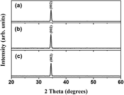
We reported the fabrication of a highly sensitive and fast switchable humidity sensor based on ZnO–TiO2 core–shell nanorods that were synthesized using hydrothermal solution and atomic layer deposition. These nanorods were thermally treated under various physical conditions to improve their sensing performance. The structural investigation revealed that the crystal and microstructure changed with the thermal treatment. Notably, the amorphous TiO2 shell layer transformed into various degrees of crystalline phase after annealing in air and a vacuum at 400 °C. Furthermore, the responses of the sensors fabricated from the ZnO–TiO2 nanorods, with and without thermal annealing, to relative humidity (RH) changes were proportional with the increase in humidity. Among various samples, ZnO–TiO2 nanorods thermally treated in a vacuum exhibited the highest humidity selectivity in response to the cyclic changes in humidity from 11% RH to 33–95% RH at room temperature. Possible mechanisms for the enhancement of sensor performance have been discussed based on structural modifications caused by the thermal treatments.


Introduction

Binary semiconductor oxides have versatile physical properties and are used in various functional sensor devices.1,2 Humidity sensors fabricated from one-dimensional (1D) oxide nanostructures with high surface-to-volume ratios have become a focus of intensive research owing to their higher sensitivity and faster response compared with sensors fabricated from their counterpart in the forms of thin films and bulk.3 Several binary oxides have been investigated for developing humidity sensors;4–6 among these, ZnO is the most commonly studied material for humidity sensor applications because of its chemical and thermal stability, low operation temperature, and ability to decrease the cost of the humidity sensor;7 however, the low hydrophilicity of ZnO limits the possibility of further exploring ZnO for application to humidity sensors with high sensitivities. Although the humidity selectivity of ZnO has been improved by using Ga, Ti, or KCl doping,8–10 the formation of impurity-doped ZnO nanostructures using a one-step doping process and homogeneous composition distribution remain challengeable.

Recently, an oxide heterostructure was demonstrated to improve the performance of sensors fabricated from one of the constituents. Liu et al. presented the fabrication of a highly sensitive humidity sensor based on a Ga2O3–SnO2 core–shell microribbon that was synthesized using a simple one-step chemical vapor deposition.11 ZnO nanorods were hydrothermally coated with coral-like CuO nanostructures at low temperatures. The humidity-sensing characteristics of the CuO–ZnO nanocoral diodes exhibited a remarkable linear decrease in the DC resistance by more than three orders when the relative humidity was changed from 30% to 90%.12 Gu et al. presented a sol–gel synthesized TiO2 layer on ZnO nanostructures to form a core–shell structure for humidity sensor applications. The core–shell-structure-based sensor is 1.5 or 3 orders of magnitude larger than that fabricated from single TiO2 and ZnO, respectively.13 Among several binary heterostructure material systems for humidity sensor applications, the heterostructure comprising ZnO with a hydrophilic TiO2 is more advantageous for nanostructured humidity device applications because large-scale, high-density ZnO nanostructures can be synthesized using several mature methodologies and the thin-film processes of ultrathin TiO2 film can be achieved through physical or chemical methodologies.1,14 In addition, ZnO–TiO2 sensors have been demonstrated to have more favorable humidity selectivity compared with sensors fabricated from ZnO or TiO2 alone.15,16 However, a detailed correlation between the structural information and humidity-sensing performance of the ZnO–TiO2 nanostructures remains to be studied, and this information is highly associated with the fabrication of efficient humidity sensors operated at room temperature. Unfortunately, such information is lacking in the most works on the ZnO–TiO2 based humidity sensors.13,15,16 This study obtained ZnO–TiO2 core–shell nanorods by combining hydrothermally synthesized ZnO nanorods and an atomic-layer-deposited, ultrathin TiO2 layer. The nanorods were thermally treated under various physical conditions, and their crystal structure, microstructure, and electric properties were evaluated to determine their potential application in humidity sensors that operate at various relative humidity levels. The enhanced humidity-sensing characteristics of the ZnO–TiO2 nanorods based on the microstructural modifications are discussed below.

Experiments

Hydrothermally synthesized high density ZnO nanorods on the 200 nm-thick SiO2/Si (100) substrates were used as templates for growth of ZnO–TiO2 core–shell nanorods. The synthesis of vertically aligned ZnO nanorods consisted of two steps corresponding to the formation of ZnO seed layer and the growth of nanorods. In the first, the ZnO seed layer was deposited on SiO2/Si substrate by magnetron sputtering. Subsequently, the substrates were perpendicularly suspended in a solution containing equimolar (0.05 M) aqueous solutions of zinc nitrate hexahydrate (Zn(NO3)2·6H2O) and hexamethylenetetramine (C6H12N4). The hydrothermal reaction temperature was fixed at 95 °C and the duration for crystal growth is 9 h. At the end of the growth period, the substrates were removed from the solution, then immediately rinsed with de-ionized water to remove any residual salt from the surface, and dried in air at room temperature. Deposition of approximately 15 nm-thick TiO2 ultra-thin films was performed using an atomic layer deposition (ALD) system at 250 °C. The as-synthesized ZnO–TiO2 core–shell nanorods are further post-annealed in air and vacuum ambients at 400 °C for 30 min to obtain various crystalline qualities and microstructures of the nanorods.

Sample crystal structures were investigated by X-ray diffraction (XRD) using Cu Kα radiation. The morphologies of the as-synthesized samples were characterized by scanning electron microscopy (SEM), and high-resolution transmittance electron microscopy (HRTEM) was used to investigate the detailed microstructures of the samples. Room temperature-dependent photoluminescence (PL) spectra were obtained using the 325 nm line of a He–Cd laser. The electrical characteristics of the sensors were tested as a function of relative humidity (RH) with an applied voltage of 5 V in a home-built testing chamber referring to the work of Hong et al.17 A computer was used to collect the signals from the sensor in the testing circuit. The different RH levels were generated by referencing various saturated salt solutions in closed chamber at room temperature.4 The RH levels for the humidity sensor test herein were controlled to be approximately 11%, 33%, 55%, 75%, 85%, and 95% and a hygrometer was used to monitor RH levels in the test chamber. The humidity selectivity test of the samples was performed with the sample initially stored in the dry ambient (11% RH); subsequently, the sensor was upon exposure to one of the higher selected RH levels (33%–95%). Finally, the sensor was restored in the 11% RH environment again to finish a test run.

Results and discussion

Fig. 1(a)–(c) presents the XRD patterns of the as-synthesized ZnO–TiO2 nanorods with and without thermal treatment. A considerably intense (002) Bragg reflection originated from the hexagonal wurtzite ZnO, as observed from the XRD patterns; these patterns revealed identical diffraction features for the ZnO–TiO2 nanorods annealed in air and vacuum ambient. However, the XRD patterns revealed no marked Bragg reflections from the TiO2 phases with and without thermal treatment. This may be because the as-deposited TiO2 ultrathin layer is amorphous, and the diffraction signal from the TiO2 phase after thermal annealing may be too low to be detected. Fig. 2(a) presents a cross-sectional SEM image of the as-synthesized ZnO–TiO2 nanorods. Well-aligned ZnO–TiO2 nanorod arrays vertically grew on the substrate and were closely packed. The average nanorod length was approximately 2 μm, and the diameter was in the range of 80–120 nm. Fig. 2(b) presents a top-view SEM micrograph of the as-synthesized ZnO–TiO2 nanorods. The top hexagonal facets of the ZnO–TiO2 nanorods indicate the inherent growth morphology of the single hexagonal ZnO crystal. However, the information provided by the SEM micrographs was insufficient to distinguish the surface microstructural changes of the nanorods with thermal treatments in air and vacuum ambient, as shown in Fig. 2(c) and (d). Nevertheless, the effects of thermal annealing on the detailed microstructures of ZnO–TiO2 nanorods were further examined by using TEM.
image file: c4ra05301j-f1.tif
Fig. 1 (a) XRD patterns of the ZnO–TiO2 nanorods with and without thermal annealing: (a) without thermal annealing, (b) annealed in air, (c) annealed in a vacuum.

image file: c4ra05301j-f2.tif
Fig. 2 (a) Cross-sectional SEM image of the as-synthesized ZnO–TiO2 nanorods. (b) SEM image of the nanorods without thermal annealing. (c) SEM image of the nanorods annealed in air. (d) SEM image of the nanorods annealed in a vacuum.

Fig. 3(a) presents a low-magnification TEM image of a single as-synthesized ZnO–TiO2 nanorod. The ZnO core was uniformly covered by the TiO2 shell layer and had a thickness of approximately 15 nm. In addition, no clear groove features or surface clusters were observed. Moreover, no clear, ordered lattice fringes were observed in the TiO2 layer on the high-resolution (HR) image. The as-deposited TiO2 layer was amorphous and its surface was flat. Fig. 3(b) shows a TEM image of a single ZnO–TiO2 nanorod annealed in air at 400 °C. The HR image in Fig. 3(b) indicates that the TiO2 layer exhibited locally ordered lattice fringes with dense shell layers. Furthermore, the interplanar spacing evaluated from the lattice fringes indicated the formation of the crystalline anatase TiO2 phase. The thermal annealing procedure induced the transformation of the amorphous TiO2 layer into the crystalline phase, with a distinct interface boundary between ZnO and TiO2. The crystalline TiO2 revealed a relatively rugged surface compared with that of the amorphous TiO2 layer without the thermal annealing process. Fig. 3(c) presents a low-magnification and HR image of a single ZnO–TiO2 nanorod annealed in a vacuum at 400 °C. Locally ordered lattice fringes were still observed in the nanorod annealed in a vacuum; however, the ordered size of lattice fringes in the TiO2 layer for the nanorod annealed in a vacuum was inferior to that of nanorod annealed in air. This indicated that various thermal annealing ambients trigger the amorphous TiO2 to transform with various crystalline qualities at 400 °C. The surface of the TiO2 shell layer was not completely smooth but locally rough compared with the surfaces of those that were as-deposited and annealed in air.


image file: c4ra05301j-f3.tif
Fig. 3 Low-magnification and HR TEM images of a single ZnO–TiO2 nanorod with and without thermal annealing: (a) without thermal annealing, (b) annealed in air, (c) annealed in a vacuum.

Two emission band features from the PL spectra of the ZnO–TiO2 nanorods were observed with various thermal treatments (Fig. 4). The ZnO–TiO2 nanorods without thermal treatment revealed a strong UV emission at approximately 3.3 eV and a negligible visible emission band at approximately 2.0 eV. Notably, the sharp and predominant UV emission band corresponded to the near-band-edge emission and originates from the recombination of free electrons and holes.18 Moreover, the weak peak located at 2.0 eV was associated with oxygen vacancies near the oxide surface.19 The transitions involving defect levels, including the recombination of photogenerated holes with singly ionized oxygen vacancies, have been posited to originate from the visible emission band in semiconductor oxides.20,21 When ZnO–TiO2 nanorods were further annealed in air ambient, the UV emission band intensity slightly decreased with increasing visible emission band intensity; however, a significant decrease of UV emission band intensity and increase in the visible emission band intensity of ZnO–TiO2 nanorods were achieved through vacuum thermal annealing. In comparison, vacuum thermal annealing resulted in a relatively large-sized defect-related emission in the ZnO–TiO2 nanorods. Similar thermal annealing effects were reported in the formation of crystal defects in oxides.22 It is believed that oxides annealed in oxygen-deficient ambient caused oxygen to easily escape from the oxide surface, and thus, oxygen vacancies are easily formed during thermal annealing.23 As the concentration of oxygen vacancies increased, the corresponding defect luminescence appeared stronger for the ZnO–TiO2 nanorods annealed in vacuum.


image file: c4ra05301j-f4.tif
Fig. 4 Room-temperature PL spectra of the ZnO–TiO2 nanorods with and without thermal annealing.

Fig. 5 shows gas response curves of the ZnO–TiO2 nanorods upon exposure to 5 ppm NO2 operating at 300 °C. The response is defined as the ratio of the electrical resistance of the sample in the target gas (Rt) to that in the absence of target gas curves (Ra). Surface chemisorption of NO2 molecules onto the n-type oxide nanostructures causes an electron depletion layer in the oxides, resulting in an increase in sensor resistance.24 The gas responses of the ZnO–TiO2 nanorods without thermal annealing, with air annealing and vacuum annealing are respectively 4.6, 6.3, and 7.8. The ZnO–TiO2 nanorods annealed in a vacuum exhibited the best gas sensing performance in this work. The oxidizing gas sensing performance of n-type oxides are highly associated with surface area-to-volume ratio and crystal defects of the nanomaterials.25,26 The high surface area-to-volume ratio and crystal defect density of the materials make them highly sensitive in gas detection. The gas sensing performance for the ZnO–TiO2 nanorods is consistent with the results of structural analyses.


image file: c4ra05301j-f5.tif
Fig. 5 Gas sensing response (Rt/Ra) curves of the ZnO–TiO2 nanorods upon exposure to 5 ppm NO2 operating at 300 °C: (a) without thermal annealing, (b) annealed in an air ambient, (c) annealed in a vacuum ambient.

The resistance variation of the ZnO–TiO2 nanorod-based sensors was monitored by alternately exposing the sensor to 11% RH (base humidity) and those higher than 11% RH (33%, 55%, 75%, 85%, and 95% RH). The typical response characteristics of the sensor resistance to the low (11% RH)–high (85% RH)–low (11% RH) cycle are firstly presented in Fig. 6(a)–(c). Responses were defined as Rb/Rh, wherein Rb is the sensor resistance at 11% RH, and Rh is that at the test RH level. Sensor responses for the samples upon exposure to 85% RH for the nanorods without thermal annealing, those annealed in air, and those annealed in vacuum were 13, 28, and 39, respectively. When the sensor was exposed to 85% RH, the sensor resistance rapidly decreased and then gradually reached a relatively stable value. Subsequently, when the sensor was switched to an 11% RH level, the resistance rapidly increased and eventually reached an original stable value. At a higher RH level of 85%, the enhancement of the humidity response of the nanorods annealed in vacuum and air was three- and two-fold higher, respectively, compared with that of nanorods without thermal annealing. A marked enhancement in humidity sensor performance was observed through thermal annealing. The cyclic humidity sensing response curves to various RH levels of the nanorods with various thermal annealing processes were further shown in Fig. 6(d) and (e). The as-synthesized ZnO–TiO2 core–shell nanorods through hydrothermal and sol–gel methods have shown a reproducible sensing response.13 In this study, the ZnO–TiO2 nanorods prepared with ALD-grown TiO2 shell layers also exhibited reproducible sensing responses when the nanorods were subjected to various thermal annealing processes. The humidity sensing response of the sensors made from the ZnO–TiO2 nanorods with thermal annealing increased with the RH level. Fig. 6(f) summarizes the humidity response values of the sensors based on nanorods with and without thermal annealing for selected RH levels ranging between 33% and 95%. In comparison, the sensor based on the ZnO–TiO2 nanorods annealed in vacuum revealed the highest degree of humidity response at various RH levels among the samples. The humidity hysteresis characteristic of the ZnO–TiO2 nanorods annealed in a vacuum was exhibited in Fig. 6(g). The moisture absorption started from 33% RH and ended at 95% RH, and then dehumidified to 33% RH again. The water vapour adsorption and desorption dependent resistance change curves are slightly noncoincident. The maximum humidity hysteresis is approximately 3.5% under 55% RH for the sensor; this is comparable to the other ZnO–TiO2 based sensors.13,15,16 The experimental result exhibited the ZnO–TiO2 nanorods annealed in a vacuum is of high reliability for making a humidity sensor. Taken together, the sensor response increased with increasing RH levels. The charge transfer between water molecules and nanorods is proportional to the number of water molecules that can be adsorbed on the surface of the nanorods for a higher RH level. Consequently, a higher resistance variation was observed for nanorod sensors exposed to a higher RH level. In this study, no clear response saturation was evident for the humidity-sensing test upon exposure to high humidity levels of 85% and 95% RH. In addition, humidity-sensing response and recovery speed were evaluated for ZnO–TiO2 nanorods with and without thermal annealing. The time span for the response and recovery was defined as the time to achieve 90% of the total resistance change during the test between the base humidity (11% RH) and a higher humidity (33%–95% RH). However, no clear correlation or marked difference was observed for response and recovery speeds under various RH levels for samples treated with various thermal annealing procedures. The response and recovery speeds ranged from 10–40 s and 5–20 s, respectively, for samples at 33%–95% RH levels. In general, the recovery speed was faster than the response speed for various samples exposed to a specific RH level in this study. For instance, at 85% RH, the response times were 20, 18, and 17 s, whereas the recovery times were 5, 16, and 10 s for ZnO–TiO2 nanorods without thermal annealing, those annealed in air, and those annealed in a vacuum, respectively, as shown in Fig. 6(a)–(c). The sensor response and recover speeds herein are superior to the previous work on the similar material system.13 A possible effect of humidity on the resistance change of the nanorods is shown in Fig. 7. It is supposed that when the samples were initially stored in a low humidity environment (11% RH), less water molecules chemisorbed onto the surfaces of the nanorods. In contrast, more water molecules were adsorbed onto the surfaces of the nanorods with an increased RH level to 33%. Many released free electrons from the adsorption of water molecules increased the conductivity of the nanorods. A more defective nature of the vacuum annealed nanorods will show a higher degree of resistance variation than those without thermal annealing (Fig. 7(a) and (b)). When the vacuum annealed nanorods exposed to 55% RH, a water molecule layer of chemisorptions onto the surfaces of the nanorods occurred and induced a marked water dissociation process and ionic conductivity in the nanorods (Fig. 7(c)). When the nanorods were switched to expose to high RH levels (>55% RH) from the base humidity, physisorptions of water molecules might dominate the adsorption process during the humidity sensing test at room temperature (Fig. 7(d)). A substantially increased ionic conductivity of the nanorods with RH level was observed; therefore, a large resistance variation was observed. At high RH levels, the van der Waals interaction between the adsorbate was weak, and, thus, the speed of recovery was faster than that of response when the sample was restored to a low humidity of 11% RH.27 Taken together, the short response and recovery time spans together with a high response to various RH levels for the samples indicate that the ZnO–TiO2 nanorods system may have potential use in humidity sensors in contrast to postulations of previous work.28 Oxygen vacancies in the TiO2 surface layer have been demonstrated to dissociate H2O through the transfer of one proton to a nearby oxygen atom, forming two hydroxyl groups for every vacancy.29 Zhang et al. reported that the size of RH% affects the mechanisms of humidity sensors fabricated from the n-type semiconductor oxides. At low humidity, surface defects of the oxides promote water dissociation into the oxide crystals. Liquid water condensed on the oxide surface at high humidity, efficient electrolytic conduction, and protonic transport may occur for oxides with high surface-to-volume ratios.30,31 The water molecular adsorption rate depends on the size of the ZnO nanoparticle surface area; a large surface area accounts for efficient humidity selectivity.32 The grainy and rough surfaces of the nanostructure-based sensor create abundant adsorption sites for various molecular species and may enhance the sensor receptor function.33 Recently, doping-induced defects on the surface of ZnO grains were postulated to act as water molecule adsorption sites that could transfer charge between the adsorbate and nanomaterials.34 Based on structural analyses, the relatively high surface oxygen vacancy density and surface area of the ZnO–TiO2 nanorods annealed in a vacuum may account for the high humidity selectivity performance in this study.


image file: c4ra05301j-f6.tif
Fig. 6 Humidity sensing response (Rb/Rh) curves of the ZnO–TiO2 nanorods sensor operated at room temperature upon exposure to 85% RH: (a) nanorods without thermal annealing, (b) nanorods annealed in air, (c) nanorods annealed in a vacuum. (d)–(e) Cyclic humidity sensing curves to various RH levels respectively for the ZnO–TiO2 nanorods annealed in air and vacuum ambients. (f) The summarized response values of the various sensors exposed to difference RH levels. (g) Hysteresis characteristic of the ZnO–TiO2 nanorods annealed in a vacuum.

image file: c4ra05301j-f7.tif
Fig. 7 Correlation between the humidity and nanorods resistance variation. The nanorods were initially stored at 11% RH and subsequently switched to a higher RH level. The size of red arrow represents the degree of charge carrier transport process in the TiO2. The blue arrow exhibits contribution of marked ionic conductivity originated from substantial water dissociation process at a high RH level. (a) Nanorods without thermal annealing exposed to 33% RH. (b) Nanorods annealed in vacuum exposed to 33% RH. (c) Nanorods annealed in vacuum exposed to 55% RH. (d) Nanorods annealed in vacuum exposed to high RH levels (>55% RH).

Conclusions

ZnO–TiO2 nanorods were annealed in air and vacuum ambient at 400 °C; the amorphous TiO2 shell layer transformed into the crystalline anatase phase at 400 °C. Thermal treatments resulted in microstructural modifications to the nanorods. The detailed TEM images and PL spectra analyses revealed the formation of crystal defects and the roughening of the surface features of the nanorods following thermal annealing. On the other hand, increased surface crystal defect size and surface irregularities were observed for nanorods annealed in a vacuum. These structural imperfections account for the high humidity selectivity of the sensor fabricated from nanorods annealed in vacuum; in addition, its humidity responses tested at 55% and 85% RH were six and three orders of magnitude larger than that of nanorods fabricated without thermal treatment, respectively. The experimental results show that simple thermal annealing processes improved the humidity sensing response of the sensors. The experimental results could be applied to the ZnO–TiO2 nanocomposites with various synthetic methodologies and morphologies.

Acknowledgements

This work is supported by the National Science Council of Taiwan (Grant no. NSC 102-2221-E-019-006-MY3) and National Taiwan Ocean University (Grant no. NTOU-RD-AA-2012-104012).

References

  1. Y. C. Liang, W. K. Liao and X. S. Deng, J. Alloys Compd., 2014, 599, 87 CrossRef CAS PubMed.
  2. Y. C. Liang and W. K. Liao, RSC Adv., 2014, 4, 19482 RSC.
  3. Y. Zhang, W. Fu, H. Yang, Q. Qi, Y. Zeng, T. Zhang, R. Ge and G. Zou, Appl. Surf. Sci., 2008, 254, 5545 CrossRef CAS PubMed.
  4. Y. Lu, Z. Wang, S. Yuan, L. Shi, Y. Zhao and W. Deng, RSC Adv., 2013, 3, 11707 RSC.
  5. M. Parthibavarman, V. Hariharan and C. Sekar, Mater. Sci. Eng., C, 2011, 31, 840 CrossRef CAS PubMed.
  6. J. Xie, H. Wang, Y. Lin, Y. Zhou and Y. Wu, Ceram. Int., 2013, 39, 3621 CrossRef CAS PubMed.
  7. X. Zhou, J. Zhang, T. Jiang, X. Wang and Z. Zhu, Sens. Actuators, A, 2007, 135, 209 CrossRef CAS PubMed.
  8. N. Han, Y. Tian, X. Wu and Y. Chen, Sens. Actuators, B, 2009, 138, 228 CrossRef CAS PubMed.
  9. P. M. Faia and C. S. Furtado, Sens. Actuators, B, 2013, 181, 720 CrossRef CAS PubMed.
  10. Q. Qi, T. Zhang, S. Wang and X. Zheng, Sens. Actuators, B, 2009, 137, 649 CrossRef CAS PubMed.
  11. K. Liu, M. Sakurai and M. Aono, J. Mater. Chem., 2012, 22, 12882 RSC.
  12. A. Zainelabdin, G. Amin, S. Zaman, O. Nur, J. Lu, L. Hultman and M. Willander, J. Mater. Chem., 2012, 22, 11583 RSC.
  13. L. Gu, K. Zheng, Y. Zhou, J. Li, X. Mo, G. Patzke and G. Chen, Sens. Actuators, B, 2011, 159, 1 CrossRef CAS PubMed.
  14. M. Wang, C. Huang, Y. Cao, Q. Yu, Z. Deng, Y. Liu, Z. Huang, J. Huang, Q. Huang, W. Guo and J. Liang, J. Phys. D: Appl. Phys., 2009, 42, 155104 CrossRef.
  15. S. Jagtap and K. R. Priolkar, Sens. Actuators, B, 2013, 183, 411 CrossRef CAS PubMed.
  16. W. P. Tasi, J. G. Kim and J. H. Oh, Sens. Actuators, B, 2003, 96, 477 CrossRef.
  17. H. S. Hong and G. S. Chung, Sens. Actuators, B, 2014, 195, 446 CrossRef CAS PubMed.
  18. Q. X. Zhao, M. Willander, R. E. Morjan, Q. H. Hu and E. Campbell, Appl. Phys. Lett., 2003, 83, 165 CrossRef CAS PubMed.
  19. Y. C. Liang, C. Y. Hu and H. Zhong, Appl. Surf. Sci., 2012, 261, 633 CrossRef CAS PubMed.
  20. S. B. Zhang, S. H. Wei and A. Zunger, Phys. Rev. B: Condens. Matter Mater. Phys., 2001, 63(7), 075205 CrossRef.
  21. K. Vanheusden, W. L. Warren, C. H. Seager, D. R. Tallant, J. A. Voigt and B. E. Gnade, J. Appl. Phys., 1996, 79, 7983 CrossRef CAS PubMed.
  22. J. Zhang, Y. Liu, Z. Wei and J. Zhang, Appl. Surf. Sci., 2013, 265, 363 CrossRef CAS PubMed.
  23. Y. C. Liang, Electrochem. Solid-State Lett., 2010, 13, G91 CrossRef CAS PubMed.
  24. C. Wang, X. Li, B. Wang, J. Ma, Y. Cao, Y. Sun and G. Lu, RSC Adv., 2014, 4, 18365 RSC.
  25. Y. Mao, S. Ma, X. Li, C. Wang, F. Li, X. Yang, J. Zhu and L. Ma, Appl. Surf. Sci., 2014, 298, 109 CrossRef CAS PubMed.
  26. L. Zhang, J. Zhao, J. Zheng, L. Li and Z. Zhu, Appl. Surf. Sci., 2011, 258, 711 CrossRef CAS PubMed.
  27. X. Chen, J. Zhang, Z. Wang, Q. Yan and S. Hui, Sens. Actuators, B, 2011, 156, 631 CrossRef CAS PubMed.
  28. S. P. Chang, S. J. Chang, C. Y. Lu, M. J. Li, C. L. Hsu, Y. Z. Chiou, T. J. Hsueh and I. Chen, Superlattices Microstruct., 2010, 47, 772 CrossRef CAS PubMed.
  29. R. Schaub, P. Thostrup, N. Lopes, E. Laegsgaard, I. Stensgaard, J. K. Norskov and F. Besenbacher, Phys. Rev. Lett., 2001, 87, 266104 CrossRef CAS.
  30. Y. Zhang, K. Yu, D. Jiang, Z. Zhu, H. Geng and L. Luo, Appl. Surf. Sci., 2005, 242, 212 CrossRef CAS PubMed.
  31. C. Lai, X. Wang, Y. Zhao, H. Fong and Z. Zhu, RSC Adv., 2013, 3, 6640 RSC.
  32. A. Erol, S. Okur, B. Comba, O. Mermer and M. C. Arıkan, Sens. Actuators, B, 2010, 145, 174 CrossRef CAS PubMed.
  33. T. Panczyk, T. P. Warzocha, P. Szabelski and W. Rudzinski, Langmuir, 2008, 24, 8721 CrossRef PubMed.
  34. X. Peng, J. Chu, B. Yang and P. X. Feng, Sens. Actuators, B, 2012, 174, 258 CrossRef CAS PubMed.

This journal is © The Royal Society of Chemistry 2014
Click here to see how this site uses Cookies. View our privacy policy here.