DOI:
10.1039/C4RA06102K
(Paper)
RSC Adv., 2014,
4, 50858-50865
Fabrication and evaluation of nanofibrous membranes with photo-induced chemical and biological decontamination functions
Received
23rd June 2014
, Accepted 17th September 2014
First published on 17th September 2014
Abstract
Hydrophilic nanofibrous membranes with photo-induced self-cleaning functionality were successfully prepared by covalently incorporating a photo-active anthraquinone derivative onto the surfaces of poly(vinyl alcohol-co-ethylene) (PVA-co-PE) nanofibrous membranes. The presence of ester bonds between anthraquinone structures and nanofiber surfaces, as evident from ATR-FTIR results, confirmed the desired surface functionalization. SEM images revealed that the prepared membranes possessed uniform nanofibers and ultrafine porous structures that remained intact after surface functionalization. Under UVA irradiation, these photo-active compound-modified nanofibrous membranes are able to generate reactive oxygen species (ROS), to which are attributed the observed self-decontamination functions. The generation of ROS was evaluated via quantitative determination of hydrogen peroxide on the membrane surfaces by using an iodometric titration method. The prepared hydrophilic nanofibrous membranes exhibited powerful photo-induced chemical and biological decontamination functions, including over 99.999% reduction against both Escherichia coli and Staphylococcus aureus, and a total decontamination of aldicarb, a carbamate pesticide, under light irradiation within a short contact time.
Introduction
The protection of health care professionals, chemical and agriculture production workers and military personnel from pathogenic microorganisms and toxic chemicals has inspired research activities on the development of protective functional materials.1–7 In an effort to produce lightweight, breathable and comfortable protective clothing materials, there is a growing interest in exploring nanofibrous membranes as physical barriers to both biological hazards and chemical toxins owing to their intrinsic properties in terms of ultrafine pore size, highly open porous structure and extraordinarily high surface area. Such structural features also impart excellent resistance to convective gas flow as well as high vapor transmission rate, making nanofibrous membranes ideal materials for protective applications.8–13 However, as a result of the absence of active functional groups, conventional nanofibrous membranes usually can serve only as barriers against large-size microorganisms and particles, but still could carry them on their outer surfaces, which may further contribute to secondary contaminations.14,15 Therefore, it is highly desirable to introduce chemical and biological decontaminating functions into nanofibrous membranes to offer improved personal protection.
Considerable efforts have been devoted to achieving chemical and biological decontamination functionalization of nanofibrous membranes.14 Among these technologies, photo-active agents have attracted great attention in recent years, since they are capable of generating reactive oxygen species (ROS), such as hydroxyl radicals, singlet oxygen and superoxide. These on-site-produced ROS are oxidative agents that can kill microorganisms and oxidatively decompose certain toxic chemicals.16–22 Such properties make photo-active agents applicable in water and air purification, and in chemical and biological protective clothing materials.23 Specifically, inorganic photo-sensitizers, such as titanium oxide, zinc oxide and magnesium oxide, were incorporated onto different textiles and nanofibrous membranes via both chemical and physical processes to offer desired photo-active properties.24–28 Unfortunately, physically embedded photo-active substances can be substantially released from material matrixes during certain usage periods, which may shorten their lifetime as functional materials. In addition, since only the functional groups on material surfaces can effectively produce ROS, incorporation of these functional agents underneath matrixes may also weaken their photo-induced functions.29 Therefore, covalent immobilization of these functional agents onto material surfaces, particularly those with ultrahigh surface area, such as nanofibrous materials, offers a potential pathway to achieve effective and durable photo-active functionality. Recently, anthraquinone derivatives were found to be able to generate reactive oxygen under UVA or visible light irradiation.30,31 Conventional textile materials modified with these photo-active compounds exhibited photo-induced antibacterial and decoloration properties.32,33 It can be envisioned that through covalent attachment of anthraquinone moieties onto the surfaces of nanofibrous membranes, novel self-decontaminating materials can be achieved.
To fulfill this goal, three hydrophilic nanofibrous membranes made from poly(vinyl alcohol-co-ethylene) (PVA-co-PE) polymers with ethylene content of 27, 32, and 44 mol% were individually fabricated, whose surfaces were subsequently functionalized with activated anthraquinone-2-carboxylic acid via a facile esterification reaction. The photo-induced antibacterial activities of these prepared membranes were evaluated via challenging the membranes against both Escherichia coli (E. coli, Gram-negative bacterium) and Staphylococcus aureus (S. aureus, Gram-positive bacterium). Moreover, their chemical detoxification properties under UVA irradiation were investigated by decomposing a carbamate pesticide, aldicarb, using various aldicarb concentrations and contact times.
Experimental
1. Materials
PVA-co-PE (ethylene content 27, 32 and 44 mol%) and anthraquinone-2-carboxylic acid (AQC) were purchased from Sigma-Aldrich (Milwaukee, WI, USA). Cellulose acetate butyrate (CAB; butyryl content 35–39%), pyridine, acetic anhydride, N,N-carbonyldiimidazole (CDI), acetone and N,N-dimethylformamide (DMF) were supplied by Acros Chemical (Pittsburgh, PA, USA). All chemicals were used as received.
2. Preparation of PVA-co-PE nanofibrous membranes
PVA-co-PE nanofibers were prepared according to a previously published procedure.34 Briefly, CAB as a sacrificial matrix was mixed with PVA-co-PE in a blend ratio of CAB/PVA-co-PE = 80/20, which was gravimetrically fed into a Leistritz co-rotating twin-screw (18 mm) extruder (model MIC 18/GL 30D, Nurnberg, Germany) at a feed rate of 12 g min−1. The blends were extruded into composite fibers through a two-strand (2 mm in diameter) rod die, hot-drawn by a take-up device with a drawing ratio of 25 (the area of cross section of the die to that of the extrudates) and air-cooled to room temperature. Then, PVA-co-PE nanofibers were obtained via Soxhlet extraction of acetone to remove CAB from the CAB/PVA-co-PE composite fibers. The prepared PVA-co-PE nanofibers were subsequently made into suspensions and deposited onto a polyester monofilament fabric as a releasing surface. After evaporation of solvents, PVA-co-PE nanofibrous membranes were obtained.
3. Determination of hydroxyl groups
Theoretic hydroxyl group content of PVA-co-PE polymer was calculated based on individual molar ratio of ethylene content, and practical hydroxyl group content of PVA-co-PE nanofiber surfaces was evaluated via an acetic anhydride/pyridine titration method.35 Typically, 60 mg PVA-co-PE nanofibrous membranes was immersed into 10 mL of dry pyridine which was previously mixed with 0.2 mL acetic anhydride. After shaking for 12 hours at 50 °C, 2 mL distilled water was added to convert the excess acetic anhydride into acetic acid, which was titrated with 0.1 M NaOH. The titration curves were recorded via a pH meter (848 Titration Plus, Metrohm, Switzerland). Blank solutions without PVA-co-PE nanofibrous membranes were also tested as controls.
4. Surface functionalization of PVA-co-PE nanofibrous membranes
AQC compound was covalently attached onto PVA-co-PE nanofibrous membranes according to Scheme 1. In detail, 0.5 mmol of AQC compound was firstly activated with an equal amount of CDI in 20 mL DMF. The mixture was heated to 60 °C for 4 hours, followed by mixing with 30 mg of PVA-co-PE nanofibrous membranes. After gently shaking overnight, the functionalized membranes were thoroughly washed with DMF (20 mL × 2), acetone (20 mL × 2), and distilled water (20 mL × 2), successively. The reaction residual and washing solutions were carefully collected together, diluted to 1 L and measured using a UV-vis spectrophotometer (Evolution 600, Thermo, USA) at 319 nm. The AQC concentrations of the final solutions were calculated based on a previously established standard calibration curve. The density of immobilized AQC on membrane surfaces was determined by the difference of AQC amounts before and after esterification reactions.
 |
| Scheme 1 AQC immobilization onto PVA-co-PE nanofibrous membranes (step 1: AQC activation by CDI; step 2: esterification between activated AQC and PVA-co-PE nanofibrous membranes). | |
5. Characterizations
Surface morphologies of pristine and surface-functionalized PVA-co-PE nanofibrous membranes were observed by using scanning electron microscopy (SEM) (XL 30-SFEG, FEI/Philips, USA) at 5 kV accelerating voltage with gold sputter coated samples. Attenuated total reflectance-Fourier transform infrared (ATR-FTIR) spectra were measured from 1900 to 500 cm−1 at a resolution of 4 cm−1 with a Nicolet 6700 spectrometer (Thermo Fisher Scientific, USA).
6. Determination of hydrogen peroxide formed on membrane surfaces
An iodometric titration method was applied to determine the amount of generated hydrogen peroxide (H2O2) on membrane surfaces.36 Briefly, 100 mg of membrane was immersed into 20 mL of deionized water, and then 10 mL of 0.2 M sulfuric acid and 10 mL of 5% potassium iodide were added to the mixture. After exposure to UVA radiation for 1 hour, the mixture was titrated against a 0.001 N sodium thiosulfate solution. The end point of the titration was determined by monitoring the changes occurring at a redox electrode. Pristine membrane was evaluated as a control. The amount of produced H2O2 can be calculated based on the following equation:
where V2 and V1 are the titration volumes (mL) for the sample and control, respectively, N is the normality of Na2S2O3 and W is the weight of membrane samples (g).
7. Antibacterial assessment
Antibacterial properties of surface-functionalized PVA-co-PE nanofibrous membranes were evaluated by using a modified American Association of Textile Chemists and Colorists (AATCC) Test Method 100 against both E. coli (K-12, Gram-negative bacterium) and S. aureus (ATCC#12600, Gram-positive bacterium). Typically, 0.5 mL bacterial suspension with bacterial concentration of 105 colony-forming units per milliliter (CFU mL−1) was applied onto the surface of the AQC-functionalized PVA-co-PE nanofibrous membranes (50 mg) which were placed in a sterilized glass container. The container was irradiated under UVA light (wavelength: 365 nm; light intensity: 1.3–2.0 mW cm−2) for 30 min and 60 min, successively, followed by adding 50 mL sterilized distilled water and vigorously shaking for 2 minutes. 0.1 mL of the solution was extracted from the container and exponentially diluted to 105 times serially. Subsequently, 0.1 mL of each diluted bacterial suspension was evenly loaded onto a nutrient agar plate and incubated at 37 °C for 18 h. The same method was also applied to pristine PVA-co-PE nanofibrous membranes as controls. The percentage reduction of bacteria was calculated according to the following equation:
where A and B are the number of viable bacteria colonies on the agar plates from the pristine and modified membranes, respectively.
8. Detoxification test
Aldicarb aqueous solution was prepared in concentrations of 0.2 and 0.02 mM. 60 mg of pristine or AQC-functionalized PVA-co-PE nanofibrous membranes were immersed into each aldicarb solution. After irradiation under UVA light (wavelength: 365 nm; light intensity: 1.3–2.0 mW cm−2) for 3 hours, a volume of 10 μL of each sample was injected into a Waters e2695 HPLC system, equipped with a Waters 2998 photodiode array detector (Waters Co., Milford, MA, USA) for chromatographic studies. Chromatographic separation of aldicarb and degraded products was achieved on a reverse phase C18 column (5 μm particle size, 4.6 mm by 250 mm). HPLC mobile phase was prepared with 60% Milli-Q water and 40% acetonitrile which both contained formic acid (0.1%, v/v) and the flow rate of the mobile phase was set at 0.33 mL min−1. The absorbance wavelength was measured ranging from 190 to 800 nm. The HPLC system was coupled to a Waters Micromass ZQ 2000 (ESI-MS) mass spectrometer. The mass spectra were obtained under a capillary voltage of 3000 V, sample cone voltage of 60 eV, source temperature of 125 °C, desolvation temperature of 350 °C and desolvation gas flow of 250 L h−1. Micromass MassLynx software (version 4.1) was used for instrument operation and result analysis.
Results and discussion
1. Fabrication of nanofibrous membranes
PVA-co-PE, a random copolymer of vinyl alcohol and ethylene segments readily available in different blending ratios, possesses good mechanical properties, high thermal stability, hydrophilicity and water insolubility.22 Most importantly, the tertiary C–H bonds of the secondary alcohol units of PVA-co-PE polymer could serve as good hydrogen donors that are required in the generation of ROS according to the photo-chemistry of anthraquinone derivatives.30 In this study, PVA-co-PE polymers with ethylene molar ratio of 27, 32 and 44% were fabricated into nanofibrous membranes, and their surface morphologies were observed via SEM. As shown in Fig. 1(a–c), all three types of membranes have a non-woven structure with a large amount of open pores and the nanofiber sizes range from 50 to 350 nm. It is noticeable that the membrane made of PVA-co-PE with 44 mol% ethylene content shows a rougher fiber surface, which can be explained by imperfect coalescence of dispersed PVA-co-PE spheres in the CAB matrix during the fabrication process.37
 |
| Fig. 1 SEM images of pristine PVA-co-PE nanofibrous membranes with (a) 27 mol%, (b) 32 mol%, (c) 44 mol% ethylene units, and AQC-functionalized PVA-co-PE nanofibrous membranes with (d) 27 mol%, (e) 32 mol%, (f) 44 mol% ethylene units (scale bar = 5 μm). | |
The feasibility of surface functionalization of nanofibrous membranes is mainly determined by the accessibility and the amount of surface reactive sites. It has been reported that several functional agents have been covalently incorporated into PVA-co-PE based materials via chemical modifications of their secondary hydroxyl groups.38–41 In theory, PVA-co-PE polymers containing 27, 32 and 44 mol% ethylene contents have amounts of hydroxyl groups of 18.4, 17.5 and 15.2 mmol g−1, respectively. However, the actual values for PVA-co-PE nanofibrous membranes will be much lower since only the surface hydroxyl groups are accessible. The actual amounts of hydroxyl groups are determined as 8.9 and 7.7 mmol g−1 membrane for PVA-co-PE membranes with 27 and 32 mol% ethylene contents, indicating this value decreases as the ethylene contents increase and the vinyl alcohol contents decrease. On the contrary, PVA-co-PE membrane with 44 mol% ethylene content shows a higher actual hydroxyl group content (9.0 mmol g−1 membrane) than the other two, which may be a result of its high nanofiber surface roughness (Fig. 1(c)).
2. Surface functionalization of nanofibrous membranes
In order to incorporate a self-cleaning property into the PVA-co-PE nanofibrous membranes, an organic photo-active compound, AQC, was covalently attached onto membrane surfaces. To facilitate this functionalization, a coupling agent, CDI, was first used to activate AQC compounds via converting their carboxylic acid groups into imidazole carboxylic ester, which then enables their subsequent esterification reactions with hydroxyl groups on membrane surfaces. The AQC-functionalized membranes with 27, 32 and 44 mol% ethylene contents are referred to as PVA-co-PE27-AQC, PVA-co-PE32-AQC and PVA-co-PE44-AQC, respectively. The successful immobilization of AQC compounds was confirmed via ATR-FTIR analysis, and the results for AQC compound, pristine PVA-co-PE (27 mol% ethylene content) membrane and PVA-co-PE27-AQC membrane are presented in Fig. 2. The spectrum of the functionalized membrane shows two new peaks around 1654 and 1592 cm−1, which are attributed to carbonyl stretching and aromatic C
C stretching of anthraquinone structures. And the results are consistent with the spectrum of AQC compound. In addition, the new peak around 1761 cm−1 is associated with carbonyl stretching of a new ester bond formed between AQC molecules and membrane surfaces.42,43 Identical ATR-FTIR results were observed for all three types of membrane samples owing to their same polymer structure and subsequent surface functionalizations. The amount of immobilized AQC on membrane surfaces was further evaluated by a UV/visible method. As shown in Fig. 3, PVA-co-PE44-AQC membranes possesses the highest AQC loading (0.24 mmol g−1 membrane), followed by PVA-co-PE27-AQC and PVA-co-PE32-AQC membranes (0.20 and 0.17 mmol g−1 nanofiber, respectively). Due to practicality concerns, the stability of these membranes during chemical functionalizations was also evaluated via SEM. No significant difference of their surface morphologies was found (Fig. 1(d–f)), suggesting that the introduction of photo-active compounds did not affect the physical morphology of the nanofibrous membranes.
 |
| Fig. 2 ATR-FTIR spectra of AQC compound, pristine PVA-co-PE nanofibrous mat with 27 mol% ethylene units, and AQC-functionalized PVA-co-PE nanofibrous membranes with 27 mol% ethylene units (PVA-co-PE27-AQC). | |
 |
| Fig. 3 The amount of immobilized AQC compound and measured (formed) H2O2 on PVA-co-PE nanofibrous membranes. | |
3. Photo-induced activity of AQC-immobilized nanofibrous membrane
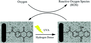
Previous studies demonstrated that, in the presence of hydrogen donors and oxygen, anthraquinone moieties were able to generate ROS under UVA irradiation, which then triggers photo-induced activities of inactivating bacteria, decolorizing textile colorants and even initiating radical polymerization.30–33 Herein, we believe that the mechanism of photo-induced reactions of these immobilized AQC structures is the same as those of the other anthraquinone derivatives reported before due to their structural similarity and the presence of desired hydrogen donor (weak C–H bonds of vinyl alcohol units of PVA-co-PE polymer) (Scheme 2). Specifically, the mechanism of this photo-reaction involves three steps: (1) under UVA irradiation, immobilized AQC structures are excited to their singlet states, which subsequently transfer into triplet states through an intersystem crossing process; (2) the triplet excited AQC structures will abstract hydrogen atoms from hydrogen donors, the C–H bond of secondary alcohol of the PVA-co-PE polymer backbone, to produce ketyl radicals; and (3) these radicals will react with oxygen to generate superoxide radical, peroxide radical and hydroxyl radical, which eventually transfer into hydrogen peroxide (H2O2).
 |
| Scheme 2 The mechanism of photo-induced reactions of immobilized AQC on PVA-co-PE nanofibrous membranes. | |
To quantitatively evaluate the photo-activities of AQC-immobilized membranes, the amount of formed H2O2 was measured by using an iodometric titration method, and the results are presented in Fig. 3. As expected, PVA-co-PE32-AQC membrane exhibited the lowest amount of H2O2 (27 ppm) due to the lowest amount of immobilized AQC. Interestingly, there was no significant difference of H2O2 values between PVA-co-PE27-AQC (43 ppm) and PVA-co-PE44-AQC (42 ppm), even though PVA-co-PE44-AQC membrane has a higher amount of immobilized AQC. As mentioned above, the C–H bonds of secondary alcohol of the PVA-co-PE polymer act as hydrogen donors to enable the generation of ketyl radicals. Thus, higher amounts of vinyl alcohol groups in the PVA-co-PE polymer will facilitate the reaction pathway to produce more ROS. As a result, both membranes generated a similar level of H2O2.
4. Photo-induced antibacterial property
The biocidal activities of the three AQC-functionalized PVA-co-PE nanofibrous membranes were evaluated against both E. coli and S. aureus according to a modified AATCC 100 method. As shown in Fig. 4 and Table 1, all three membranes could kill more than 80% of E. coli and 70% of S. aureus after exposure to UVA light (365 nm) for 30 min. When the irradiation time further increased to 1 hour, no colony of viable bacteria was found on the agar plates for both E. coli and S. aureus that were exposed onto the AQC-functionalized nanofibrous membranes. Meanwhile, proliferated colonies of the two bacteria were observed for all pristine membranes under various irradiation durations. Interestingly, the overall reduction rate of S. aureus was faster than that of E. coli which may be explained by the difference in bacteria structures. It has been reported that a longer time is usually required to let biocides penetrate into the cell wall of E. coli compared to S. aureus since the cell wall of Gram-negative bacteria is overlaid with a supplementary barrier, which does not exist in Gram-positive bacteria.44
 |
| Fig. 4 Antibacterial test results of E. coli (top) and S. aureus (bottom) treated with pristine PVA-co-PE nanofibrous membranes (left) and PVA-co-PE27-AQC (right) after 60 min contact time. | |
Table 1 Bacterial reduction (%) caused by AQC-functionalized nanofibrous membranes for different UVA exposure times
Bacterium |
S. aureus |
E. coli |
Exposure time |
30 min |
60 min |
30 min |
60 min |
PVA-co-PE27-AQC |
87.623 |
>99.999 |
80.443 |
>99.999 |
PVA-co-PE32-AQC |
86.893 |
>99.999 |
78.012 |
>99.999 |
PVA-co-PE44-AQC |
87.920 |
>99.999 |
82.375 |
>99.999 |
In an effort to study the durability of the membrane photo-induced antibacterial functions, all tested samples were sterilized and thoroughly washed before repeated antimicrobial experiments. Benefiting from the chemical bonding between AQC molecules and membrane surfaces, no noticeable decrease of their antibacterial efficiencies was found after three repeat tests, suggesting that these photo-induced antibacterial properties are durable and reusable.
5. Photo-induced chemical detoxification property
Aldicarb, namely 2-methyl-2-(methylthio)propanal-O-(N-methylcarbamoyl)oxime, is a carbamate pesticide that has been widely used as a nematicide in agricultural production.45 It has been reported that aldicarb can be oxidized and converted into less toxic compounds in the presence of photosensitizers.45 In this study, aldicarb was employed as a target toxin to evaluate the photo-induced self-decontamination functions of the AQC-functionalized PVA-co-PE nanofibrous membranes. Pure aldicarb solution and aldicarb solutions immersed with pristine membranes or AQC-functionalized membranes were all exposed to UVA irradiation for certain contact times, and then were analyzed via HPLC and mass spectrometry to identify any decomposition or oxidation products.
When the aldicarb concentration was 0.2 mM (Fig. 5(a)), the peak (retention time at 13.14 min) was identified as aldicarb due to two natural fragment ions, [M − OCONHCH3]+ at m/z 116 and [M + H2O]+ at m/z 208, found from its mass spectrum (Fig. 6(a)). For the aldicarb solutions treated with AQC-functionalized membranes, the average area of this peak apparently decreased and the reduction rate followed the order: PVA-co-PE32-AQC (20%) < PVA-co-PE27-AQC (25%) < PVA-co-PE44-AQC (26%). Consistent with the amounts of generated hydrogen peroxide, PVA-co-PE32-AQC exhibited the weakest photo-induced oxidative functions. The peak (retention time at 5.41 min) is identified as aldicarb sulfoxide, an oxidized product of aldicarb. Though it was overlapped with certain impurity also present in the pure aldicarb solution, the average areas of this peak significantly increased from the solutions treated with AQC-functionalized membranes. The mass spectrum is presented in Fig. 6(b), with two characteristic peaks, [M + H]+ ion at m/z 207 and [M + H2O]+ at m/z 224, consistent with the literature.46
 |
| Fig. 5 HPLC results of pure aldicarb, and aldicarb solutions treated with pristine and AQC-functionalized PVA-co-PE nanofibrous membranes with (a) aldicarb concentration of 0.2 mM and (b) aldicarb concentration of 0.02 mM. | |
 |
| Fig. 6 Mass spectrometry results of (a) aldicarb at RT 13.14 min, and (b) aldicarb sulfoxide at RT 5.41 min. | |
When the aldicarb concentration was 0.02 mM (Fig. 5(b)), the characteristic peak of aldicarb totally disappeared for the solutions treated with AQC-functionalized membranes after 3 hours of UVA irradiation, and the peak intensity of aldicarb sulfoxide was visibly enlarged, indicating the pesticide was successfully oxidized and detoxified. No significant changes were observed in the solution of pure aldicarb and that treated with pristine membranes. These results demonstrated the powerful oxidative functions of AQC-functionalized PVA-co-PE nanofibrous membranes under UVA irradiation.
Conclusions
In this study, photo-induced self-decontaminating nanofibrous membranes were successfully prepared. Three PVA-co-PE polymers containing various ethylene contents were fabricated into nanofibrous membranes, which were subsequently surface functionalized with a photo-active compound, anthraquinone-2-carboxylic acid. The morphologies of uniform nanofibers and the open porous structures of the prepared membranes were unchanged after the chemical reactions, and the immobilization of photo-active agents was confirmed by ATR-FTIR analysis and quantitatively evaluated. PVA-co-PE44-AQC membrane with a highly rough surface offered a higher hydroxyl group density and more immobilized AQC on its surface. PVA-co-PE27-AQC membrane with the lowest ethylene content provided more hydrogen donors of C–H bonds of secondary alcohol units to facilitate hydrogen peroxide generation. These AQC-functionalized PVA-co-PE nanofibrous membranes exhibited excellent photo-induced antibacterial activities and efficient pesticide decontamination function against aldicarb, which demonstrated their great potential to serve as self-decontaminating materials.
Acknowledgements
This research was financially supported by National Textile Center (S06-CD01) and Defense Threat Reduction Agency (HDTRA1-08-1-0005). The authors are grateful to Jastro-Shields Graduate Student Research Fellowship Award at the University of California, Davis.
References
- S. K. Obendorf, AATCC Rev., 2010, 10, 44–50 CAS.
- H. L. Schreuder-Gibson, Q. Truong, J. E. Walker, J. R. Owens, J. D. Wander and W. E. Jones, MRS Bull., 2003, 28, 574–578 CrossRef CAS.
- O. B. Koper, I. Lagadic, A. Volodin and K. J. Klabunde, Chem. Mater., 1997, 9, 2468–2480 CrossRef CAS.
- G. W. Wagner, O. B. Koper, E. Lucas, S. Decker and K. J. Klabunde, J. Phys. Chem. B, 2000, 104, 5118–5123 CrossRef CAS.
- A. F. Bedilo, M. J. Sigel, O. B. Koper, M. S. Melgunova and K. J. Klabunde, J. Mater. Chem., 2002, 12, 3599–3604 RSC.
- S. Rajagopalan, O. Koper, S. Decker and K. J. Klabunde, Chem.–Eur. J., 2002, 8, 2602–2607 CrossRef CAS.
- S. Sundarrajan, A. R. Chandrasekaran and S. Ramakrishna, J. Am. Ceram. Soc., 2010, 93, 3955–3975 CrossRef CAS.
- P. W. Gibson, H. L. Schreuder-Gibson and D. Rivin, Colloids Surf., A, 2011, 187–188, 469–481 Search PubMed.
- P. W. Gibson, H. L. Schreuder-Gibson and D. Rivin, AIChE J., 1999, 45, 190–195 CrossRef CAS.
- H. L. Schreuder-Gibson, P. W. Gibson, K. Senecal, M. Sennett, J. Walker, W. Yeomans, D. Ziegler and P. P. Tsai, J. Adv. Mater., 2002, 34, 44–55 Search PubMed.
- S. S. Lee and K. S. Obendorf, Fibers Polym., 2007, 8, 501–506 CrossRef.
- S. S. Lee and K. S. Obendorf, J. Appl. Polym. Sci., 2006, 102, 3430–3437 CrossRef CAS.
- S. S. Lee and K. S. Obendorf, Text. Res. J., 2007, 77, 696–702 CrossRef CAS.
- S. Sundarrajan, A. Venkatesan and S. Ramakrishna, Macromol. Rapid Commun., 2009, 30, 1769–1774 CrossRef CAS PubMed.
- I. P. Parkin and R. G. Palgrave, J. Mater. Chem., 2005, 15, 1689–1695 RSC.
- K. Tan and S. K. Obendorf, J. Membr. Sci., 2007, 305, 287–298 CrossRef CAS.
- K. Tan and S. K. Obendorf, J. Membr. Sci., 2007, 289, 199–209 CrossRef CAS.
- L. Chen, L. Bromberg, A. Hatton and G. Rutledge, Polymer, 2007, 48, 4675–4682 CrossRef CAS.
- L. Chen, L. Bromberg, A. Hatton and G. Rutledge, Polymer, 2008, 49, 1266–1275 CrossRef CAS.
- L. Chen, L. Bromberg, H. L. Schreuder-Gibson, J. Walker, A. Hatton and G. Rutledge, J. Mater. Chem., 2009, 19, 2432–2438 RSC.
- L. Chen, L. Bromberg, J. A. Lee, H. Zhang, H. Schreuder-Gibson, P. Gibson, J. Walker, P. Hammond, A. Hatton and G. Rutledge, Chem. Mater., 2010, 22, 1429–1436 CrossRef CAS.
- J. Zhu, Q. Bahramian, P. Gibson, H. Schreuder-Gibson and G. Sun, J. Mater. Chem., 2012, 22, 8532–8540 RSC.
- M. R. Hoffmann, S. T. Martin, W. Y. Choi and D. W. Bahnemann, Chem. Rev., 1995, 95, 69–96 CrossRef CAS.
- E. Santala, M. Kemell, M. Leskel and M. Ritala, Nanotechnology, 2009, 20, 035602 CrossRef PubMed.
- J. A. Lee, K. C. Krogman, M. L. Ma, R. M. Hill, P. T. Hammond and G. C. Rutledge, Adv. Mater., 2009, 21, 1252–1256 CrossRef CAS.
- H. Q. Liu, J. X. Yang, J. H. Liang, Y. X. Huang and C. Y. Tang, J. Am. Ceram. Soc., 2008, 91, 1287–1291 CrossRef CAS.
- S. Sundarrajan and S. Ramakrishna, J. Mater. Sci., 2007, 24, 8400–8407 CrossRef.
- N. L. Lala, R. Ramaseshan, B. J. Li, S. Sundarrajan, R. S. Barhate, Y. J. Liu and S. Ramakrishna, Biotechnol. Bioeng., 2007, 42, 8400–8407 Search PubMed.
- J. Zhu and G. Sun, J. Mater. Chem., 2012, 22, 10581–10588 RSC.
- N. Liu and G. Sun, Ind. Eng. Chem. Res., 2011, 50, 5326–5333 CrossRef CAS.
- N. Liu and G. Sun, Dyes Pigm., 2011, 91, 215–224 CrossRef CAS.
- N. Liu and G. Sun, ACS Appl. Mater. Interfaces, 2011, 3, 1221–1227 CAS.
- J. Y. Zhuo and G. Sun, ACS Appl. Mater. Interfaces, 2013, 5, 10830–10835 CAS.
- D. Wang, G. Sun and B. S. Chiou, Macromol. Mater. Eng., 2007, 292, 407–414 CrossRef CAS.
- J. Luo, C. Pardin, W. D. Lubell and X. X. Zhu, Chem. Commun., 2007, 21, 2136–2138 RSC.
- N. Liu, G. Sun and J. Zhu, J. Mater. Chem., 2011, 21, 15383–15390 RSC.
- D. Wang, W. L. Xu, G. Sun and B. S. Chiou, ACS Appl. Mater. Interfaces, 2011, 3, 2838–2844 CAS.
- M. E. Avramescu, W. F. C. Sager, M. H. V. Mulder and M. Wessling, J. Membr. Sci., 2002, 210, 155–173 CrossRef CAS.
- J. Zhu, J. Yang and G. Sun, J. Membr. Sci., 2011, 385–386, 269–276 CrossRef CAS.
- J. Zhu and G. Sun, React. Funct. Polym., 2012, 72, 839–845 CrossRef CAS.
- J. Zhu and G. Sun, ACS Appl. Mater. Interfaces, 2014, 6, 925–932 CAS.
- L. A. Gribov, O. B. Zubkova and A. A. Sigarev, J. Struct. Chem., 1993, 34, 147–154 CrossRef.
- I. N. Juchnovski and T. M. Kolev, Spectrosc. Lett., 1986, 19, 1183–1193 CrossRef.
- J. Zhu and G. Sun, AATCC Rev., 2011, 11, 59–64 Search PubMed.
- X. Fei, P. F. Gao, T. Shibamoto and G. Sun, Arch. Environ. Contam. Toxicol., 2006, 51, 509–514 CrossRef CAS PubMed.
- V. Dixit, J. C. Tewari and S. K. Obendorf, J. Chromatogr. A, 2009, 1216, 6394–6399 CrossRef CAS PubMed.
|
This journal is © The Royal Society of Chemistry 2014 |
Click here to see how this site uses Cookies. View our privacy policy here.