A novel and environmentally friendly colorimetric method for detection of cystine in human urine using unmodified gold nanoparticles†
Li-Qiang Lu*a,
Qian Gaoa,
Chi Songa,
Xi-Ke Tiana and
An-Wu Xub
aNano-Mineral Materials and Application Engineering Research Center of Ministry of Education, Faculty of Materials Science and Chemistry, China University of Geosciences, Wuhan 430074, P. R. China. E-mail: llqdick@cug.edu.cn
bDivision of Nanomaterials and Chemistry, Hefei National Laboratory for Physical Sciences at Microscale, Department of Chemistry, University of Science and Technology of China, Hefei 230026, P. R. China
First published on 9th June 2014
Abstract
Cystine was reduced by ascorbic acid to cysteine, which induced the aggregation of unmodified gold nanoparticles. The accompanied color change was distinguishable and perceivable by the naked eye. This facile assay method was successfully applied to the detection of cystine in human urine.
Cystinuria is a congenital metabolic disease, which stems from the defective transport of cystine and dibasic amino acids through the epithelial cells of the renal tubule and intestinal tract, resulting in the recurrent formation of cystine stones in the renal tract.1 Cystinuria could be characterized by high concentrations of cystine in human urine (usually >400 mg L−1).2 Several methods for the analysis of cystine have been developed during the past decades, including iodimetry,3 liquid chromatography,4 electrochemistry,5–7 spectrophotometry,8,9 spectrofluorimetry10 and colorimetry.11–13 Among these methods, colorimetric assay is most attractive, especially in a resource poor setting, due to its simplicity and no requirement for sophisticated instruments. The traditional colorimetric cystine detection are based on the reaction between cysteine and nitroprusside,12 where cysteine is reduced from cystine by potassium cyanide. Although this assay has been used for clinical analysis of cystine in human urine, it suffers from high blanks, unstable colors, low specificity, high sensitivity to reaction conditions, and toxicity of reductant.13 On the basis of the aforementioned facts, it is desirable to develop sensors that provide sensitive, specific and environmentally friendly detection of cystine.
Gold nanoparticles (AuNPs) have been demonstrated to be suitable for colorimetric sensors, owing to their unique optical properties related to surface plasmon resonance (SPR).14,15 A vast variety of AuNPs-based colorimetric sensors have been exploited to detect DNA,16 proteins17 and small molecules18–22. Thereinto, cetyltrimethylammonium bromide capped AuNPs,18 fluorosurfactant stabilized AuNPs19 and unmodified AuNPs20 were used for sensing cysteine. However, as far as we are aware, AuNPs-based colorimetric methods have not yet been applied for the detection of cystine.
Herein, we present a facile, rapid and sensitive colorimetric method based on unmodified AuNPs for the assay of cystine. As shown in Scheme 1, cystine which cannot induce the aggregation of AuNPs is reduced by ascorbic acid to cysteine that can readily bind to the surface of AuNPs via Au–S bond. Consequently, the AuNPs aggregation occurs due to the electrostatic interaction between the positively charged amino group and the negatively charged carboxyl group of cysteine on the surface of AuNPs, with a corresponding red to blue color change which can be easily observed by the naked eye or measured with ultraviolet-visible (UV-vis) spectroscopy.
 | ||
| Scheme 1 Schematic illustration of the strategy for the colorimetric assay of cystine based on unmodified AuNPs and ascorbic acid. | ||
AuNPs with ∼18 nm in diameter were synthesized by citrate reduction of HAuCl4 (ESI,† Experimental section) as previously reported,23 exhibiting a SPR absorption band peaked at 520 nm (Fig. 1A, black curve). The AuNPs solution could be stabilized against aggregation ascribed to the electrostatic repulsion between citrate capped AuNPs. In the presence of cystine, only a very slight decrease in the SPR absorption band of AuNPs was observed (Fig. 1A, blue curve), and the solution color remained red (Fig. 1B, left), indicating that AuNPs did not aggregate, which was also demonstrated by TEM observation (Fig. 1C). However, it has been previously discussed that cysteine can induce the aggregation of AuNPs because of the covalent bonding to AuNPs via the mercapto group and the electrostatic interaction between cysteine zwitterions on the surface of neighboring AuNPs.24 Accordingly, we designed our AuNPs-based colorimetric method to indirectly detect cystine by reducing cystine to cysteine. Ascorbic acid was chosen as the reductant since it can reduce the poorly soluble cystine to the readily soluble cyteine and has been used to treat cystinuria.25 In the cystine detection case, cystine (10 μM) was mixed with ascorbic acid (2 mM) and incubated at 40 °C for 15 min, and then AuNPs (0.5 ml) were added (ESI,† Experimental section). The SPR peak at 520 nm significantly decreased and a new distinct SPR peak at 670 nm appeared (Fig. 1A, green curve), showing the aggregation of AuNPs, which was confirmed by TEM image (Fig. 1D). The solution color change from red to blue was distinguishable and perceivable by the naked eye (Fig. 1B, right). The control experiment showed that ascorbic acid (2 mM) had no effect on the stability of AuNPs (Fig. 1A, red curve). Therefore, it is convenient to probe cystine through the color change and absorption spectra of AuNPs solutions.
 | ||
| Fig. 1 Absorption spectra (A), optical photographs (B) and TEM images (C and D) of AuNPs in the presence of cystine and in the presence of the reaction mixture of cystine and ascorbic acid. | ||
To achieve better analysis performance, optimizations of some key variables of cystine measurement were conducted, where the relative absorbance A650/A520 that is the ratio of the extinction coefficients at 650 nm and 520 nm was used to express the molar ratio of the aggregated to the dispersed AuNPs. The amount of AuNPs was optimized to 0.5 ml where the ratio of A650/A520 reachs its maximum (ESI, Fig. S1†). The concentration of the ascorbic acid, reaction temperature and reaction time of cystine reduction were optimized to 2 mM (ESI, Fig. S2†), 40 °C (ESI, Fig. S3†) and 15 min (ESI, Fig. S4†), respectively, for further experiments.
Under the optimum conditions, different concentrations of cystine were tested and UV-visible spectra of the solutions were recorded. As shown in Fig. 2A, absorbance at 520 nm decrease while absorbance at 650 nm increase with sequentially increasing concentrations of aliquots of cystine. The ratios of A650/A520 is linearly related to the cystine concentrations ranging from 1 μM to 8 μM, and the calibration equation, A = 0.18055c−0.04126, is obtained with a correlation coefficient of 0.991 (Fig. 2B). In addition, Fig. 2C clearly shows that the color of AuNPs solution is gradually changed from wine red to blue with the increase of cystine concentration. The detection limit of cystine is down to 1 μM with the naked eye, which is much better than the previously reported electrochemical5 and colorimetric13 determination of cystine. The result indicates that the proposed assay method can conveniently determine cystine with high sensitivity.
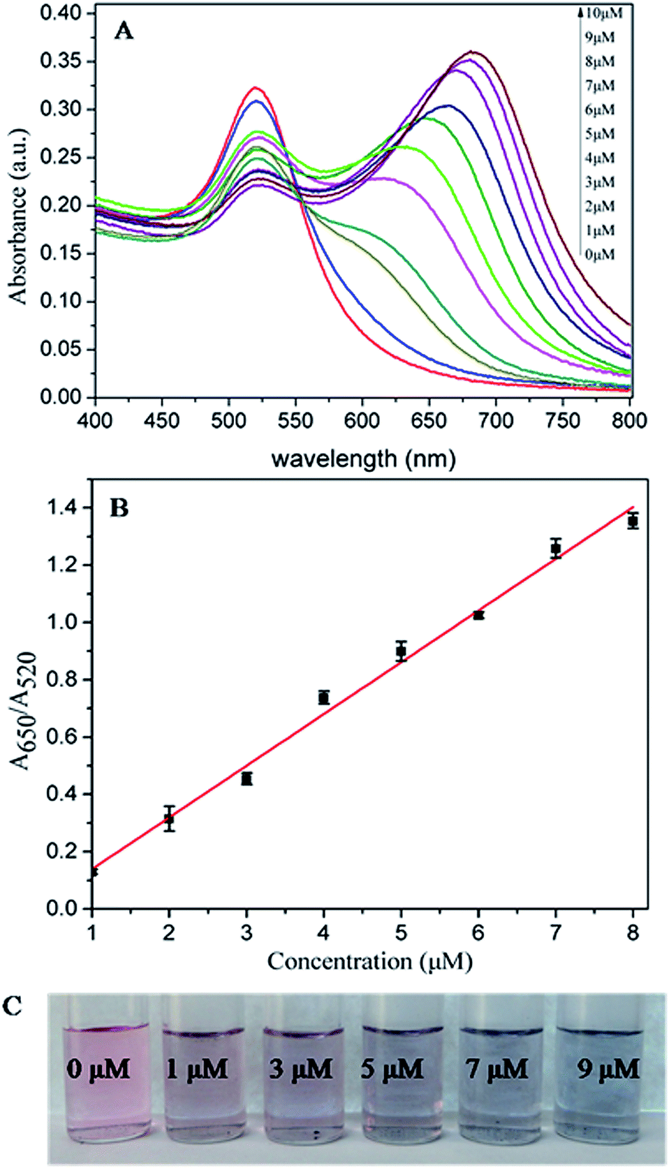 | ||
| Fig. 2 (A) Absorbance response for different concentrations of cystine. (B) Calibration curve for the detection of cystine. The error bars denote standard deviation from three independent measurements. (C) Color change of AuNPs solution with the increase of cystine concentration. | ||
Several other substances perhaps existing in the urine were employed to assess the selectivity of the present colorimetric cystine assay method. As illustrated in Fig. 3A, cystine, homosyteine and cysteine result in red-to-blue color changes while the solutions remain red in the presence of other objects. The corresponding relative absorbance data show that other substances cause a negligible increase of A650/A520 at a 2-fold molar excess (Fig. 3C). As known in previous reports, the concentrations of cysteine (98 ± 35 μmol L−1) in urine from cystinurics are much lower than that of cystine (1185 ± 451 μmol L−1),26 and the homocysteine is normally found at relatively low concentration (2–14 μmol L−1) in human urine.27 Therefore, the interference of cysteine and homocysteine could be ignored in diluted patients urine sample. In coexistence interference experiments, only iodide makes a decrease of absorption ratio value (Fig. 3D) with a purple solution (Fig. 3B). It is known that the concentrations of iodide in healthy human urine are about 86.8 ± 19.0 μg L−1.28 Thus, the interference of iodide could also be ignored in urine samples from cystinurics.
 | ||
| Fig. 3 (A) Color change of AuNPs solution in the presence of 10 μM cystine or 20 μM other substances. (B) Color change of AuNPs solution in the presence of 10 μM cystine and 20 μM other substances. (C) The relative absorption value for 10 μM cystine or 20 μM other substances. (D) The relative absorption value for 10 μM cystine and 20 μM other substances. | ||
Based on the positive results, we next studied the application of the colorimetric assay method in real urine samples. Prior to analysis, the urine sample was diluted 1![[thin space (1/6-em)]](https://www.rsc.org/images/entities/char_2009.gif) :100 (v/v) with deionized water to avoid the aggregation of AuNPs induced by the high concentration of ions, protein and small molecules. A series of obtained urine samples were prepared by spiking them with standard solution of cystine over the range from 1 μM to 9 μM. The measuring results of cystine in urine samples were shown in Fig. 4A. There is a good linear relationship between the ratio of A650/A520 and the concentration of cystine with a correlation coefficient of 0.995 (Fig. 4B). The lowest detectable concentration of cystine in urine sample was also 1 μM. Validation of the proposed method was examined by spiking urine samples collected from two healthy children (volunteer I: 0.5 years old, volunteer II: 2 years old) with cystine concentrations of 2 μM, 5 μM and 8 μM. The analytical results for the urine samples are shown in Table 1. As can be seen in Table 1, the recoveries of cystine range from 99.5% to 106.8% with RSDs ranging from 1.27% to 5.74%, thereby proving that this colorimetric assay is an accurate and sensitive method for the analysis of cystine in human urine.
:100 (v/v) with deionized water to avoid the aggregation of AuNPs induced by the high concentration of ions, protein and small molecules. A series of obtained urine samples were prepared by spiking them with standard solution of cystine over the range from 1 μM to 9 μM. The measuring results of cystine in urine samples were shown in Fig. 4A. There is a good linear relationship between the ratio of A650/A520 and the concentration of cystine with a correlation coefficient of 0.995 (Fig. 4B). The lowest detectable concentration of cystine in urine sample was also 1 μM. Validation of the proposed method was examined by spiking urine samples collected from two healthy children (volunteer I: 0.5 years old, volunteer II: 2 years old) with cystine concentrations of 2 μM, 5 μM and 8 μM. The analytical results for the urine samples are shown in Table 1. As can be seen in Table 1, the recoveries of cystine range from 99.5% to 106.8% with RSDs ranging from 1.27% to 5.74%, thereby proving that this colorimetric assay is an accurate and sensitive method for the analysis of cystine in human urine.
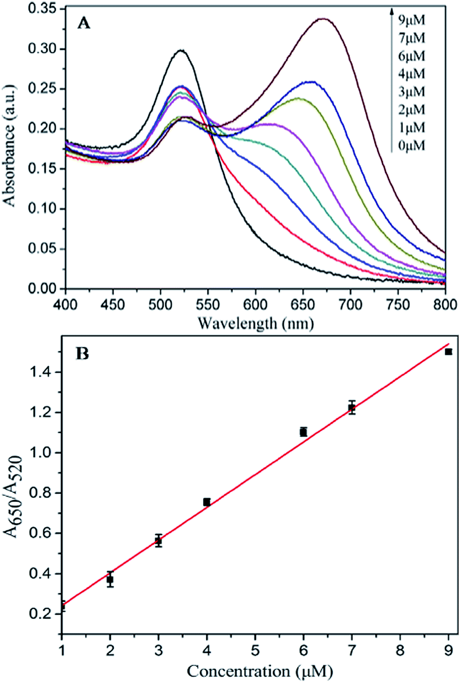 | ||
| Fig. 4 (A) Absorbance response for different concentrations of cystine in urine sample. (B) Calibration curve for the detection of cystine. The error bars denote standard derivation from three independent measurements. | ||
| Sample | Added (μM) | Detected (μM) | Recovery (%) | RSD (%, n = 6) |
|---|---|---|---|---|
| Volunteer I | 2 | 1.994 | 99.7 | 3.43 |
| Volunteer II | 2.079 | 101 | 3.02 | |
| Volunteer I | 5 | 4.975 | 99.5 | 2.09 |
| Volunteer II | 5.34 | 106.8 | 5.74 | |
| Volunteer I | 8 | 8.2 | 102.5 | 1.27 |
| Volunteer II | 8.29 | 103.6 | 2.12 |
In summary, we have developed a novel and simple strategy based on unmodified AuNPs for the colorimetric detection of cystine. Cystine is effectively reduced by nontoxic ascorbic acid to cysteine, inducing the aggregation of unmodified AuNPs through the efficient electrostatic interaction between cysteine-bonded AuNPs. The color detection limit of the proposed method is down to 1 μM with the naked eye, which is more sensitive than previously reported colorimetric methods. The practicability of this method was validated through analyses of real urine samples. Compared with the existing methods for measurement of cystine in human urine, the colorimetric assay method demonstrated here is advantageous in terms of its simplicity and nontoxicity, and is thus highly anticipated to assist in the diagnosis of human cystinuria.
Acknowledgements
The authors acknowledge the geological survey project of China Geological Survey (12120113015300) and the special funding support from the Fundamental Research Funds for the Central Universities (CUG090108).Notes and references
- M. J. Calonge, P. Gasparini, J. Chillarón, M. Chillón, M. Gallucci, F. Rousaud, L. Zelante, X. Testar, B. Dallapiccola, F. Di Silverio, P. Barceló, X. Estivill, A. Zorzano, V. Nunes and M. Palacín, Nat. Genet., 1994, 6, 420–425 CrossRef CAS PubMed.
- S. R. Karl and C. M. James, Saudi J Kidney Dis Transplant, 2003, 14(1), 351–357 Search PubMed.
- R. W. Virtue and H. B. Lewis, J. Biol. Chem., 1934, 104, 415 CAS.
- Y. Wang, X. J. Kang and W. H. Ge, Chromatographia, 2007, 65, 527–532 CAS.
- P. Mikus, P. Kubacak, I. Valaskova and E. Havaranek, Pharmazie, 2003, 58, 111–113 CAS.
- G. Li, J. Yang, X. Zheng, M. Meng and J. Cao, Microchim. Acta, 2010, 168, 277–282 CrossRef CAS.
- M. S. Damle, L. A. A. Newton, M. M. Villalba, R. Leslie and J. Davis, Electroanalysis, 2010, 22, 2491–2495 CrossRef CAS.
- J. Chrastil, Analyst, 1989, 114, 1133–1136 RSC.
- J. Chrastil, Analyst, 1990, 115, 1383–1384 RSC.
- A. A. Ensafi, B. Rezaei and S. Nouroozi, J. Braz. Chem. Soc., 2009, 20, 288–293 CrossRef CAS PubMed.
- Y. Morioka and K. Kobayashi, Biol. Pharm. Bull., 1997, 20, 825–827 CAS.
- I. W. Grote, J. Biol. Chem., 1931, 93, 25–30 CAS.
- K. Shinohara, J. Biol. Chem., 1935, 109, 665–679 CAS.
- M. Hu, J. Chen, Z. Li, L. Au, G. V. Hartland, X. Li, M. Marquez and Y. Xia, Chem. Soc. Rev., 2006, 35, 1084–1094 RSC.
- S. K. Ghosh and T. Pal, Chem. Rev., 2007, 107, 4797–4862 CrossRef CAS PubMed.
- H. Li and L. J. Rothberg, J. Am. Chem. Soc., 2004, 126, 10958–10961 CrossRef CAS PubMed.
- H. Wei, B. Li, J. Li, E. Wang and S. Dong, Chem. Commun., 2007, 3735–3737 RSC.
- J. Wang, Y. F. Li, C. Z. Huang and T. Wu, Anal. Chim. Acta, 2008, 626, 37–43 CrossRef CAS PubMed.
- Q. Xiao, F. Shang, X. Xu, Q. Li, C. Lu and J. M. Lin, Biosens. Bioelectron., 2011, 30, 211–215 CrossRef CAS PubMed.
- L. Li and B. Li, Analyst, 2009, 134, 1361–1365 RSC.
- G. Patel and S. Menon, Chem. Commun., 2009, 3563–3565 RSC.
- K. Ai, Y. Liu and L. Lu, J. Am. Chem. Soc., 2009, 131, 9496–9497 CrossRef CAS PubMed.
- G. Frens, Nature, Phys. Sci., 1973, 241, 20–22 CrossRef CAS.
- P. K. Sudeep, S. T. Shibu Joseph and K. George Thomas, J. Am. Chem. Soc., 2005, 127, 6516–6517 CrossRef CAS PubMed.
- B. Lux and P. May, Urol. Int., 1983, 38, 91–94 CrossRef CAS PubMed.
- H. Birwe and A. Hesse, Clin. Chim. Acta, 1991, 199, 33–42 CrossRef CAS.
- K. Kusmierek, R. Glowacki and E. Bald, Anal. Bioanal. Chem., 2006, 385, 855–860 CrossRef CAS PubMed.
- J. L. Burguera, M. R. Brunetto, Y. Contreras, M. Burguera, M. Gallignani and P. Carrero, Talanta, 1996, 43, 839–850 CrossRef CAS.
Footnote |
| † Electronic supplementary information (ESI) available: Experimental section and variables optimization. See DOI: 10.1039/c4ra04552a |
| This journal is © The Royal Society of Chemistry 2014 |