Investigation of plasma electrolytic oxidation on valve metals by means of molecular spectroscopy – a review

Stevan Stojadinović*a, Rastko Vasilića and Miljenko Perićb
aUniversity of Belgrade, Faculty of Physics, Studentski trg 12–16, 11000 Belgrade, Serbia. E-mail: sstevan@ff.bg.ac.rs; Fax: +381-11-3282619; Tel: +381-11-7158161
bUniversity of Belgrade, Faculty of Physical Chemistry, Studentski trg 12–16, 11000 Belgrade, Serbia

Received 28th April 2014 , Accepted 21st May 2014

First published on 21st May 2014


Abstract

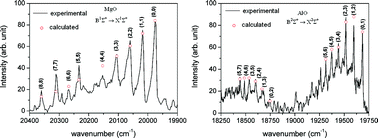
A review of results of molecular spectroscopic investigations during plasma electrolytic oxidation of valve metals is presented. Particular attention is paid to three spectral systems, B1Σ+ → X1Σ+ of MgO, and B2Σ+ → X2Σ+, and C2Π–X2Σ+ of AlO. It was shown that a reliable assignment of the observed spectral features can only be carried out by critical comparison with the data obtained from high-resolution spectroscopy, and by using the results of quantum mechanical structure calculations. Assuming the existence of partial local thermal equilibrium, we used our spectroscopic results to determine the plasma temperature. Although limited in quality, the obtained spectra are very rich, they cover large wavelength regions, and are used to obtain information about physical and chemical processes that take place in the course of plasma electrolytic oxidation of light metals and their alloys.


1. Introduction

In the present study, we review the results of investigations on molecular emission spectra recorded during plasma electrolytic oxidation of magnesium and aluminum. The goal of these investigations was to get information about the processes that take place in these systems, with the aim to become able to monitor them. Here we focus our attention on three spectral systems, B1Σ+ → X1Σ+ of MgO and B2Σ+ → X2Σ+ and C2Π–X2Σ+ of AlO. Random appearance of spectral sources (microdischarge plasmas), poor resolution, low intensities, and unfavorable signal/noise ratios, caused great difficulties in assignment of molecular bands. The assignment was carried out by comparison with the data obtained from high-resolution spectroscopy, but also by using the results of quantum mechanical structure calculations. Assuming the existence of partial local thermal equilibrium, we used our spectroscopic results to determine the plasma temperature.

The paper is organized as follows: after the introductory section, in Section 2 we describe briefly the process of plasma electrolytic oxidation and in Section 3 we give the key information about our experimental setup. In the quite extensive Section 4 we present the theoretical background of spectral investigations we carried out. The formulae collected in this section are used to explain the features of recorded spectra. The results for MgO are presented in Section 5 and those for AlO in Section 6. We conclude the paper with Section 7, where we address briefly a model that describes the systems we have been dealing with.

2. Plasma electrolytic oxidation

The process, which is the topic of the present study, is based on the anodization of valve metals (aluminum, magnesium, titanium, zirconium, tantalum, etc.) in aqueous solutions at applied potential greater than breakdown voltage (typically from 400 V to 600 V) of the original oxide film. This process is known as plasma electrolytic oxidation (PEO), microarc oxidation (MAO) or anodic spark deposition (ASD). In this work, we will use the terminology plasma electrolytic oxidation, because it has become dominant in the literature.

PEO is an economic, efficient, and environmentally benign processing technique capable of producing in situ oxide coatings on valve metals, as well as on their alloys. Oxide coatings have controllable morphology and composition, excellent bonding strength with the substrate, good electrical and thermal properties, high microhardness, high-quality wear and corrosion resistance, etc. The PEO process is coupled with the formation of plasma, as indicated by the presence of microdischarges on the metal surface, accompanied by gas evolution.1–3 The anodic gas consists predominantly of oxygen with minor fractions of other elements.4 Various processes including chemical, electrochemical, thermodynamical, and plasma-chemical reactions occur at the discharge sites, due to increased local temperature (103 K to 104 K) and pressure (∼102 MPa). These processes are responsible for modifying the structure, composition, and morphology of obtained oxide coatings.

2.1. Voltage–time characteristic

Typical voltage versus time characteristic during the anodization of valve metals is shown in Fig. 1.5,6 Depending on increasing trend of the voltage, three regions can be clearly identified. From the beginning of anodization the voltage increases linearly with time, resulting in the constant formation rate of thin, compact barrier oxide layer on the substrate. This oxide layer is produced at both metal/oxide and oxide/electrolyte interfaces as a result of migration of O2−/OH anions and metal cations Men+ (n = 2, 3, 4 or 5) across the oxide, assisted by a high electric field (∼107 V cm−1).7 Also, small amounts of anionic components of electrolyte are incorporated into the oxide at the oxide/electrolyte interface during the anodization. Barrier oxide layer is amorphous, it has ionic conductivity and high electrical resistance. Thickness of barrier oxide layer is limited to several hundred nanometers due to the dielectric breakdown under high electric field. This stage of anodization is followed by an apparent deflection from linearity in voltage–time curve, starting from so-called breakdown voltage (stage II). After the breakdown, voltage continually increases, but the voltage–time slope decreases and a large number of small-size microdischarges appear, evenly distributed over the whole sample surface. Further anodization results in relatively stable value of the voltage (stage III).
image file: c4ra03873h-f1.tif
Fig. 1 Typical voltage–time dependence during anodization of valve metals.

In the anodization process the total current density is the sum of ionic current density and electron current density.8 In the stage I, the electric field strength, for a given current density, remains constant during the anodic growth and the ionic current is two to three orders of magnitude larger than the electronic component. In order to maintain the constant electric field strength, the voltage of anodization increases linearly with time. During anodization electrons are injected into the conduction band of the anodic oxide and accelerated by the electric field, producing avalanches by an impact ionization mechanism.8 When the avalanche electronic current reaches its critical value, the breakdown occurs.9 In the stage II a relatively low voltage increase (compared with the stage I) is required to maintain the same total current density, due to the independence of the electron current density of the anodic oxide film thickness. In the stage III, the fraction of electron current density in total current density becomes dominant. In this stage, the total current density is almost independent of the anodic oxide film thickness and the voltage–time slope is close to zero.

2.2. Mechanisms of formation of PEO coatings

PEO of valve metals is a complex process combining coexisting partial processes of oxide formation, dissolution, and dielectric breakdown. The main chemical reactions at the metal/oxide interface are:2,4,10,11
 
Me → Mesolidn+ + ne, (1)
where n = 2, 3, 4 or 5,
 
Me2+ + Osolid2− → MeO, 2Me3+ + 3Osolid2− → Me2O3, Me4+ + 2Osolid2− → MeO2, 2Me5+ + 5Osolid2− → Me2O5. (2)

Also, direct ejection of metal into the electrolyte can occur through microdischarge channels during the breakdown:4

 
Meejected + nOH → Me(OH)n + ne, (3)
n = 2, 3, 4 or 5.

The main reactions at oxide/electrolyte interface are the formation of gaseous oxygen:

 
4OH → 2H2O + O2↑ + 4e, (4)
oxidation of the ejected metal:
 
Men+ + (n + 1)OH → Me(OH)(n+1), (5)
and chemical dissolution of metal oxides caused by instability of oxide films:
 
MeO + 2OH → MeO22− + H2O, Me2O3 + 2OH → 2MeO22− + H2O. (6)

Three main steps lead to formation of oxide coatings during PEO.12 In the first step, a number of separated discharge channels are formed in the oxide layer as a result of loss in its dielectric stability in a region of low conductivity. This region is heated by generated electron avalanches up to temperatures of l04 K.13 Due to the strong electric field, the anionic components of electrolyte are drawn into the channels. Concurrently, the metal is melted out of the substrate, enters the channels, and becomes oxidized. As a result of these processes, plasma chemical reactions take place in the channels. These reactions lead to an increase in pressure inside the channels. At the same time, separation of oppositely charged ions occurs in the channel due to the presence of the electric field. The cations are ejected from the channels into the electrolyte by electrostatic forces. In the next step, oxidized metal is ejected from the channels into the coating surface in contact with the electrolyte and in that way the coating thickness around the channels increases. Finally, discharge channels get cooled and the reaction products are deposited on its walls. This process repeats itself at a number of discrete locations over the coating surface, leading to increase in the coating thickness. The coating material, formed at the sites of breakdown, contains crystalline and amorphous phases, with constituent species derived both from the metal and from the electrolyte.

2.3. Optical emission spectroscopy of microdischarges

The investigation and characterization of microdischarges are important steps in understanding the PEO process. Distribution and types of microdischarges have important effects on the formation mechanism, composition, morphology, and various properties of the resultant oxide coatings. Real-time imaging of the PEO process gives information of the microdischarge spatial density, surface fraction under microdischarges, and the dimensional distribution of microdischarges at various stages of PEO.11,14–17

Given the liquid environment, optical emission spectroscopy (OES) is the best available technique for microdischarges characterization. The most popular application of OES for PEO diagnostics is spectral characterization and observation of temporal evolution of spectral lines in the visible and near UV spectral region. The main difficulty in an application of OES for PEO characterization comes from space and time inhomogeneity of microdischarges appearing randomly across the anode surface. Thus, the PEO spectra represent time integrated radiation recorded by spectrometer-detector system. The results of spectroscopic observation are even more complex to analyze if one takes into account that the radiation intensity is rather low and long exposure times are required. For this reason, high-light power spectrometers with low spectral resolution are usually employed for spectra recordings. Consequently, fine details of spectral line shape of hydrogen lines and narrow line-widths of nonhydrogenic lines are not widely used for PEO characterization.

It has been found that discharge optical emission spectra originate in general from both the species present in substrate and in electrolyte. When the substrate consists of elements with relatively low melting points (Al, Mg), the corresponding atomic and ionic lines appear independently of the type of electrolyte.18–23 On the other hand, the (non/) appearance of spectral lines of metals with high melting points (Ta, Ti, Zr) strongly depends on the electrolyte.16,24–27

Hydrogen Balmer lines Hα (656.28 nm) and Hβ (486.13 nm) can always be detected during the PEO process, and plasma broadened profiles of these lines were used for electron number density (Ne) measurement.16,17,21,22,24,25,28 Jovović et al. showed that the Balmer line Hα is very intense and strongly self-absorbed in the PEO process.17,21,22 For this reason Hα is not suitable for the spectral line shape analysis. Analysis of the Balmer line Hβ line profile during PEO of valve metals showed that the Hβ line shape can be properly fitted only if two Lorentzian profiles are used.16,17,21,22,24,25 These Lorentzian profiles correspond to electron number densities of Ne ≈ 1.0 × 1015 cm−3 and Ne ≈ 2.2 × 1016 cm−3. Spectral line shape analysis of single charge ionic lines of aluminum at 704.2 nm (ref. 21 and 22) and magnesium at 448.12 nm (ref. 21) were used for electron number density measurement in systems under consideration, and the larger electron number densities Ne ≈ (1.2–1.6) × 1017 cm−3 were obtained.

According to Hussein et al.28 three plasma discharge models have been proposed (see, Fig. 9 in ref. 28): discharging that occurs at metal/oxide interface (B) and discharging that occurs at oxide/electrolyte interface at either coating upper layer (A) or at the coating top layer (C). The highest Ne measured from single charge ion lines of aluminum and magnesium is emitted from metal plasma generated in the process of type B. Low and medium Ne are related to the processes of type A (discharge in relatively small holes near the surface of oxide layer) and of type C (discharge in the micropores at the surface of oxide layer). Microdischarges that result in evaporation of anodic material (type B) always occur during PEO of aluminum and magnesium (metals with lower melting point) regardless of the type of electrolyte. During PEO of titanium, zirconium, and tantalum (metals with high melting point) occurrence of this type of microdischarges strongly depends on the type of electrolyte.

For plasma electron temperature (Te) measurement during PEO, relative line intensities were used. This temperature is assumed equal to the electron excitation temperature, calculated from relative line intensities. The application of this approach is based on the assumption of Partial Local Thermal Equilibrium (PLTE) conditions. The discussion of the fulfillment of PLTE is given in ref. 21. For plasma electron temperature (Te) measurement during PEO of aluminum some research groups used the intensity ratio of two Al I lines at 396.2 nm and at 309.1 nm. Upon performed calculations, Te in the range of (4500–10[thin space (1/6-em)]000) K was determined.28–30 Alongside, the intensity ratio of the Hβ and the Hα lines was used also for Te measurements and Te = 3480 K was determined.31 We suspect that these results are questionable. First, the Balmer line Hα is self-absorbed, while the Hβ interferes with an AlO band.24 Secondly, on the red side of the Al I line at 309.2 nm there is another weaker line from the same multiplet overlapping with the stronger one. In addition, these lines are Stark and Doppler broadened and positioned within the OH band, which obstructs any line shape analysis. Having this in mind, Jovović et al. used O II lines, which are always present during PEO, to determine Te.21 These lines are sometimes weak but this is an advantage from the point of view of self-absorption. Relative line intensities were used for Te measurements and upon application of Boltzmann plot technique Te ≈ 40[thin space (1/6-em)]000 K was determined for aluminum.21,22 Using the same procedure, electron temperature obtained from Mg I lines is ∼4000 K,22 from Zr I lines is in the range (7500 ± 1000) K,27 and from Ti I lines in the range (3700 ± 500) K.25

In contrast to the extensive use of atomic emission spectroscopy for investigation of PEO processes and determinations of plasma parameters, there has been very little information about the appearance of molecular bands in the considered systems. Early attempts on this field32–34 were restricted to identification of band heads and their more or less provisory assignment. Posuvailo and Klapkiv32 detected the v′ = 0; v′′ = 0, v′ = 0; v′′ = 1, and v′ = 1; v′′ = 0 bands of the B2Σ+ → X2Σ+ spectral system of AlO. In our previous study on Al,34 we assigned several bands of AlH, AlO, Al2, and possibly AlH2. Posuvailo33 claimed to identify several band heads of β and γ spectral systems of ZrO. However, as shown in our recent study on PEO of Zr,27 the corresponding emission spectrum, and particularly the structure of the ZrO bands are very complex and require much more careful investigation in order to make reliable assignment of the observed features.

3. Experimental

Rectangular samples of dimensions 25 mm × 10 nm of aluminum (99.999% purity, Goodfellow) and magnesium alloy AZ31 (96% Mg, 3% Al, 1% Zn, Alfa Aesar) were used as working electrodes in experiments. The working electrodes were sealed with insulation resin leaving only active surface area of 1.5 cm2. Before the anodization, the surface of the samples was degreased and dried in a warm air stream. The PEO process took place in an electrolytic cell with flat quartz glass windows.35 Two platinum wires (5 cm long and 1 mm in diameter) were used as cathodes. During the PEO, the electrolyte circulated through the chamber–reservoir system and the temperature of the electrolyte was maintained at (20 ± 1) °C. Aluminum was anodized in 0.1 M citric acid, while magnesium alloy was anodized in 4 g L−1 Na2SiO3·5H2O and 4 g L−1 KOH. The electrolytes were prepared by using double distilled and deionized water and PA (pro analysis) grade chemical compounds. Anodizing was carried out at current density of 100 mA cm−2

Spectroscopic measurements were performed utilizing two different grating spectrometers. Spectral measurements during PEO of valve metals in a wavelength range from 380 nm to 850 nm were taken on a spectrometer system based on the intensified charge coupled device (ICCD). Optical detection system consisted of a large-aperture achromatic lens, a 0.3 m Hilger spectrometer (diffraction grating 1200 grooves per mm and inverse linear dispersion of 2.7 nm mm−1), and a very sensitive PI-MAX ICCD thermoelectrically cooled camera (−40 °C) with high quantum efficiency manufactured by Princeton Instruments.36 The CCD chip consisted of 430 × 256 active pixels, each approximately 26 μm × 26 μm. The system was used with several grating positions with overlapping wavelength range of 5 nm. Spectra were recorded in segments of 43 nm and the whole spectral range was obtained by adding one spectra interval to the previous one. The optical-detection system was calibrated using a LED based light source.37

Spectral measurements during PEO of valve metals in UV region were taken on a spectrometer system consisting of a quartz objective, Czerny–Turner spectrometer, and thermoelectrically cooled (−10 °C) CCD detector (2048 × 506 pixels, each approximately 12 μm × 12 μm) manufactured by Hamamatsu. The optical-detection system was calibrated using a hydrogen (D2) lamp. In both systems, the image of anode surface was projected with unity magnification to the entrance slit of the spectrometers.

4. Spectroscopy of diatomic molecules

4.1. Hamiltonian of a diatomic molecule

No matter how strange it might sound, the derivation of the complete Hamiltonian for a diatomic molecule is in a certain sense conceptually more demanding than that for a polyatomic molecule with non-linear equilibrium geometry. This is a consequence of the fact that one of the principal moments of inertia (that coinciding with the molecular axis) of a diatomic molecule is vanishing, what causes anomalous commutation relations between the components of angular momenta and leads to a peculiar form of the rotation part of the Hamiltonian.

Let us consider a molecule with the nuclei A and B whose masses are mA and mB, and charge numbers ZA and ZB, respectively, and with N electrons (mass = me). The total mass of the nuclei (Mn) and of the molecule (M are thus MnmA + mB, M = Mn + Nme), respectively. We start with the non-relativistic molecular Hamiltonian defined in a space-fixed coordinate system (SFS). To separate off the translational motion of the molecule, we use an intermediate frame with the axes parallel to those of the SFS and with the origin laying in the mass center of the molecule (involving also the electrons). However, we refer the electron coordinates to the mass center of the nuclei, and we call this frame nuclear center of mass system (NCMS). As the three linearly independent nuclear coordinates we chose the components X, Y, Z of the position vector, [r with combining right harpoon above (vector)], of the nucleus B with respect to that of the nucleus A, [r with combining right harpoon above (vector)] = [R with combining right harpoon above (vector)]B[R with combining right harpoon above (vector)]A. We define the components of the angular momentum operator of the nuclei, image file: c4ra03873h-t1.tif, spatial electron angular momentum, image file: c4ra03873h-t2.tif, electron spin operator, image file: c4ra03873h-t3.tif, total molecular angular momentum excluding spin, image file: c4ra03873h-t4.tif, and total (rovibronic) angular momentum, image file: c4ra03873h-t5.tif (we ignore possible presence of the nuclear spins). They satisfy the normal commutation relations,

 
[[M with combining circumflex]X, [M with combining circumflex]Y] = i[M with combining circumflex]Z, [[M with combining circumflex]Y, [M with combining circumflex]Z] = i[M with combining circumflex]X, [[M with combining circumflex]Z, [M with combining circumflex]X] = i[M with combining circumflex]Y, (7)
where image file: c4ra03873h-t6.tif is image file: c4ra03873h-t7.tif, image file: c4ra03873h-t8.tif, image file: c4ra03873h-t9.tif, image file: c4ra03873h-t10.tif, or image file: c4ra03873h-t11.tif. Further, all the components of different angular momenta (image file: c4ra03873h-t12.tif, image file: c4ra03873h-t13.tif, and image file: c4ra03873h-t14.tif) commute with one another because they act on different coordinates.

We introduce finally a molecule-fixed coordinate system (MFS) with the goal to separate, as completely as possible, the vibrational from rotational degrees of freedoms. We need the real (original) SFS no more and we rename NCMS into SFS. We choose the MFS so that its z-axis lays along the molecular axis (the direction from the nucleus A towards B) – in this way the rotation of the nuclear skeleton is reduced to the rotation of the MFS. The orientation of this axis with respect to the NCMS is defined by the Euler angles ϕ and θ, which represent thus the rotational coordinates. We fix the value of the third Euler angle χ (that determines the orientation of the x and y axes) to χ = 0. The instantaneous distance between the nuclei, r, is the coordinate whose change corresponds to the vibrations of the nuclei. We denote the spatial coordinates of the electrons in the MFS by xμ, yμ, zμ. The Hamiltonian of the molecule transforms into38

 
Ĥ = [T with combining circumflex]e + [T with combining circumflex]n + [V with combining circumflex]en + [V with combining circumflex]ee + [V with combining circumflex]nn. (8)
[T with combining circumflex]e is the electronic kinetic energy operator:
 
image file: c4ra03873h-t15.tif(9)

The nuclear kinetic energy operator has the form

 
[T with combining circumflex]n = [T with combining circumflex]vib + [T with combining circumflex]rot (10)
where
 
image file: c4ra03873h-t16.tif(11)
and
 
image file: c4ra03873h-t17.tif(12)
μmAmB(mA + mB) is the reduced mass of the nuclei. The expressions ((11)/(12)) correspond to the volume element dVn = r2[thin space (1/6-em)]sin[thin space (1/6-em)]θdrdθdϕ. The potential energy part of the Hamiltonian (8) is
 
image file: c4ra03873h-t18.tif(13)
where RAμ = |[R with combining right harpoon above (vector)]μ[R with combining right harpoon above (vector)]A| = |[R with combining right harpoon above (vector)]μ + mB[R with combining right harpoon above (vector)]/Mn|, RBμ = |[R with combining right harpoon above (vector)]μ[R with combining right harpoon above (vector)]B| = |[R with combining right harpoon above (vector)]μmA[R with combining right harpoon above (vector)]/Mn|, Rμv = |[R with combining right harpoon above (vector)]v[R with combining right harpoon above (vector)]μ|.

The components of the electronic spatial and spin angular momenta along the MFS-axes are defined in usual way. However, the components of the (rovibronic) angular momentum are38

 
image file: c4ra03873h-t19.tif(14)

The components of the electronic angular momenta along the MFS-axes have normal commutation relations, analogous to (7). On the other hand, the components of the rovibronic angular momentum image file: c4ra03873h-t20.tif along the MFS-axes fulfill the following anomalous commutation relations:38

 
[Ĵx, Ĵy] = −i[thin space (1/6-em)]cot[thin space (1/6-em)]θĴxiℏ([L with combining circumflex]z + Ŝz), [Ĵy, Ĵz] = 0, [Ĵz, Ĵx] = 0. (15)
writing eqn (9)–(12) we separated the kinetic energy operator into three parts, the first ([T with combining circumflex]e) being the electronic kinetic energy operator, the second ([T with combining circumflex]vib) involving derivatives with respect to the coordinate r, and the third one ([T with combining circumflex]rot) that contains the angular momenta. Note, however, that the separation of the electronic, vibrational, and rotational degrees of freedom is not complete: In the rotation part we also have the vibrational coordinate r as well as the electronic coordinates via the operators [L with combining circumflex]α and Ŝα. The Schrödinger equation corresponding to the kinetic energy operator ((9)–(12)) and the potential energy (13) is solved successively. In the framework of the Born–Oppenheimer approximation,39 the electronic part of the Hamiltonian, which involves the electronic kinetic energy operator (9), and the potential energy operator (13), is replaced by the corresponding eigenvalue, V(r), being a function of the coordinate r. In the lowest-order (harmonic) approximation,
 
image file: c4ra03873h-t21.tif(16)
where re represents the equilibrium value of r. If we replace the volume element dVn by the more convenient one in the present context, dVn = sin[thin space (1/6-em)]θdrdθdϕ, eqn (11) transforms into
 
image file: c4ra03873h-t22.tif(17)

The vibration–rotation problem with the Hamiltonian involving the nuclear kinetic energy operator (10) and V(r), playing the role of the potential, can be handled simultaneously for both vibrations and rotations. An alternative way is first to solve the vibrational part of the Schrödinger equation [Ĥvib = [T with combining circumflex]vib + V(r)] and after that the rotational part (Ĥrot = [T with combining circumflex]rot), where the quantity 1/r2 in eqn (12) is replaced by its electronically and vibrationally averaged counterpart 〈1/r2〉(in the lowest-order, “rigid-rotor” approximation, 〈1/r2〉 = 1/re2).

4.2. Isomorphic Hamiltonian

A linear molecule only has two rotational degrees of freedom described by the Euler angles ϕ and θ, being sufficient to define the orientation of the internuclear axis (z) in space. At the same time, the third Euler angle χ is arbitrary. The Hamiltonian ((8)–(13)) corresponds to the choice χ = 0, but in a more general treatment χ is considered as a certain function of ϕ and θ.40,41 The lack of the third Euler angles causes anomalies as unusual commutation relations, eqn (15), and a strange form of the rotational Hamiltonian, eqn (12). These problems can be solved introducing the “isomorphic Hamiltonian”.38,40–42 It has three rotational degrees of freedom (as that of a nonlinear molecule) so that the components of all angular momenta satisfy usual (or “almost” usual, i.e. up to the sign) commutation relations. On the other hand, the isomorphic Hamiltonian has a number of eigenvalues not being the eigenvalues of the real Hamiltonian. These artificial eigenvalues can, however, be easily eliminated when the Hamiltonian is represented in a suitable basis.

In order to derive the isomorphic Hamiltonian, we introduce χ as an independent variable that defines a new coordinate frame (xyz′) tied to the molecule; it is obtained from the frame (xyz), corresponding to the choice χ = 0, by rotation through χ about the z-axis. The components of the total angular momentum along the (xyz′)-axes are38,41,43

 
image file: c4ra03873h-t23.tif(18)

They commute with the components of the angular momentum along the SFS-axes, ĴX, ĴY, and ĴZ. The operators Ĵx, Ĵy, and Ĵz commute with the components of image file: c4ra03873h-t24.tif and image file: c4ra03873h-t25.tif (along the axes of the same frame), because they act onto different coordinates. However, the commutation relations between Ĵx, Ĵy, and Ĵx are41,43–46

 
[Ĵx, Ĵy] = −iĴz, [Ĵy, Ĵz] = −iĴx, [Ĵz, Ĵx] = −iĴy. (19)

They are anomalous [indeed not so extremely anomalous as (15)], because they differ from the normal commutation relations for angular momenta, like those in eqn (7), in the sign. On the other hand, the electronic angular spatial and spin momenta have normal commutation relations. The anomalous commutation relations of the type (19) have the components along the xyz′-axes of all angular momenta which involve nuclear coordinates, like image file: c4ra03873h-t26.tif and image file: c4ra03873h-t27.tif. Note that the components of the nuclear angular momentum along the MFS-axes in general do not commute with the components of the electronic angular momentum.

The components of the angular momenta along the (xyz′)-axes are related to the components of image file: c4ra03873h-t28.tif along the (xyz) axes, by

Ĵx[L with combining circumflex]xŜx = cos[thin space (1/6-em)]χ(Ĵx[L with combining circumflex]xŜx) − sin[thin space (1/6-em)]χ(Ĵy[L with combining circumflex]yŜy),

Ĵy[L with combining circumflex]yŜy = sin[thin space (1/6-em)]χ(Ĵx[L with combining circumflex]xŜx) + cos[thin space (1/6-em)]χ(Ĵy[L with combining circumflex]yŜy),
 
Ĵz[L with combining circumflex]zŜz = Ĵz[L with combining circumflex]zŜz = 0. (20)

The rotational Hamiltonian (in cm−1) has in terms of new components of the angular momenta the form

 
image file: c4ra03873h-t112.tif(21)
where B = 〈ℏ/(4πcμr2)〉v is the (electronically and) vibrationally averaged rotational constant. The isomorphic Hamiltonian is defined, taking into account the third relation in eqn (20) as
 
image file: c4ra03873h-t29.tif(22)

Note that the Hamiltonians (12) and (22) are mutually isomorphic but not identical. The border between the identity and isomorphism is crossed by skipping from eqn (21) and (22), i.e. by using the condition Ĵz[L with combining circumflex]zŜz = 0. Since Ĥisorot commutes with the operator (Ĵz[L with combining circumflex]zŜz), these two operators have common eigenfunctions, and the mentioned condition will be fulfilled only when we use the basis functions which correspond to the zero eigenvalues of the operator (Ĵz[L with combining circumflex]zŜz) – only these eigenfunctions of the isomorphic Hamiltonian will be the true wave functions of the operator (12). They are obtained as solutions of the eigenvalue problems

 
Ĵzψr = Ωψr, [L with combining circumflex]zψe = Λψe, ŜzψS = ΣψS. (23)
where Λ and Σ are projections of the operators Ω = Λ + Σ and image file: c4ra03873h-t30.tif on the z′-axis, and Ω = Λ + Σ.

We shall use further the isomorphic rotation Hamiltonian. To simplify the notation we shall delete the primes and the superscript iso. The operator (22) can be written in several alternative forms, depending on whether the relation Ĵz[L with combining circumflex]zŜz = 0 is taken into account explicitly or implicitly e.g.

 
image file: c4ra03873h-t31.tif(24)

The last term on the right-hand side of eqn (24), producing no dependence on J, shifts all rotational levels by the same amount and thus we shall consider it as a part of the electronic Hamiltonian (i.e. we neglect it when we only consider rotational structure). The terms involving the products of electronic ladder operators, [L with combining circumflex]+[L with combining circumflex]x + i[L with combining circumflex]y, [L with combining circumflex][L with combining circumflex]xi[L with combining circumflex]y, and Ĵ±/Ŝ± only have off-diagonal electronic matrix elements (i.e. they couple different electronic state). When we calculate the rotational levels in a particular electronic state, the contribution of these terms is small and we shall in general neglect them. (However, they are responsible e.g. for the Λ-splitting in Π states.) Such a simplified rotation Hamiltonian can be written as

 
image file: c4ra03873h-t32.tif(25)
we shall usually use this form.

The rotational Hamiltonian (24) obviously commutes with Ĵ2, Ŝ2, and [L with combining circumflex]2, and its approximate form eqn (25) additionally with [L with combining circumflex]z. Recall, however, that the rotational Hamiltonian only represents a part of the total molecular Hamiltonian. The complete molecular Hamiltonian involves besides Ĥrot the electronic and vibrational parts, Ĥe and Ĥvib, respectively. Since Ĥe does not commute with [L with combining circumflex]2, the quantum number L, which would correspond to [L with combining circumflex]2, is never good. Note also that in the rest of the Hamiltonian appear in general the terms that also involve the rotation coordinates, i.e. which take into account the coupling of rotations with the other degrees of freedom. Some of the couplings can be neglected, but there are several of them interfering qualitatively and quantitatively with the rotational structure of molecular spectra. The most important of these is usually the spin–orbit/rotation coupling, because the corresponding effects are on the energy scale comparable with those caused by molecular rotations. For this reason, we shall below complete the Hamiltonian by the terms describing the leading part of the spin couplings.

In multiplet (i.e. non-singlet) Σ electronic states (Λ = 0, S ≠ 0) we have the spin–rotation coupling; we add then to the Hamiltonian (24) or (25) the corresponding operator. We shall represent it in the “phenomenological form”, ref. 43

 
image file: c4ra03873h-t33.tif(26)
where γ is the (averaged) spin-rotation constant, that may be positive or negative.

In multiplet Π, Δ,… (Λ ≠ 0, S ≠ 0) electronic states the main spin effect is the spin–orbit coupling. It is usually described by the phenomenological operator43

 
image file: c4ra03873h-t34.tif(27)

The matrix elements of the second term on the right-hand side of eqn (27) are vanishing within an electronic state and do not contribute to the energy in first order. Thus, we usually use the simplified form of the spin–orbit operator,

 
image file: c4ra03873h-t35.tif(28)
ASO is the “spin–orbit constant”, which may, depending on the electron configuration in question, have + or −sign. The spin-rotation coupling is also present in the multiplet spatially degenerate states, but it is there normally much weaker than the spin–orbit one.

In the general case the only operators which commute with the Hamiltonian of a molecule are Ĵ2, Ŝ2, and the operator E* that inverts the coordinates of all particles (electrons and nuclei) in the SFS; the quantum numbers of these operators are J, S, and + or −, respectively. However, depending on the concrete situation, there may be various near (good) quantum numbers. Such quantum numbers correspond to the operators that do not commute with the complete Hamiltonian, Ĥ, but they do commute with the dominant part of it, say Ĥ1. The eigenfunctions of Ĥ1 build a suitable basis for representation of Ĥ, and the quantum numbers associated with the operators that commute with Ĥ1 label these basis functions. They represent “nearly good” quantum numbers, because the leading part of Ĥ, namely Ĥ1, is in such a basis represented by a diagonal matrix. The near quantum numbers are particularly useful for assignment of experimentally observed spectral features. Note that, strictly speaking, even J is not a good quantum number – it is such only in the case of vanishing nuclear spins. In the presence of nuclear spin, image file: c4ra03873h-t36.tif, the only good quantum number is that corresponding to the total angular momentum image file: c4ra03873h-t37.tif. However, in this case J is (usually) very nearly a good quantum number.

4.3. Basis functions

The fact that there are three operators Ĵ2, Ĵz, and ĴZ that commute with one another means that there exist sets of their common eigenstates |JΩM〉. Thus,
 
Ĵ2|JΩM〉 = J(J + 1)2|JΩM〉, ĴZ|JΩM〉 = Mℏ|JΩM〉, Ĵz|JΩM〉 = Ωℏ|JΩM〉. (29)

Non-vanishing matrix elements of the relevant operators in this basis are

 
image file: c4ra03873h-t38.tif(30)
where we introduce the “raising” and “lowering” operators in the SFS and MFS:
 
Ĵs±ĴX ± Y, Ĵm±Ĵx ± y. (31)

Note the sign differences in the expressions (30) for matrix elements of these operators. Since we do not deal with external fields, the choice of the quantum number M is arbitrary.

We shall normally use the basis functions corresponding to Hund's case (a) coupling scheme.41,43,46,47 According to it image file: c4ra03873h-t39.tif and image file: c4ra03873h-t40.tif are both “tied” to the internuclear axis (the z axis of the MFS) making the signed projections Λ and Σ, respectively, while image file: c4ra03873h-t41.tif is perpendicular to this axis. Thus the total angular momentum image file: c4ra03873h-t42.tif makes the projection M on the SFS axis Z and the projection Ω = Λ + Σ on the MFS axis z. Consequently, in this case the rotational basis functions are the symmetric top wave functions |JΩM〉. The total case (a) wave functions are labeled additionally by the electronic spatial quantum numbers n (“principal” quantum number) and Λ, the spin quantum numbers S, Σ, and the vibrational quantum number v. We write them in the product form41,43

 
|n, Λs; v; S, Σ, J, Ω, M〈 = |s〉|v〉|〉|(M)〉, (32)
where s is odd for Σ and even for all other electronic states. (The unimportant quantum number M is on the right-hand side put in parentheses.) Actually, the separation |nΛSΣ〉 = |〉|SΣ〉 is strictly valid only for one- and two-valence electronic molecules. Justification for the notation (32) is the following: Each of the operators image file: c4ra03873h-t43.tif, and image file: c4ra03873h-t44.tif, appearing in the rotational Hamiltonian only acts on one set of the coordinates and their effect on the vector |nΛSΣ〉 is the same as the effect on the factorized (in the general case, fictitious) form of this vector |〉|SΣ〉. The basis (32) is convenient because the key operators [L with combining circumflex]z, Ĵ2, Ĵz, Ŝ2, and Ŝz are all diagonal in this coupling scheme.

Another frequently used basis consists of Hund's case (b) functions, where the intermediate nearly good quantum number N is used (its projection on the z-axis is Λ) and the spin quantum number Σ is omitted:

 
|n, Λs; v; N, Λ, S, J, M〉 = |s〉|v〉|SJNΛ(M)〉. (33)

The fact that the components along the MFS-axes of some of the momenta we use image file: c4ra03873h-t45.tif have anomalous commutation relations, whereas the commutation relations for the other class of operators image file: c4ra03873h-t46.tif are normal, causes certain problems. The momenta image file: c4ra03873h-t47.tif, image file: c4ra03873h-t48.tif, image file: c4ra03873h-t49.tif, when expressed in terms of their MFS components are actually not proper angular momenta, because the angular momentum operator is defined as a vector operator whose components satisfy commutation relations of the type (7). Thus, when working with these operators we cannot directly use a number of relations derived for proper momenta (e.g. those based on the application of the Wigner–Eckart theorem, the Clebsh–Gordan coefficients etc.).48 Further, the composite momentum from the standpoint of the theory of addition of angular momenta, image file: c4ra03873h-t50.tif [see eqn (33)], is the difference and not the sum of “partial momenta” image file: c4ra03873h-t51.tif and image file: c4ra03873h-t52.tif. These problems can generally be solved in three ways. (a) One can work with the components of all momenta along the SFS-axes – all of them satisfy normal commutation relations – and only after carrying out all algebraic manipulation, the results are expressed in terms of the MFS components.45,46 (b) The other possibility is to work consequently with the MFS components, bearing in mind anomalies and restrictions mentioned above. (c) The third way was proposed by Van Vleck:44 Instead of momenta [P with combining circumflex]x, [P with combining circumflex]y, [P with combining circumflex]z, with anomalous commutation relations like those for Ĵx, Ĵy, Ĵz, eqn (19), we can use the “reverse” momenta

 
[P with combining circumflex]rx = −[P with combining circumflex]x, [P with combining circumflex]ry = −[P with combining circumflex]y, [P with combining circumflex]rz = −[P with combining circumflex]z, (34)
which have normal commutation relations,
 
[[P with combining circumflex]rx, [P with combining circumflex]ry] = iℏ[P with combining circumflex]rz, [[P with combining circumflex]ry, [P with combining circumflex]rz] = iℏ[P with combining circumflex]rx, [[P with combining circumflex]rz, [P with combining circumflex]rx] = iℏ[P with combining circumflex]ry. (35)

(Indeed, Van Vleck preferred to invert the momenta with normal commutation relations, [L with combining circumflex]rx = −[L with combining circumflex]x, etc. so that all momenta along the MFS-axes consistently had anomalous commutation relation.) Consequently, all momenta along the MFS-axes ([L with combining circumflex]x, Ŝx, [R with combining circumflex]rx, [N with combining circumflex]rx, Ĵrx,…) have normal commutation relations.

Each wavefunction associated with an energy level may be classified as even or odd according to whether it remains unchanged or changes sign on the operation E*, carrying out inversion of SFS spatial coordinates of all electrons and nuclei. Since the molecular Hamiltonian is invariant under this symmetry operation, it has non-vanishing matrix elements only between the state vectors of the same parity. This operation does not affect the vibrational coordinates. It does not act directly on the spin coordinates, but in the case when the spin angular momenta are quantized along the MFS-axes [Hunds's basis (a)], there is an indirect effect on the spin functions because the transformation from SFS to MFS involves the Euler angles, being functions of the SFS nuclear coordinates. It can be shown that the effect of E* on Hund's (a) basis functions is41,43

E*|s〉|v〉|〉|(M)〉 = (−1)p|n, −Λ〉|v〉|S, −Σ〉|J, −Ω, M〉,
 
p = JS + s. (36)

For Hund's (b) basis,

 
E*|s〉|v〉|N, Λ, S, J, M〉 = (−1)N+s|n, −Λ〉|v〉|N, −Λ, S, J, M (37)

It follows that the simple case (a) or case (b) basis functions are not eigenfunctions of E*. The appropriate combinations which do have a definite parity are in the case (a)41

 
image file: c4ra03873h-t53.tif(38)

The ±(−1)JS+s phase factor in eqn (38) causes the parity labels to alternate as J increases, so that the lower of the near-degenerate pair for a given J might be + and the upper −, the designation becomes opposite for the next J value and so on. To avoid this alternation, another parity labeling convention, e/f, has been introduced.49 For integral J, e-levels have parity (−1)J and f-levels (−1)J+1; for half-integral J, e-levels have parity (−1)J−1/2 and f-levels (−1)J+1/2. Following this convention, all lower components of parity doublets have the same label, e.g. e, and all the upper components have the opposite label, in this case f.

4.4. Transitions

4.4.1. Transition moments. We consider the transition between two states of a diatomic molecule, whose quantum numbers are denoted by prime (upper state) and two prime marks (lower state).50,51 We label the corresponding state vectors in the sense of Hund's (a) case. The intensity of the transition between these states is proportional to the square of the transition moment defined as
 
R,′′ = 〈nΛ′; v′; SΣ′; JΩM′|Â|n′′Λ′′; v′; S′′Σ′′; J′′Ω′′M′′〉 (39)
where  is the operator which determines the mechanism of the transition. In the case of “optical” transitions, we consider here,  is the (vector) dipole operator, [small mu, Greek, circumflex], defined by
 
image file: c4ra03873h-t54.tif(40)
where qi is the charge, and [r with combining right harpoon above (vector)]i radius vector of the ith particle and the sum runs over all charged particles (electrons, nuclei). The proportionality factor between the intensity of the transition and |R,′′|2 involves, beside natural constants and statistical weights, in the case of absorption the energy difference, ΔE, between the levels in question, and at emission ΔE4.

In the framework of usually applied non-relativistic, Born–Oppenheimer, and rigid rotator/infinitesimal vibrator approximations the state vectors are assumed in the form of products of partial state vectors for individual degrees of freedom, like on the right-hand side of eqn (32). If we are only interested in “allowed” transitions, the spin variables can be excluded from consideration since in this case transitions are only possible when both states in question have the same spin function (S′ = S′′, Σ′ = Σ′′). We start with the expression for the components of the transition moment, [small mu, Greek, circumflex]F, (F = X, Y, Z) along the axes of the SFS,

 
R′,′′F = 〈JΩ′|〈v′|〈nΛ′|[small mu, Greek, circumflex]F|n′′Λ′′〉|v′′〉|J′′Ω′′〉, (41)
where we factorize the state vector, delete the spin parts and arbitrary quantum M′ = M′′ = M. Since the electronic and vibrational variables are naturally defined within the MFS, we express now the SFS components of the dipole operator in terms of their MFS counterparts,
 
image file: c4ra03873h-t55.tif(42)
where g = x, y, or z, and λFg are the elements of the rotation matrix which involve sine and cosine functions of three Euler angles ϕ, θ, and χ. Since (only) the rotation functions depend on the Euler angles (and only on them), eqn (41) may be transformed into
 
image file: c4ra03873h-t56.tif(43)
The expression
 
nΛ′|μg|n′′Λ′′〉 ≡ Re′,e′′g, (44)
is the g-component of the “electric transition moment”. It represents the generalization of the usual dipole moment within a single electronic state (when n′ = n′′ and Λ′ = Λ′′), and unlike the latter it may have both parallel and perpendicular components with respect to the internuclear axis. This quantity is a function of the bond length, r, (i.e. of the vibrational coordinate), and we represent it by a Taylor expansion
 
image file: c4ra03873h-t57.tif(45)
where re is the equilibrium bond length. Inserting (45) into (43) we obtain
 
image file: c4ra03873h-t58.tif(46)

4.4.2. Electronic transitions. With help of the formula (46) we can discuss all selection rules for optical transitions in diatomic molecules. However, we are dealing in the present study with electronic transitions (n′ ≠ n′′ and/or Λ′ ≠ Λ′′) and consider only this case. In the first, Franck–Condon (F–C), approximation we only retain the first term in square parentheses of eqn (46),
 
image file: c4ra03873h-t59.tif(47)

This expression will be nonvanishing if the electric transition moment does not equal zero. This is the case when the product of the irreducible representations of the electronic wave functions equals the irreducible representation of the x, y, or z-components of the dipole operator. In the case of heteronuclear diatomics, the z-component of this operator belongs to the Σ+ and x- and y-components building together the basis for a Π irreducible representation of the point group C∞v. This results in the selection rule ΔΛ = 0, ±1. Additional selection rule is that the transitions Σ+ ↔ Σ+ and Σ ↔ Σ are allowed, while the transitions Σ+ ↔ Σ are forbidden. In the case of homonuclear diatomics, the z-component of this operator belongs to the Σu+ and x- and y-components to the Πu irreducible representation of the point group D∞v. This yields the additional selection rule that the electronic transition is allowed only if one of the combining states has g and the other u symmetry.

4.4.3. Vibrational structure of electronic transitions. The square of the scalar product 〈v′|v′′〉 in eqn (47), representing the overlap of the vibrational wave functions of the combining state,
 
image file: c4ra03873h-t60.tif(48)
is the Franck–Condon factor (FCF). We also define the vibrational transition moment (VTM), the vector quantity with the components along the MFS-axes,
 
image file: c4ra03873h-t61.tif(49)

In the F–C approximation, image file: c4ra03873h-t62.tif. The magnitude of |VTM|2 or (sometimes significantly less reliably) FCF determines the intensity distribution within a (rotationally unresolved) band system, which consists of bands, each one arising by rotational transitions between a pair of vibrational levels of two electronic states. The most prominent features in band systems are progressions (characterized in absorption by a fixed vibrational quantum number v′′ and variable quantum number v′, and fixed v′ and variable v′′ in emission) and sequences, v′–v′′ = const. If the equilibrium bond lengths and vibrational frequencies are similar in both electronic states, the most intense bands in an experimentally recorded spectrum correspond to the transitions v′–v′′ = 0. A long progression with the maximum at a quite large value of |v′–v′′| indicates that the equilibrium bond lengths in the combining electronic states are appreciably different. While the bands in a progression are separated from one another by several hundreds to few thousands cm−1 (corresponding to the wave numbers of vibrational fundamentals), the separation of the bands in a sequence is usually between several tens and several hundreds cm−1, reflecting the difference in the values of vibrational frequencies in the two electronic states in question.

The F–C approximation is thus mathematically based on the assumption that the electric transition moment is a constant quantity. This corresponds to physical picture of the “vertical” electronic transitions, i.e. the transitions at unchanged distance between the nuclei. If we go beyond the F–C approximation, we have to take into account the dependence of the electric transition moment on the internuclear distance [the second term in of eqn (45)/(46)]. In a diatomic molecule (where the unique vibrational coordinate, r, is totally-symmetric) this only quantitatively influences the transition probabilities; in polyatomic linear molecules it may make the transitions, forbidden in the F–C approximation, “vibronically” allowed.

Measured electronic-vibration terms (in cm−1) are usually represented by the formula

 
image file: c4ra03873h-t63.tif(50)
where Te is the electronic term value and is the vibrational wave number. Te is the energy difference between the minima of the potential curves for the upper and lower electronic state; it is actually a quantity that is not directly measured and plays in eqn (50) just the role of a fitting parameter. Note that Herzberg used instead of the symbol ωe (as well as ω) and called it “vibrational frequency measured in cm−1”.47 (The subscript “e” in ωe stands for “equilibrium”.) On the other hand, we retain the notation (ωexe) and (ωye) for the experimentally determined anharmonicity parameters expressed in cm−1 and the parentheses should indicate that we shall always handle these products as single quantities.

We have used the experimentally derived formula (50) for assignment of bands observed in our spectra. However, for determination of the plasma temperature we also need the FCFs and/or VTMs. In order to calculate these quantities we have to solve the vibrational Schrödinger equation for two electron states in question and to use the so obtained wave functions to compute the required quantities. If one wishes to avoid explicit ab initio calculations of the potential energy curves for the electronic states in question, he has first to find a way how to extract the potential energy function that, combined with the corresponding kinetic energy operator, gives the energy eigenvalues as close as possible to those presented by the formula (50). We have solved this problem combing the quantum-mechanical perturbative and variational approaches.

We assume the vibrational Hamiltonian in the form

 
H = H0 + V′, (51)
where
 
image file: c4ra03873h-t64.tif(52)
and
 
V′ = k3x3 + k4x4 +…; (53)
where x ≡ (rre). Applying the perturbation theory we can relate the force constants k, k3, k4… to the coefficients in the expansion (50).52 We shall use below only the first two of them:
 
k = μω2 = 4μπ2c22, (54)
and
 
image file: c4ra03873h-t65.tif(55)

In variational calculations of the eigenvalues and eigenfunctions of an anharmonic oscillator, as well as of the FCFs and VTMs for particular combinations of vibrational levels of two electronic state, the following procedure is applied:53 one of the electronic states, a, (typically ground state) is chosen as the referent one. The basis functions (being the eigenfunctions of a suitably chosen harmonic oscillator) are centered with respect to its equilibrium bond length, re. If the potential energy curve is ab initio computed, it is fitted to a polynomial of the type (53). The potential energy curve of the other state, b, is fitted by polynomial series in the same variable as for the reference state,

 
Vb = kb0 + kb1(rre) + kb2(rre)2 + kb3(rre)3 +… (56)
where re is (as before) the equilibrium bond length in the ground electronic state. In this case we have also a constant and a linear term in (rre), because the two states in question have different equilibrium bond lengths and their minima are separated in energy. Of course, we may use instead of ab initio computed potentials their experimentally derived counterparts. In variational calculations the eigenfunctions for the v′′th level of the electronic state a and the v′th level of the state b are obtained in the form
 
image file: c4ra03873h-t66.tif(57)
where |ψ(0)i〉 are the basis functions, and cv′′i and dvj are the expansion coefficients. The advantage of the choice of the same basis for both states becomes now clear. The FCF for the two vibrational states in question is simply
 
image file: c4ra03873h-t67.tif(58)

When the bond-length dependence of the electric transition moment is taken into account, it is fitted to the form

 
image file: c4ra03873h-t68.tif(59)
and the VTM is computed as
 
image file: c4ra03873h-t69.tif(60)

The intensity of the spectral band appearing as a consequence of the emission transition between the vibrational level v′ of the electronic state e′ and the vibrational level v′′ of the electronic e′′ is determined by the formula

 
image file: c4ra03873h-t70.tif(61)
where Ev′e′ and Ee′′v′′ are the energies of these two states, Nv is the number of molecules in the upper state, k is the Boltzmann constant, and T the temperature. Since the wave number differences between all bands we used in the present study for determination of temperature, namely the members of the band sequences v′–v′′ = 0, −1, −2, are negligibly small compared to the energy of the electronic transition, the quantity Ev′e′Ee′′v′′ can in eqn (61) be assumed constant and then the number of the molecules in the upper state is
 
image file: c4ra03873h-t71.tif(62)

In the F–C approximation, it is assumed that the electric transition moment is constant, and in this case we replace in the formula (62) |[R with combining right harpoon above (vector)]e′v′,e′′v′′|2 by the FCF.

Assuming a partial local thermal equilibrium of the system, the ratio of the number of molecules Ne′v in the vibrational state v′ and in the lowest vibrational state v′ = 0 of the same electronic species, Ne′0 is determined by

 
image file: c4ra03873h-t72.tif(63)

Combining the formulae (62) and (63), we obtain for the sequence v′–v′′ = −Δv

 
image file: c4ra03873h-t73.tif(64)
or, in the F–C approximation,
 
image file: c4ra03873h-t74.tif(65)

We use below the formulae (64) and/or (65) to determine the plasma temperature. This approach has the advantage that it does not require the knowledge of the Einstein transition probabilities and that the measured peaks appear in a narrow spectral range such that the problems caused by wavelength dependence of the measurement sensitivity are avoided.

4.4.4. Rotational structure of electronic transitions. The general selection rule for rotational transitions is
 
J′ − J′′ = 0, ±1, (66)
with the restriction that the transition J = 0 ↔ J = 0 is forbidden. However, the presence of electronic spatial and spin angular momenta introduced additional, not rigorous selection rules. In transitions involving Hund's (a)-type states (large spin–orbit constant, typical for molecules with relatively heavy nuclei and nonvanishing electronic spatial and spin angular momenta), the projection of the total electron angular momentum (involving both the spatial and spin contributions) on the internuclear axis, ΩΛ + Σ (being at the same time the projection of the total angular momentum) is nearly conserved. In this case we have the additional selection rule ΔΩ = 0. In the other extreme case of relatively week spin effects, like in Σ electronic states or in molecules with light atoms, particularly at strong rotation [Hund's case (b)] the ΔΩ = 0 selection rule is replaced by ΔN = 0, ±1 (with the restriction that ΔN = 0 does not occurs in Σ–Σ electronic transitions). A more detailed discussion or the rotational structure of electronic spectra will be given below for each spectral system considered in the present study.

4.5. Sources of experimental data

The classical sources of information about molecular spectra represent the monographs by Herzberg47,54–56 and Pearse and Gaydon.57 In Herzberg's (H) volumes I47 and III53 are collected all relevant experimental data up to fifties/sixties of the preceding century. In the fourth volume, published in 1979 in collaboration with Huber (HH),56 one can find the data on diatomic molecules reported until the end of 1975. The fourth (to our knowledge the last one) edition of the book by Pearse and Gaydon (PG) is printed in 1976, i.e. almost at the same time as the HH's compilation of data on diatomics. This edition contains the data published until the end of 1974. Thus, at searching for information about molecular spectra, one has practically only to look at experimental papers published after appearance of the mentioned monographs, and to complete in this way the information collected by H/HH and PG. These books has become such a standard source of information on molecular spectra, that many authors of new studies do not cite explicitly the authors of old experimental works but they simply quote these studies as “H or PG and the references therein”. We will in general follow this praxis in the present review.

While the three volumes by Herzberg47,54,55 involve also complete theory underlying molecular spectra, as well as numerical values for all molecular parameters like vibrational frequencies and anharmonicity constants, rotational constants, internuclear distances, etc., the book by PG only involves the results of direct spectral measurements, i.e. positions of band heads and their relative intensities. Thus, the books by H and PG are in a sense complementary. It should be noted that in the H's books the transition energies are given in wave numbers for vacuum, whereas they are collected by PG in wavelengths, as measured in air. Therefore, a PG wavelength is not exactly the inverse of the corresponding H's wave number.

4.6. On the interplay between molecular spectroscopy and quantum chemistry

One of the greatest spectroscopists, D. A. Ramsay, presented at a Conference (Breukelen, Holland) in 1976 an interesting table (Table 1, first and second column with error margins). It showed what experimentalists expected from the theoreticians at that time (i.e. forty years ago): Not necessarily data of spectroscopic accuracy but rather results with definite error margins that would help answer some important questions, which could not be answered on the basis of experiments alone. So, e.g., the theoretical information about the occurrence and mutual interactions of various electronic states in a certain energy range was very valuable for the experimentalists; the help of theoreticians was sought when it was not possible to assign unambiguously the spectral lines observed. Ramsay gave two sets of error margins, which he named “acceptable” and “good”. It is clear that the error limits quoted in Table 1 as “acceptable” hardly possessed any predictive value – they only could have been of some help in the interpretation of general features of spectra. On the other hand, the situation was different with the “good” theoretical results: In a number of cases it has been shown that results of such accuracy have lead to reliable predictions of spectra not observed before.
Table 1 Expected accuracy of quantum chemical calculationsa
Quantity Acceptable (1976) Good (1976) State of the art (1995) State of the art (2014)
a r ≡ equilibrium bond length; θ ≡ equilibrium bond angle; ΔE ≡ vertical electronic transition energy; D ≡ dissociation energy; I ≡ ionization potential; ω ≡ vibrational frequency; μ ≡ dipole moment; f ≡ oscillator strength.b ≡ stretching frequency.c ≡ bending frequency. For further explanation see the text.
r, Δr 0.05 Å 0.01 Å 0.01 Å 0.01 Å
θ 5 degrees 1 degrees 1–2 degrees  
ΔE, D, I 0.5 eV 0.1 eV 0.05 eV 0.05 eV
ω 250 cm−1 50 cm−1 100–200,b 50c cm−1 10b cm−1
μ 0.5 D 0.1 D    
f 250% 50%    


About twenty years ago (i.e. in 1995) one of the authors of the present study showed in a review paper51 another table with the accuracy of ab initio results for a number of triatomic molecules involving the atoms from the first three periods of the Periodic table. It is included in Table 1 in the column “state of the art (1995)”. The results were obtained in the eighties and nineties of the preceding century, and concerned the pairs of lowest-lying electronic species correlating with a degenerate (Π or Δ) electronic state at the linear molecular geometry. The calculations of the one-dimensional potential energy curves have been performed employing the atomic orbital basis sets involving s and p functions for hydrogen atoms, and s, p, and d functions for heavier atoms. Only in some cases f-type basis functions have been used. A typical number of contracted Gaussian basis functions employed in these calculations was, say, 50. The final results were obtained by means of quite modest multi-reference (single and) double excitation configuration interaction method (MRDCI) calculations. As seen from Table 1, the overall accuracy was comparable to that qualified by Ramsay as “good”.

Nowadays the ab initio calculations are carried out at a much higher level of sophistication, i.e. by employing tremendously more computer and human time (last column in Table 1). In the first of the two ab initio studies we will discuss below, published in 1999 (Table 3), only the oxygen atom was described by 80/109, and in the second one from 2010 (Table 7) even with 145 contracted Gaussians. At the first glance the accuracy of the results is, except for the vibrational frequencies, the same as before. However, it should be noted that the error margins for the electronic energies quoted in the last column concern great number of electronic states, many of them being highly excited.

The content of Table 1 justifies the strategy we apply at assignment of our spectral results and their use for determination of the plasma parameters, like the temperature and electron number density. In any case when we have at disposal the results of direct high-resolution spectroscopic measurements, like positions of spectral lines/bands, we use them to assign our spectra. The reason for that is not only that these results are more accurate than their ab initio counterparts: Even if the theoretical results for e.g. electronic energy differences and vibrational frequencies were exact, it would not be easy to assign with help of them unresolved structure of our molecular bands, where we only can identify the (sub-)band heads. We have similar situation with the rotational constants and equilibrium bond lengths. Although these quantities are not directly measured, their precise values can (in diatomic molecules) be easily determined based on the rotationally resolved spectra. On the other hand, the ab initio computed rotational constants are less accurate, because of their quadratic dependence on the equilibrium bond lengths, the latter quantities not yet being computed accurately enough by ab initio methods. On the contrary, there are some quantities that are easier to calculate than to measure. Such is, e.g. the electric transition moment, and particularly its dependence on the instantaneous bond length. If we have accurate potential energy curves and equilibrium bond lengths (and these quantities can be extracted from experimental findings) it is then easy, e.g. as described in Subsection 4.4.3, to compute accurate intensity distributions.

5. Spectrum of MgO

5.1. Spectral systems of MgO

Selected data for MgO, taken from HH,56 are presented in Table 2.
Table 2 A part of the table with molecular constants for 24Mg16O given by HH52,56
State Te (cm−1) ωe (cm−1) ωexe (cm−1) re (Å) Observed transitions v00 (cm−1)
G1Π [40[thin space (1/6-em)]259.8]     [1.834] G → A 36[thin space (1/6-em)]3654
G → X 39[thin space (1/6-em)]8686
F1Π (37[thin space (1/6-em)]922) [696]   [1.7728] F → X 37[thin space (1/6-em)]8791
E1Σ+ (37[thin space (1/6-em)]722) [705]   [1.829] E → A 34[thin space (1/6-em)]180
E → X 37[thin space (1/6-em)]6835
C1Σ 30[thin space (1/6-em)]080.6 632.4 5.2 [1.8729] C → A 26[thin space (1/6-em)]500.94
e3Σ         (e ← a)  
D1Δ 29[thin space (1/6-em)]851.6 632.5 5.3 1.8718 D → A 26[thin space (1/6-em)]272.04
d3Δi (29[thin space (1/6-em)]300) (650)   (1.87) (d ↔ a) 26[thin space (1/6-em)]867
c3Σ+ (28[thin space (1/6-em)]300)       (c ← a) 25[thin space (1/6-em)]900
B1Σ+ 19[thin space (1/6-em)]984.0 824.08 4.76 1.7371 B → A 16[thin space (1/6-em)]500.29
B ↔ X 20[thin space (1/6-em)]003.57
A1Π 3563.3 664.44 3.91 1.8640    
a3Πi (2400) (650)   (1.87)    
X1Σ+ 0 785.06 5.18 1.7490    


In the book by PG57 there are data about five spectral systems of MgO, namely of a strong green, a weaker red, and several ultra-violet systems. The red system, consisting of single-headed bands degraded to the violet, appears in the wavelength range 690–470 nm and is assigned to the B1Σ+–A1Π electronic transition. The data were taken from ref. 58–60. The information about the green system, assigned to B1Σ+–X1Σ+, was taken from Mahanti58 and Lagerqvist.61 This spectrum appears in standard sources like arc and flames, but also in the sun-spots. The bands are embedded in the wavelength region 521–476 nm. The system is dominated by a very marked (0, 0) sequence and the bands are degraded to the violet. The data about the violet systems of MgO, appearing between 396 and 364 nm originate from several studies.62–66 Two systems of red-degraded bands C1Σ–A1Π and D1Σ–A1Π, and a violet-degraded 3Δ–3Π system were detected. Besides, the identification of three other red-degraded ultra-violet systems appearing at 265.2 nm (E1Σ+–X1Σ+), 263.7 nm (F1Π–X1Σ+), and 255.6 nm (G1Π–X1Σ+) was reported.67–69 A violet-degraded band, assigned to G1Π–A1Π, was detected at 275.01 nm.

Newer experimental studies, including optical spectroscopy, laser-magnet resonance, laser-induced fluorescence, two-color resonance-enhanced two-photon ionization studies, and vibrationally resolved photoelectron spectroscopy70–82 have been cited in the comprehensive theoretical study by Maatouk et al.83

5.2. Quantum chemical studies on MgO

The MgO molecule has been subject of several ab initio studies.75,83–96 We shall rely our discussion on the most exhaustive one, carried out by Maatouk et al.83 These benchmark electronic structure calculations were performed using the MOLPRO program suite.97 The procedure involved complete active self-consistent field (CASSCF)98 approach with the cc-pV5Z atomic orbital basis sets,99,100 followed by the internally contracted multi-reference configuration interaction (MRCI) method.101,102 The potential energy curves, electric transition moments, and spin–orbit matrix elements were computed for a large number of singlet, doublet, and quintet electronic states. Selected results of these calculations are presented in Table 3. They are compared with their experimentally derived counterparts, in order to present to the reader the state of the art of modern ab initio computations.
Table 3 Comparison of the results of ab initio electronic structure calculations on MgO83 with the corresponding experimentally derived quantities52
State Te (cm−1) (Te)exp (cm−1) ωe (cm−1) (ωe)exp (cm−1) ωexe (cm−1) (ωexe)exp (cm−1) re (Å) (re)exp (Å)
a Ref. 56, and references therein.b Ref. 80.c Ref. 79.d Ref. 82.e Ref. 72.f Ref. 103.g Ref. 70.h Ref. 74.i Ref. 73.j Ref. 77.k Ref. 81.l Ref. 71.
33Δ 52[thin space (1/6-em)]321   340.8   1.85   2.367  
33Σ 51[thin space (1/6-em)]748   337.5   2.03   2.356  
21Σ              
41Π 44[thin space (1/6-em)]987.9   891.8   1.31   2.172  
15Π 41[thin space (1/6-em)]390.0   141.2   5.42   2.577  
G1Π 40[thin space (1/6-em)]364.1 40[thin space (1/6-em)]259.8a 621.4   2.59   1.869 1.834a
21Δ 39[thin space (1/6-em)]173.6   601.3   85.71   2.650  
E1Σ+ 39[thin space (1/6-em)]113.1 37[thin space (1/6-em)]722a 698.1 705a 10.95 4.18b 1.837 1.829a
37[thin space (1/6-em)]719b 714.2b 1.83b
33Π 38[thin space (1/6-em)]050.9 39[thin space (1/6-em)]967c 880.5   59.25   1.921  
F1Π 37[thin space (1/6-em)]322.6 37[thin space (1/6-em)]922a 699.2 696a 5.12   1.786 1.772a
37[thin space (1/6-em)]919c 705c   4.5c 1.766c
  711d 6.9d   1.77d
23Σ 31[thin space (1/6-em)]520.3   798.4   28.95   1.991  
e3Σ 30[thin space (1/6-em)]076 31[thin space (1/6-em)]250a         a
C1Σ 29[thin space (1/6-em)]516.1 30[thin space (1/6-em)]080.6a 626.9 632.4a 4.19 5.2a 1.886 1.873a
D1Δ 29[thin space (1/6-em)]228.2 29[thin space (1/6-em)]851.6a 625.1 632.5a 4.27 5.3a 1.886 1.8718a
29[thin space (1/6-em)]835.4e 631.6e 5.2e 1.8606e
d3Δ 28[thin space (1/6-em)]930.5 29[thin space (1/6-em)]300a 653.5 650a 4.34   1.875 1.87a
29[thin space (1/6-em)]466.2e 655.2e 4.9e 1.8710e
23Π 28[thin space (1/6-em)]218.4   283.4   1.61   2.799  
c3Σ+ 27[thin space (1/6-em)]703.0 28[thin space (1/6-em)]300a 642.4   4.60   1.880  
B1Σ+ 19[thin space (1/6-em)]332.7 19[thin space (1/6-em)]984.0a,f,j 808.2 824.08a 3.79 4.76a 1.753 1.737a
19[thin space (1/6-em)]982.6g
b3Σ+ 7726.6   673.7   4.37   1.807  
A1Π 3078.5 3563.3af 654.3 664.4a 4.03 3.91a 1.879 1.864a
3561.9g      
3563.8377h 664.3929h 3.9293h 1.864325h
3560.1i 664.3i 3.8i 1.8636i
3563j 664.4765j 3.9264j  
3558.50124k 664.4360k 3.92853k  
a3Π 1645.4 2400a 644.8 650a 5.3   1.885 1.87a
2492.5c 691.1c 4.0c  
2623g 648g 3.9g  
  648.3l 3.9l  
2620.6i 650.2i 4.2i 1.8687i
2618.9453k 650.18028k 4.2k  
X1Σ+ 0 0 769.0 785.06af 4.45 5.18af 1.766 1.749a
0 785.2183j 5.1327j
0 785.14g 5.07g
0 785.262621k 5.12379k


We consider here the results for the X1Σ+ and B1Σ+ electronic states (and the neighboring species), being involved in the electronic transition we shall discuss below.

In the ground electronic state, X1Σ+, and not far from the equilibrium geometry [F–C region], the MgO molecule has two dominating electronic configurations, … 5σ2141 and … 5σ224. At large MgO distances, the latter one becomes predominating. In the F–C region, the lowest-lying excited electronic states (embedded only about 0.2–0.4 eV above the ground state) of MgO are a3Π and A1Π, both of them corresponding to the… 5σ2231 electronic configuration. All three electronic species are strongly mixed by rotational or spin–orbit couplings. The next two excited states are b3Σ+ and B1Σ+. The b3Σ+ state has… 5σ2141 as the dominant configuration, while the B1Σ+ state, with the vertical energy of about 2.5 eV (corresponding roughly to 20[thin space (1/6-em)]000 cm−1) is in the F–C region dominated by the same electronic configurations as the ground state. Consequently, the equilibrium bond lengths and vibrational frequencies are similar in the B1Σ+ and X1Σ+ states, but these spectroscopic parameters are quite different from their counterparts in the other electronic species mentioned. Upon enlarging bond length, the B1Σ+ electronic state is continuously more dominated by the electronic configurations… 5σ224 and … 5σ2222, and thus at these geometries it differs considerably from the X1Σ+ state. Because of that, the electric transition moment between these two species shows a strong dependence on the bond length, as seen in Fig. 5 of ref. 86. The other electronic states of MgO lie in the F–C region at considerably higher energies (>3.5 eV).

5.3. B1Σ+ → X1Σ+ system of MgO

5.3.1. Rotational structure of the electronic transition. In the simplest case of singlet Σ electronic states (Λ = 0, S = 0), image file: c4ra03873h-t75.tif, and the isomorphic rotation Hamiltonian ((24)/(25)) reduces to
 
image file: c4ra03873h-t76.tif(67)

This Hamiltonian is analogous to the angular part of the Hamiltonian for the hydrogen atom. In this case the rotation wave functions, whose general form is |JΩM〉, reduce to |J0M〉 [the quantum number Ω(=K) = Λ + Σ equals in the present case zero], i.e. to spherical harmonics. The eigenvalues (in cm−1) of the Hamiltonian (67) are

 
= BN(N + 1) = BJ(J + 1). (68)

The rotation levels are 2N + 1 degenerate because the wave functions depend on the quantum number M too, taking for a given N all integer values between −N and N. In Σ states the effect of the spatial inversion on the electronic wave functions is the same as that of the reflection in the planes involving the molecular axis, E∗Σ± = σvΣ± = ±Σ±. The parity of rotational levels is (−1)J, i.e. in 1Σ+ states the overall parity of the levels with even J is positive, whereas the levels with odd J have negative parity. Consequently, all the levels of a 1Σ+ state are e-levels.

The selection rule for rotational transitions when both electronic states are of 1Σ symmetry is J′ = J′′ + 1 (“R branch”) and J′ = J′′ − 1 (“P branch”). Let us take J′′ = J. When the effects of anharmonicity can be neglected, then we have for the R branch J′ = J + 1, and the term values are

 
= 0 + F′(J′) − F′′(J′′) = 0 + B′(J + 1)(J + 2) − B′′J(J + 1) = 0 + 2B′ + (3B′ − B′′)J + (B′ − B′′)J2, (69)
where 0 is a constant for the given electronic–vibrational transition, the “band origin”. For the P branch, J′ = J′′ − 1 = J − 1, and the term values are thus
 
= 0 + F′(J′) − F′′(J′′) = 0 + B′(J − 1)J − B′′J(J + 1) = 0 − (B′ + B′′)J + (B′ − B′′)J2. (70)
when B′ > B′′, as in the present case, the contribution of the linear term in J in eqn (70) is negative, while the contribution of the quadratic tem is positive. At small J values the linear term dominates, but the difference in the contribution of the linear and quadratic term becomes continuously smaller with increasing J, at a certain J value, J = Jh, they equalize, and after that the quadratic term becomes dominant. Around the J = Jh value (vertex of the parabola) the rotational lines are crowded and build a “band head”. It appears at a wave number lower than that of the band origin, 0, i.e. at the „red side“ with respect to 0. The band is “shaded” (“degraded”) towards the violet (i.e. towards shorter wave lengths). The position of the band head is found when the condition d/dJ = 0 is fulfilled. In the case when B′ > B′′, e.g., differentiating eqn (70) we obtain
 
image file: c4ra03873h-t77.tif(71)

Replacing in eqn (70) J with Jh given by eqn (71), we obtain for the term difference between the bad head and the (real or extrapolated) band origin

 
image file: c4ra03873h-t78.tif(72)

5.3.2. Identification of bands. In our study on PEO of magnesium,107 we recorded an optical emission spectrum in the wavelength range from 370 nm to 850 nm. It consisted of a number of atomic and ionic lines that originated either from magnesium alloy electrode or from the electrolyte. Besides, we identified several bands, the most pronounced of them appearing in the spectral range between 500 and 420 nm (19[thin space (1/6-em)]950–24[thin space (1/6-em)]000 cm−1). In this narrower wavelength range we recorded a series of spectra corresponding to different time delays with respect to the beginning of the PEO process. A typical spectrum is displayed in Fig. 2. It appears as a broad peak with clearly pronounced structure. The most intense sub-peak is at 19[thin space (1/6-em)]976 cm−1, and the other sub-peaks are blue-shifted with respect to it. It turned out that the overall intensity of this broad peak significantly varied with time, but relative intensities of local peaks within it showed quite small variations. The results for relative intensity of the local peaks we used were obtained by averaging over about 30 recorded spectra. An inspection of the data collected by HH56 and PG57 indicated that these bands could belong to the B1Σ+ → X1Σ+ spectral system of MgO.
image file: c4ra03873h-f2.tif
Fig. 2 A part of the emission spectrum recorded during PEO of Mg. Anode luminescence contribution is subtracted. The peaks are assigned to (v′, v′′) bands of the B1Σ+ → X1Σ+ band system of MgO. Circles denote intensities of peaks obtained in the simulation procedure described in text.52,104

The most important experimental studies on the B1Σ+–X1Σ+ spectral system of MgO were discussed in two of our previous studies.52,104 We repeat here the key points.

Ghosh et al.105 recorded about thirty rotationally unresolved violet-degraded emission bands in the wave number region between 19[thin space (1/6-em)]000 cm−1 and 21[thin space (1/6-em)]000 cm−1 and assigned them to the sequences v′–v′′ = 0, ±1, with the vibrational quantum number v up to ten. Interestingly, neither HH nor PG cited this important reference. Mahanti58 and Lagerqvist and Uhler60,103 carried out a vibrational and rotational analysis of the bands of this system and assigned it to B1Σ+–X1Σ+. This analysis was confirmed by Pešić,64,106 who measured isotopic 24MgO18/26MgO16 shifts. In several studies the rotational constants for the X1Σ+ and B1Σ+ states were precisely determined (Table 3). It has been found that they are very similar in these two electronic states, that for the upper state being slightly larger. Similarity of the equilibrium bond lengths and vibrational frequencies, as well as the fact that the B–X system involves the 1Σ species, determines the general features of the spectrum. It is dominated by the v′ − v′′ = 0 band sequence, the bands have P and R branches, and the head of each P branch is quite far from the corresponding band origin. The position of the band heads (h) with respect to the band origins (0) can be estimated using the rotational constants B′′ = 0.5743 cm−1 and B′ = 0.5822 cm−1.56 By means of the formula (72) we obtain h0 ≅ 40 cm−1.

In Table 4 (Exp. (1)) we present the positions of the band heads of the v′–v′′ = 0 sequence measured by Ghosh et al.105 The term values relative to the position of the (0–0) band are given in parentheses below the absolute term values. Like in all later studies, these bands were fitted to the formula of type (50), quadratic in the vibrational quantum number. The parameters derived by adapting the original formula by Ghosh et al. to the form (50), as well as the term values computed by means of it, are presented in column Fit 1 of Table 4. The experimental results by Lagerqvist,60 and Pešić106 (for 24Mg18O) are given in columns Exp. 2 and Exp. 3, respectively, and those taken by Pearse and Gaydon57 from the original references by Mahanti58 and Lagerqvist60 in column Exp. 4. Pešić106 fitted the position of both band origins and heads (columns Fit 2 and Fit 3, respectively) using the parameters given on the top of column Fit 3 for origins, and a set of parameters corrected to account for the difference h0 for calculating the positions of the band heads. In column Fit 4 are presented the results of calculations of band origins by means of the most reliable set of parameters, adopted by HH.56 Comparing the results of direct measurements with those obtained by calculating the band positions with the formulae of type (50) we conclude that they agree reasonably with one another only for small vibrational quantum numbers (v′ = v′′ ≤ 4). No formula quadratic in the vibrational quantum number v, applied thus far, has given a good reproduction of the measured band positions for higher v values. The explanation of this fact is simple: Looking at the differences in wave numbers of the successive bands observed (e.g. in column Exp. 4) we find that they are 42, 44, 46, 50, 51, 58, and 47 cm−1, i.e. they follow quadratic dependence only for first few terms. This fact will be kept in mind in the following discussion.

Table 4 Positions (in cm−1) of the v′–v′′ = 0 band heads (h) and origins 0 of the B1Σ+–X1Σ+ spectral system of MgO. In parentheses are given the term values relative to the position of the 0–0 band52,104
  Exp. 1a Exp. 2b Exp. 3c Exp. 4d Exp. 5e Fit 1a Fit 2c Fit 3c Fit 4e Fit 5f
a Ref. 105.b Ref. 60.c Ref. 106.d Ref. 57.e Ref. 56.f Our study, Ref. 52 and 104.
′′           721.96   758.38 785.06  
(ωexe)′′           5.96   4.83 5.18  
Te           19[thin space (1/6-em)]950.23   19[thin space (1/6-em)]983.96 19[thin space (1/6-em)]984.0  
          754.06   796.08 824.08  
(ωexe)′           3.06   4.44 4.76  
v′–v′′ h h h h h h h 0 0 0
0–0 19[thin space (1/6-em)]966 19[thin space (1/6-em)]965 19[thin space (1/6-em)]967 19[thin space (1/6-em)]971 19[thin space (1/6-em)]976 19[thin space (1/6-em)]967 19[thin space (1/6-em)]966 20[thin space (1/6-em)]003 20[thin space (1/6-em)]004 20[thin space (1/6-em)]004
(0) (0) (0) (0) (0) (0) (0) (0) (0) (0)
1–1 20[thin space (1/6-em)]007 20[thin space (1/6-em)]007 20[thin space (1/6-em)]008 20[thin space (1/6-em)]013 20[thin space (1/6-em)]018 20[thin space (1/6-em)]005 20[thin space (1/6-em)]008 20[thin space (1/6-em)]241 20[thin space (1/6-em)]044 20[thin space (1/6-em)]044
(41) (42) (41) (42) (42) (38) (41) (38) (40) (40)
2–2 20[thin space (1/6-em)]049 20[thin space (1/6-em)]051 20[thin space (1/6-em)]049 20[thin space (1/6-em)]057 20[thin space (1/6-em)]060 20[thin space (1/6-em)]049 20[thin space (1/6-em)]049 20[thin space (1/6-em)]081 20[thin space (1/6-em)]084 20[thin space (1/6-em)]084
(83) (86) (82) (86) (84) (82) (83) (78) (81) (81)
3–3 20[thin space (1/6-em)]093 20[thin space (1/6-em)]097 20[thin space (1/6-em)]092 20[thin space (1/6-em)]103 20[thin space (1/6-em)]106 20[thin space (1/6-em)]098 20[thin space (1/6-em)]092 20[thin space (1/6-em)]121 20[thin space (1/6-em)]126 20[thin space (1/6-em)]127
(127) (132) (125) (132) (130) (131) (125) (118) (122) (123)
4–4 20[thin space (1/6-em)]146   20[thin space (1/6-em)]137 20[thin space (1/6-em)]153 20[thin space (1/6-em)]153 20[thin space (1/6-em)]153 20[thin space (1/6-em)]134 20[thin space (1/6-em)]162 20[thin space (1/6-em)]168 20[thin space (1/6-em)]171
(180)   (170) (182) (177) (186) (168) (159) (164) (167)
5–5 20[thin space (1/6-em)]200     20[thin space (1/6-em)]204 20[thin space (1/6-em)]231 20[thin space (1/6-em)]215   20[thin space (1/6-em)]203 20[thin space (1/6-em)]211 20[thin space (1/6-em)]216
(234)     (233) (255) (248)   (200) (208) (213)
6–6 20[thin space (1/6-em)]257     20[thin space (1/6-em)]262 20[thin space (1/6-em)]268 20[thin space (1/6-em)]281   20[thin space (1/6-em)]245 20[thin space (1/6-em)]255 20[thin space (1/6-em)]265
(291)     (291) (292) (314)   (243) (252) (261)
7–7 20[thin space (1/6-em)]304     20[thin space (1/6-em)]309 20[thin space (1/6-em)]310 20[thin space (1/6-em)]354   20[thin space (1/6-em)]289 20[thin space (1/6-em)]300 20[thin space (1/6-em)]316
(338)     (338) (334) (387)   (286) (297) (312)
8–8 20[thin space (1/6-em)]347       20[thin space (1/6-em)]360 20[thin space (1/6-em)]433   20[thin space (1/6-em)]333 20[thin space (1/6-em)]346 20[thin space (1/6-em)]372
(381)       (384) (466)   (330) (342) (368)
9–9 20[thin space (1/6-em)]388         20[thin space (1/6-em)]517   20[thin space (1/6-em)]377 20[thin space (1/6-em)]393 20[thin space (1/6-em)]434
(422)         (550)   (374) (389) (430)


The results of our measurements104 are presented in column Exp. 5. The accuracy of the band head positions is estimated to ±5 cm−1. The agreement with the results of previous more precise gas-phase spectral measurements is within this error margin.

5.3.3. Computation of Franck–Condon factors and vibrational transition moments. In the calculations of the FCFs and VTMs for the B1Σ+–X1Σ+ system of MgO we used a set of experimentally derived molecular parameters56 and the ab initio computed electric transition function.83 We assumed the potential energy part of the Hamiltonian in the form of a polynomials of third order in the coordinate x ≡ (rre). In this subsection we express the potential energy and the bond length in atomic units (me ≡ 1, qe ≡ 1, ħ ≡ 1); thus the energy is given in hartree (1 hartree = 27.211 eV) and the bond length in bohr (1 bohr = 0.529177 Å). The force constants k2 and k3 are determined as described in Subsection 4.4.3. Employing the molecular parameters from ref. 56 we obtained:
 
V(X1Σ+) = 0.111991x2 − 0.07426x3, V(B1Σ+) = 0.0910532 + 0.12331(x + 0.0225)2 − 0.07844(x + 0.0225)3, (73)
where Δre = −0.0225 bohr is the difference between the equilibrium bond lengths in the excited and ground electronic state. Based on Fig. 5 of ref. 83 the electric moment for the transition between the B1Σ+ and X1Σ+ states was assumed in the form
 
Re (au) = −1.2 + 0.7x. (74)

The vibrational Schrödinger equation corresponding to the potentials (73) was solved variationally, with the basis consisting of eigenfunctions of the harmonic oscillator approximating the X1Σ+ state.

The results for the band origins obtained in these calculations are presented in column Fit 5 of Table 4. For low vibrational quantum numbers (v′ = v′′ = 0–3) they coincide with the numbers in column Fit. 4, generated employing the formula of type (50) with the same set of molecular parameters. The agreement becomes continuously poorer with increasing v′ = v′′ ≥ 4, reflecting the restricted reliability of the perturbative approach used to determine the force constants that appear in the formula (73). However, as stated above, the levels with v′ ≥ 4 are in any case unsatisfactorily described by the formulae of type (50) and they were not used for estimation of the plasma temperature.

The computed FCFs and (squared) VTMs (in atomic units) for the levels up to v = 4 are given in Table 5. For comparison, we give the FCFs computed by Prasad and Prasad107 and those quoted by Ikeda et al.70 Our results either agree well with those from the previous studies or lie between the values published in ref. 70 and 107. The ratio |VTM|2/FCF decreases uniformly with increasing vibrational quantum number within the v′–v′′ = 0 sequence, reflecting the decrease of the absolute value of the electric transition moment with increasing bond length.

Table 5 FCFs (first row for each quantum number v′′), and squared VTMs (third row) for transitions between vibrational levels of the X1Σ+ and B1Σ+ electronic states of MgO (our study104). Second row: FCFs computed in previous studiesa52,104
v′′ v
0 1 2 3 4
a Ref. 107.b Ref. 70.
0 0.9826 0.0170 0.0004    
0.983a 0.017a 0.000a 0.0003b
1.421 0.0089 0.0002
1 0.0173 0.9464 0.0351 0.0011  
0.017a 0.948a 0.033a 0.0375b 0.001a
0.0480 1.344 0.0180 0.0001
2   0.0364 0.9067 0.0544 0.0024
0.035a 0.911a 0.901b 053a 0.0576b 0.0018b
0.0969 1.263 0.0274 0.0011
3   0.0002 0.0573 0.8632 0.0751
0.000a 0.053a 0.0606b 0.881a 0.857b 0.0804b
0.0017 0.1471 1.179 0.0371
4     0.0005 0.0801 0.8154
0.0010b 0.061a 0.0827b 0.806b
0.0039 0.1981 1.090


5.3.4. Estimation of vibrational temperature. The relative population of vibrational levels in the B1Σ+ electronic state of MgO, Nv, was estimated by means of the procedure described in Subsection 4.4.3. We obtained it for the levels v′ = 0–3 as the ratio of the measured intensity of the peaks corresponding to the (0–0), (1–1), (2–2), and (3–3) band heads and the corresponding squared VTMs. In Fig. 3b are displayed the values for ln(Nv/Nv = 0) as function of the vibrational term values of the B1Σ+ electronic state. If we had a plasma in thermal equilibrium, these points should lie on a straight line, whose slope determines the plasma temperature (as −hc/kT). Having in mind that the accuracy of measured relative intensities is roughly 10%, and that the vibrational levels of the B1Σ+ state can also depopulate through transitions to those of the A1Π electronic state (red system of MgO), we considered the deviations of particular points from the straight line in Fig. 3b negligible. Thus, we found it justified to conclude that we have quasi-equilibrium conditions, at least for vibrational motions. The slope of this line corresponds to T ≈ 11[thin space (1/6-em)]500 K. In Fig. 3b we also present the result obtained when the |VTM|2 are replaced by the corresponding FCFs. In this case we obtained T ≈ 9800 K. The difference between these two temperature values points at the importance of taking into account the variation of the electric transition moment with the bond length. Accounting for the limited accuracy of our experimental results, as well as of the electric transition moment function employed, we estimated the temperature of our plasma to be T = 11[thin space (1/6-em)]000 ± 2000 K. Assuming T = 11[thin space (1/6-em)]000 K and using the vibrational transition moments for v′ = v′′ = 0–8, we simulate in Fig. 2 the complete v′–v′′ = 0 band sequence of the B1Σ+–X1Σ+ spectral system of MgO. While some discrepancies between the experimental and simulated results are obvious, the general agreement can be considered as satisfactory.
image file: c4ra03873h-f3.tif
Fig. 3 (a) Computed FCFs (F–C) and squared VTMs (TM) for the (v′ = v′′) bands of the B1Σ+ → X1Σ+ band system of MgO as functions of vibrational term values of the B1Σ+ electronic state; (b) logarithm of the relative population of v′ = 0, 1, 2 and 3 vibrational levels as function of the corresponding term values.52,104
5.3.5. Estimation of rotational temperature. The plasma temperature was also estimated by means of the emission spectrum of OH.52 In the range between 31[thin space (1/6-em)]000 and 33[thin space (1/6-em)]000 cm−1 we recorded four groups of unresolved rotational lines with the maxima at 32[thin space (1/6-em)]364, 32[thin space (1/6-em)]484, 32[thin space (1/6-em)]597, and 32[thin space (1/6-em)]622 cm−1. They were assigned to the heads of the Q2, Q1, R2, and R1 sub-branches corresponding to the A2Σ+ (v′ = 0)–X2Π (v′′ = 0) transition (Fig. 4). Employing the approach proposed by de Izarra,108 we estimated the temperature of 3500 ± 500 K. The significant difference between this temperature and that referred in Subsection 5.3.3 could be an indication for non-existence of thermal equilibrium between vibrational and rotational degrees of freedom in our plasma. However, as discussed in ref. 52, it can also be explained in terms of the two-plasma-zones model, that will be briefly discussed in Conclusions.
image file: c4ra03873h-f4.tif
Fig. 4 A2Σ+ (v′ = 0)–X2Π (v′′ = 0) luminescence spectrum of OH between 31[thin space (1/6-em)]000 and 33[thin space (1/6-em)]000 cm−1.52,104

6. Spectrum of AlO

6.1. Spectral systems of AlO

The part of the data for AlO we need below, as collected by HH,56 is presented in Table 6. HH constructed this table based on about 50 studies published from 1927 to 1975. All spectra observed until 1975 involved doublet electronic states.
Table 6 A part of the table with molecular constants for 27Al16O given by HH56
State Te (cm−1) ωe (cm−1) ωexe (cm−1) re (Å) Observed transitions v00 (cm−1)
F2Σ+ [47[thin space (1/6-em)]677.3]     [1.8164] F → A 41[thin space (1/6-em)]843.52
41[thin space (1/6-em)]972.36
E2Δi 45[thin space (1/6-em)]562 (503)   [1.8444] E ↔ A 39[thin space (1/6-em)]979.81
45[thin space (1/6-em)]431 39[thin space (1/6-em)]977.17
D2Σ+ 40[thin space (1/6-em)]266.7 819.6 5.8 1.7234 (D → B) (19[thin space (1/6-em)]552)
D ↔ A 34[thin space (1/6-em)]841.23
  34[thin space (1/6-em)]970.09
D↔X 40[thin space (1/6-em)]187.2
C2Πr 33[thin space (1/6-em)]153 856 6   (C → B) (12[thin space (1/6-em)]457)
33[thin space (1/6-em)]079   (12[thin space (1/6-em)]383)
  C ↔ X 33[thin space (1/6-em)]092
    33[thin space (1/6-em)]018
B2Σ+ 20[thin space (1/6-em)]688.95 870.05 3.52 1.6670 B ↔ X 20[thin space (1/6-em)]635.22
A2Πi 5470.6 728.5 4.15 [1.7708] A ↔ X 5346
5341.7 5217
X2Σ+ 0 979.23 6.97 1.6179    


PG57 gave information about six spectral systems of AlO. The green B2Σ+–X2Σ+ system, appearing in the wavelength range 541–433 nm, is described in most detail. It was stated that it occurred in a variety of sources including arcs and flames, and that it appeared in form of marked sequences of red-degraded single-headed bands. PG presented twenty-one band heads taken from Lagerqvist et al.109 and Tyte and Nicholls.110 Five ultra-violet spectral systems of AlO were briefly described in the book by PG. The information about them was based on ref. 111–116. These systems occur in emission from arcs, hollow-cathode discharges, and microwave excitation. The bands of the C2Π–X2Σ+ (extended from 332 to 287 nm), E2Σ+–A2Π (around 250 nm), D2Σ+–X2Σ+ (280–230 nm), and F2Π–A2Π (at 238.0 nm) systems are degraded to the red, and are double-headed, except of the F2Π–A2Π, which are double double-headed. The bands of the D2Σ+–A2Π system (300–280 nm) are double-headed and degraded to the violet.

Comparing the data collected by HH and PG, we state that HH included in their book the A2Σ+–X2Σ systems in the (infra-red) region at about 1900 nm, only indirectly mentioned by PG. The reason might be that the first ref. 117 used by HH appeared between third and fourth edition of PG's book, and the second one118 even later.

The third comprehensive source of literature data on the spectra of AlO represents the theoretical paper by Zenouda et al.119 The electronic ground state was well characterized by infra-red optical double resonance spectroscopy,120 by purely rotational transitions121–123 and fine structure splitting.124 The infra-red A2Σ+–X2Σ+ system was used to determine accurate spectroscopic constants for both electronic species in question.125–128 The blue-green B2Σ+–X2Σ+ system was investigated in ref. 128–135. Precise rotational constants were determined132 for many vibrational levels. A series of studies were devoted to the C2Σ+–X2Σ+ system.136–141 The systems D2Σ+–X2Σ+,136 C2Π–A2Π,137,138 D2Σ+–A2Π,139 and E2 Δ–A2Π136 were also investigated. Low-lying electronic states of AlO were studied by photoelectron spectroscopy, too.142,143 Potential energy functions for the X2Σ+, A2Π, X2Σ+, B2Σ+, and D2Σ+ states and FCFs for the corresponding transitions were computed by means of the RKR method.133,144,145

6.2. Quantum chemical studies on AlO

The early ab initio calculations on the AlO molecule146–151 were carried out employing similar approaches and presented the results of similar type (equilibrium geometries, vibrational frequencies, excitation energies, transition moments) as their MgO counterparts.75,82–96 We skip immediately to the benchmark study by Zenouda et al.119 Like the study by Maatouk et al.83 on MgO, it was carried out using the MOLPRO program package97 in its somewhat older performance. The computations were performed at the CASSCF and the MRCI level of sophistication, involving basis sets of quintuple zeta quality.99 The potential curves and electric dipole/transition moments for the ground state and all doublet and quartet excited states up to 50[thin space (1/6-em)]000 cm−1 were calculated. We concentrate on the states we are interested in, namely on X2Σ+, B2Σ+, and C2Π.

The completely populated 1s, 2s, 2p AOs of Al and 1s, 2s of O build the molecular orbitals (MOs) 1σ–5σ, and 1π of AlO. These orbitals are found to be completely populated in all Slater determinants significantly contributing to the electronic states computed in the framework of the study. As expected, the most important role in building of lower-lying (i.e. up to 50[thin space (1/6-em)]000 cm−1) states of AlO play the MOs involving the 3s, 3p AOs of Al and 2p of O. The seven AOs, 3sσ, 3pσ, 3pπ of Al and 2pσ, 2pπ of O built the next four σ and two π orbitals. It was found that the most important electron configurations are those in which seven valence electrons (three originating from the Al atom, a and four from oxygen) are distributed among the following MOs: the 6σ bonding orbital, built by the 2pσ AO of oxygen and admixtured by 3s and 3pσ of Al; the 2π orbital representing mainly the 2pπ orbital of oxygen; the non-bonding 7σ orbital being a combination of the 3s and 3p orbitals of Al; the 3π orbital built predominantly by 3pπ orbital of Al.

The dominant configurations for the X2Σ+ and B2Σ+ electronic states are 6σ241 (Al2+O2−) and 6σ142 and 6σ2311 (Al+O). The two main configurations for the D2Σ+ state are 6σ2311 and 6σ241. The main configuration for the A2Π state is 6σ232. The C2Π state is strongly multiconfigurational with two dominant configurations 6σ241 and 6σ1141. The main configuration of the C′2Π state is 6σ2221. A consequence of an avoided crossing between the C and C′ states is an energy barrier of 0.68 eV above the dissociation asymptote for the C2Π state. Zenouda et al. claimed that this fact explained why excited vibrational levels up to v′ = 10 had been observed,138 even though the highest of them are located above the dissociation limit.

In the F–C region, the largest electric transition moment involving these species is that for the X2Σ+–B2Σ+ transition. It was found to be between approximately 0.7 and 0.4 a.u. in a rather broad F–C region; it decreases with increasing bond length, becoming negligible when the Al–O distance overestimates by roughly 1 Å its equilibrium value. The electric moment for the C2Π–X2Σ+ transition was found to be fairly large (0.5 a.u.), indeed like all those involving the ground and the low-lying excited doublet electronic states.

The results of the ab initio study by Zenouda et al. are compared in Table 7 with the corresponding experimental findings.

Table 7 Comparison of theoretical119 and experimentally derived molecular structure parameters for doublet electronic states of AlO
State Te (cm−1) (Te)exp (cm−1) ωe (cm−1) (ωe)exp (cm−1) ωexe (cm−1) (ωexe)exp (cm−1) re (Å) (re)exp (Å)
a Ref. 114.b Ref. 136.c Ref. 115.d Ref. 140.e Ref. 111.f Ref. 132.g Ref. 127.
F2Σ+ 48[thin space (1/6-em)]895 47[thin space (1/6-em)]677a 850       1.78 1.816a
C′2Π 47[thin space (1/6-em)]380   770       2.19  
e4Π 47[thin space (1/6-em)]320   590       1.79  
E′2Σ 47[thin space (1/6-em)]225   490       1.865  
E2Δ 46[thin space (1/6-em)]250 45[thin space (1/6-em)]431b 496 503b     1.858 1.844b
d4Π 41[thin space (1/6-em)]190   768       1.72  
D2Σ+ 40[thin space (1/6-em)]685 40[thin space (1/6-em)]268c 833 817.5c     1.726 1.727c
C2Π 32[thin space (1/6-em)]875 33[thin space (1/6-em)]108d 846 856e     1.679 1.671d
G′2Σ 32[thin space (1/6-em)]351   716       1.791  
G2Δ 31[thin space (1/6-em)]852   719       1.786  
c4Σ 30[thin space (1/6-em)]678   690       1.788  
b4Δ 29[thin space (1/6-em)]350   699       1.785  
a4Σ+ 27[thin space (1/6-em)]222   723       1.776  
B2Σ+ 20[thin space (1/6-em)]192 20[thin space (1/6-em)]689f 869 870.44f 4.18 3.668f 1.677 1.667
A2Π 5050 5460g 720 729.7g 4.18 4.88g 1.777 1.7678g
X2Σ+ 0 0 977 979.5g 6.8 7.08g 1.623 1.6179g


6.3. B2Σ+ → X2Σ+ spectral system of AlO

6.3.1. Rotational structure of the electronic transition. In multiplet Σ electronic states (Λ = 0, S ≠ 0) we have also the spin-rotation coupling; we add then to the Hamiltonian ((24)/(25)) the operator (26). Thus we are dealing with
 
image file: c4ra03873h-t79.tif(75)

Since this operator only involves the scalar products operators, the Hamiltonian is so symmetric that the SFS and MFS are equally appropriate for working. We choose the SFS to avoid any complications caused by anomalous commutation relations. We use here the Hund's case (b) basis functions (33). In view of eqn (37), each individual of them has a definite parity, being (−1)N for Σ+, and (−1)N+1 for Σ electronic states. In this basis the first operator on the right-hand side of eqn (75) has only diagonal matrix elements equal BN(N + 1). To calculate the matrix elements of the second operator, we use the relation

 
image file: c4ra03873h-t80.tif(76)
and transform the second operator on the right-hand side of eqn (75) into
 
image file: c4ra03873h-t81.tif(77)

Also this operator is diagonal in the chosen basis. Its matrix elements are γ[J(J + 1) − N(N + 1) − S(S + 1)]/2. For each value of the quantum number N there are two close-lying levels of the same parity, corresponding to J = N ± 1/2. The J = N + 1/2 and J = N − 1/2 levels are called F1 and F2, respectively. Their term values are

 
image file: c4ra03873h-t82.tif(78)
and
 
image file: c4ra03873h-t83.tif(79)

The magnitude of the splitting (except for the N = 0, J =± 1/2 level that is not split) is

 
(J = N + 1/2) − (J = N − 1/2) = (N + 1/2)γ. (80)

This shows that the magnitude of the spin-rotation splitting linearly increases with N.

The selection rule for rotational transitions when both electronic states are of 2Σ symmetry is the same as for 1Σ–1Σ electronic transitions, namely J′ = J′′ + 1 (“R branch”) and J′ = J′′ − 1 (“P branch”). Additionally, we have here not strict, but very pronounced N′ = N′′ ± 1 selection rule. In the case B2Σ+ → X2Σ+ electronic transition of AlO, we have B′ < B′′ and thus red degraded bands with the head in the R-branch at (see eqn (69))

 
image file: c4ra03873h-t84.tif(81)
and
 
image file: c4ra03873h-t85.tif(82)

6.3.2. Identification of bands. In our recent study on Al152 we investigated the emission spectrum recorded in the wave number range between 18[thin space (1/6-em)]000 and 20[thin space (1/6-em)]000 cm−1. It consisted of two broad peaks with clearly pronounced structure, extending from roughly 18[thin space (1/6-em)]200 to 18[thin space (1/6-em)]800 cm−1 and from 19[thin space (1/6-em)]000 to 19[thin space (1/6-em)]700 cm−1 with the maxima at approximately 18[thin space (1/6-em)]500 and 19[thin space (1/6-em)]500 cm−1, respectively. Thus, they appeared in the spectral region where the appearance of the bands of the B2Σ+–X2Σ+ emission transition of AlO was expected. We estimated the uncertainty of measured spectral lines/peak maxima to be 5 cm−1, and of their relative intensities (after subtracting the anodic luminescence contribution) to roughly 10%.

In order to assign the observed spectral features, we constructed a Deslandres table for the B2Σ+–X2Σ+ system of AlO (Table 8). We used for that the band origins published by Saksena et al.135 Using the rotational constants from that reference, B′′ = 0.64165 and B′ = 0.60897, and eqn (81), with J replaced by N [see eqn (78)/(79) for Σ states], we estimated in the lowest-order approximation the quantum number N corresponding to the head of the R branch to be 18. The position of the band head with respect to the band origin is thus, according to eqn (82), about 12 cm−1 blue-shifted. At our resolution of the spectrum we did not observed the spin-rotation splitting, eqn (80).

Table 8 The band positions (in cm−1) of the green system, B2Σ+–X2Σ+ of AlO. The term values, Tv′′, Tv are taken from ref. 135 (we completed these results with the values labeled by asterisk). The numbers with superscript 0 represent the positions of the band origins, and those with superscript h of the recorded band heads (mean values for R1 and R2 sub-branches). The numbers without superscript are the band positions measured in our study152
Tv′′ Tv 20[thin space (1/6-em)]6350 21[thin space (1/6-em)]4980 22[thin space (1/6-em)]3540 23[thin space (1/6-em)]2030 24[thin space (1/6-em)]0440 24[thin space (1/6-em)]8770 25[thin space (1/6-em)]7040 26[thin space (1/6-em)]5230 27[thin space (1/6-em)]3350 28[thin space (1/6-em)]1390
v 0 1 2 3 4 5 6 7 8 9
v′′
00 0 20[thin space (1/6-em)]6350 21[thin space (1/6-em)]4980 22[thin space (1/6-em)]3540 23[thin space (1/6-em)]2030 24[thin space (1/6-em)]0440 24[thin space (1/6-em)]8770 25[thin space (1/6-em)]7040 26[thin space (1/6-em)]5230 27[thin space (1/6-em)]3350 28[thin space (1/6-em)]1390
20[thin space (1/6-em)]646h 21[thin space (1/6-em)]508h 22[thin space (1/6-em)]362h
20[thin space (1/6-em)]640 21[thin space (1/6-em)]500  
9650 1 19[thin space (1/6-em)]6700 20[thin space (1/6-em)]5330 21[thin space (1/6-em)]3890 22[thin space (1/6-em)]2370 23[thin space (1/6-em)]0780 23[thin space (1/6-em)]9120 24[thin space (1/6-em)]7380 25[thin space (1/6-em)]5580 26[thin space (1/6-em)]3690 27[thin space (1/6-em)]1740
19[thin space (1/6-em)]682h 20[thin space (1/6-em)]544h 21[thin space (1/6-em)]398h 22[thin space (1/6-em)]246h
19[thin space (1/6-em)]680 20[thin space (1/6-em)]550 21[thin space (1/6-em)]390  
19170 2 18[thin space (1/6-em)]7180 19[thin space (1/6-em)]5820 20[thin space (1/6-em)]4370 21[thin space (1/6-em)]2860 22[thin space (1/6-em)]1270 22[thin space (1/6-em)]9610 23[thin space (1/6-em)]7870 24[thin space (1/6-em)]6060 25[thin space (1/6-em)]4180 26[thin space (1/6-em)]2220
18[thin space (1/6-em)]733h 19[thin space (1/6-em)]594h   21[thin space (1/6-em)]295h 22[thin space (1/6-em)]135h
18[thin space (1/6-em)]730 19[thin space (1/6-em)]590 20[thin space (1/6-em)]440 21[thin space (1/6-em)]280  
28540 3 17[thin space (1/6-em)]7810 18[thin space (1/6-em)]6440 19[thin space (1/6-em)]5000 20[thin space (1/6-em)]3480 21[thin space (1/6-em)]1890 22[thin space (1/6-em)]0230 22[thin space (1/6-em)]8500 23[thin space (1/6-em)]6690 24[thin space (1/6-em)]4810 25[thin space (1/6-em)]2850
18[thin space (1/6-em)]660h 19[thin space (1/6-em)]513h 21[thin space (1/6-em)]199h 22[thin space (1/6-em)]032h
18[thin space (1/6-em)]660 19[thin space (1/6-em)]510 21[thin space (1/6-em)]190  
37780 4 16[thin space (1/6-em)]8580 17[thin space (1/6-em)]7210 18[thin space (1/6-em)]5770 19[thin space (1/6-em)]4250 20[thin space (1/6-em)]2660 21[thin space (1/6-em)]1000 21[thin space (1/6-em)]9260 22[thin space (1/6-em)]7460 23[thin space (1/6-em)]5570 24[thin space (1/6-em)]3620
18[thin space (1/6-em)]593h 19[thin space (1/6-em)]439h   21[thin space (1/6-em)]935h
18[thin space (1/6-em)]590 19[thin space (1/6-em)]435 20[thin space (1/6-em)]270  
46870 5 15[thin space (1/6-em)]9490 16[thin space (1/6-em)]8120 17[thin space (1/6-em)]6670 18[thin space (1/6-em)]5160 19[thin space (1/6-em)]3570 20[thin space (1/6-em)]1910 21[thin space (1/6-em)]0170 21[thin space (1/6-em)]8360 22[thin space (1/6-em)]6480 23[thin space (1/6-em)]4530
18[thin space (1/6-em)]533h 19[thin space (1/6-em)]371h
18[thin space (1/6-em)]530 19[thin space (1/6-em)]365
55820 6 15[thin space (1/6-em)]0530 15[thin space (1/6-em)]9160 16[thin space (1/6-em)]7720 17[thin space (1/6-em)]6240 18[thin space (1/6-em)]4620 19[thin space (1/6-em)]2960 20[thin space (1/6-em)]1220 20[thin space (1/6-em)]9410 21[thin space (1/6-em)]7530 22[thin space (1/6-em)]5570
18[thin space (1/6-em)]480h 19[thin space (1/6-em)]310h
18[thin space (1/6-em)]475 19[thin space (1/6-em)]305
64630 7 14[thin space (1/6-em)]1720 15[thin space (1/6-em)]0350 15[thin space (1/6-em)]8910 16[thin space (1/6-em)]7390 17[thin space (1/6-em)]5810 18[thin space (1/6-em)]4140 19[thin space (1/6-em)]2410 20[thin space (1/6-em)]0600 20[thin space (1/6-em)]8720 21[thin space (1/6-em)]6760
18[thin space (1/6-em)]435h 19[thin space (1/6-em)]257h
18[thin space (1/6-em)]430 19[thin space (1/6-em)]250
73300* 8 13[thin space (1/6-em)]3050* 14[thin space (1/6-em)]1680* 15[thin space (1/6-em)]0240* 15[thin space (1/6-em)]8730* 16[thin space (1/6-em)]7140* 17[thin space (1/6-em)]5470* 18[thin space (1/6-em)]3740* 19[thin space (1/6-em)]1930* 20[thin space (1/6-em)]0050* 20[thin space (1/6-em)]8090*
18[thin space (1/6-em)]385 19[thin space (1/6-em)]205
81830* 9 12[thin space (1/6-em)]4520* 13[thin space (1/6-em)]3150* 14[thin space (1/6-em)]1710* 15[thin space (1/6-em)]0200* 15[thin space (1/6-em)]8610* 16[thin space (1/6-em)]6940* 17[thin space (1/6-em)]5210* 18[thin space (1/6-em)]3400* 19[thin space (1/6-em)]1520* 19[thin space (1/6-em)]9560*
18[thin space (1/6-em)]350 19[thin space (1/6-em)]160
90230* 10 11[thin space (1/6-em)]6120* 12[thin space (1/6-em)]4750* 13[thin space (1/6-em)]3310* 14[thin space (1/6-em)]1800* 15[thin space (1/6-em)]0210* 15[thin space (1/6-em)]8540* 16[thin space (1/6-em)]6810* 17[thin space (1/6-em)]5000* 18[thin space (1/6-em)]3120* 19[thin space (1/6-em)]1160*
18[thin space (1/6-em)]320 19[thin space (1/6-em)]115
98480* 11 10[thin space (1/6-em)]7870* 11[thin space (1/6-em)]6500* 12[thin space (1/6-em)]5060* 13[thin space (1/6-em)]3550* 14[thin space (1/6-em)]1960* 15[thin space (1/6-em)]0290* 15[thin space (1/6-em)]8560* 16[thin space (1/6-em)]6750* 17[thin space (1/6-em)]4870* 18[thin space (1/6-em)]2910*
18[thin space (1/6-em)]295


We completed the data from Table 3 of the original ref. 135 by computing the term values for the quantum numbers v′′ = 8–11 by means of the molecular parameters from Table 5 of that paper. The positions of the experimentally observed band heads (mean values for the R1/R2 branches) are also presented in Table 8. The results of our study match very reasonably the v′–v′′ = −1 and −2 sequences of the B2Σ+–X2Σ+ system. Moreover, we detected several bands [(v′, v′′) = (7, 8),…, (11, 12) and (6, 8),…, (10, 12); not all of them are presented in Table 8] that have not been analyzed experimentally thus far. They involve the ground state levels v′′ = 8–12 which were suspected to be heavily perturbed and/or predissociated.109,135,153

6.3.3. Computation of Franck–Condon factors and vibrational transition moments. The FCFs for the transitions between vibrational levels of the B2Σ+ and X2Σ+ electronic states of AlO have been computed in several studies. A comparison of the results obtained by various authors was made in a study by Londhe et al.145 However, only the study by Sato et al.134 addressed the problem of variation of the electric transition moment with the bond length. The authors found that the electric moment for the transition between these two species dramatically decreases with the elongation of the Al–O bond. This conclusion was confirmed in ab initio calculations by Zenouda et al.119

We calculated the FCFs and VTMs for the B2Σ+–X2Σ+ system of AlO in the same way as for the B1Σ+–X1Σ+ system of MgO, using a set of experimentally derived molecular parameters132 and the ab initio computed electronic transition function.119 The results of our calculations of the FCFs and squared VTMs (in atomic units) for the bands we are interested in are presented in Table 9 and Fig. 6(a). For comparison, we give in column FCFexp the FCFs computed by Londhe et al.145 The agreement between these results and ours is quite good (two slightly different sets of molecular parameters are used in these two studies).

Table 9 v′–v′′ = −1 and −2 sequences of the B2Σ+–X2Σ+ luminescence spectrum of AlO. T: band positions measured in our study.152 FCF: Franck–Condon factors computed in the present study by employing molecular parameters from ref. 132. FCFexp: F–C factors published in ref. 145. |VTM|2: Vibrational transition moments (squared) computed using molecular parameters from ref. 132 and ab initio computed electronic transition moment function.119 I: measured band intensities (in arbitrary units)152
v′, v′′ T FCF FCFexp |VTM|2 I I/FCF I/FCFexp I/VTM
0, 1 19[thin space (1/6-em)]680 0.235 0.243 0.112 74 315 → 1.00 305 → 1.00 660 → 1.00
1, 2 19[thin space (1/6-em)]590 0.3315 0.352 0.1545 81 244 → 0.77 230 → 0.75 524 → 0.79
2, 3 19[thin space (1/6-em)]510 0.357 0.384 0.162 86 241 → 0.76 224 → 0.73 530 → 0.80
3, 4 19[thin space (1/6-em)]435 0.352 0.373 0.155 63 179 → 0.57 169 → 0.55 406 → 0.62
4, 5 19[thin space (1/6-em)]365 0.340 0.342 0.1445 56 165 → 0.52 164 → 0.54 388 → 0.59
5, 6 19[thin space (1/6-em)]305 0.332 0.304 0.1355 47 141 → 0.45 155 → 0.51 347 → 0.53
6, 7 19[thin space (1/6-em)]250              
7, 8 19[thin space (1/6-em)]205              
8, 9 19[thin space (1/6-em)]160              
9, 10 19[thin space (1/6-em)]115              
0, 2 18[thin space (1/6-em)]730 0.0354 0.032 0.0151 11 311 → 1.00 344 → 1.00 728 → 1.00
1, 3 18[thin space (1/6-em)]660 0.0806 0.077 0.0334 20 248 → 0.80 260 → 0.76 599 → 0.82
2, 4 18[thin space (1/6-em)]590 0.124 0.126 0.0496 30 242 → 0.78 238 → 0.69 605 → 0.83
3, 5 18[thin space (1/6-em)]530 0.160 0.171 0.0618 30 187 → 0.60 175 → 0.51 485 → 0.67
4, 6 18[thin space (1/6-em)]475 0.189 0.211 0.0695 30 159 → 0.51 142 → 0.41 432 → 0.59
5, 7 18[thin space (1/6-em)]430 0.208 0.245 0.0725 31 149 → 0.48 127 → 0.37 427 → 0.59
6, 8 18[thin space (1/6-em)]385              
7, 9 18[thin space (1/6-em)]350              
8, 10 18[thin space (1/6-em)]320              
9, 11 18[thin space (1/6-em)]295              


image file: c4ra03873h-f5.tif
Fig. 5 A part of the emission spectrum recorded during PEO of Al. Anode luminescence contribution is subtracted. The peaks are assigned to v′–v′′ = −1 and −2 band sequences of the B2Σ+ → X2Σ+ system of AlO. Circles denote intensities of peaks obtained in the simulation procedure described in text.152

image file: c4ra03873h-f6.tif
Fig. 6 (a) Computed relative population of v′ = 0, 1, 2, 3, 4, and 5 vibrational levels (from left to right) of the B2Σ+ electronic state of AlO. Measured intensity distributions within recorded band progressions are combined with the FCFs (F–C) and VTMs (TM) calculated in our study 152, as well as with the FCFs (FCFexp) published in ref. 145. (b) Logarithm of the relative population of v′ = 1,…, 5 vibrational levels as function of the corresponding term values. For detailed explanation see text.152
6.3.4. Estimation of vibrational temperature. In the last three columns in Table 9 are presented the ratios of the measured intensity of bands (in arbitrary units) and the computed FCFs and squared VTMs. These numbers should be proportional to the population of the vibrational levels of the B2Σ+ state. If our approach is correct, they should be equal for the transitions v′–v′′ = −1 and v′–v′′ = −2 with the same quantum number v′. We find the agreement between both sets of results satisfactory [e.g. for the v′, v′′ = 0, 1 and 0, 2 bands we have 315 versus 311 (FCFs, this study) 305 versus 344 (FCFexp), and 660 versus 728 (VTM)]. In Table 9 are also presented the values for I/FCF and I/|VTM|2 normalized to one for the v′, v′′ = 0, 1 and 0, 2 transitions. These numbers should correspond to relative population of the excited vibrational v′ levels with respect to the v′ = 0 one, Ne′v/Ne′0. They are presented graphically in Fig. 6a.

Assuming again the existence of partial (vibrational) thermal equilibrium, we can extract from the above results the value for the mean temperature of our plasma. In Fig. 6b are displayed the values for ln(Ne′v/Ne′0) as functions of the vibrational term values (G) of the B2Σ+ electronic state. In spite of some deviations of the points from the straight lines in Fig. 6b, one can extract quite unambiguously three temperature values: T ≈ 8000 K (FCF data), T ≈ 6800 K (FCFexp data), and T ≈ 9400 (VTM data). The differences in the F–C factors computed in the present study and those published by Londhe et al.145 cause a discrepancy between the corresponding temperatures of over 1000 K. On the other hand, when the variation of the electronic transition moment with the bond length is taken into account, the estimated temperature is almost 1500 K higher than that obtained by neglecting this dependence. If this dependence was combined with the F–C factors by Londhe et al., we would again obtain the temperature value of roughly 8000 K. Since we were not able to decide unambiguously which of these three results is the most correct one, we found it correct to conclude that the mean temperature of our plasma is T = 8000 ± 2000 K. In Fig. 5 we present the intensity distribution simulated by employing the temperature value of 8000 K and the FCFs computed in the present study (circles). Similar simulated spectrum is obtained when instead of the FCFs the VTMs combined with T = 9400 are employed.

Let us note that the above vibrational temperatures are very similar to the temperature we obtained very recently (the results are not yet published) employing several atomic lines of Al. This is an indication that the concept of partial “local thermal equilibrium” can be extended such that we can speak about “local thermal equilibrium” that involves at least vibrational and (electron) excitation modes.

6.4. C2Π → X2Σ+ system of AlO

6.4.1. Rotational energy levels and wave functions of Π electronic states. Spatially degenerate electronic states (Λ ≠ 0) introduce a new element in play: the molecular axis (z) as an distinguished direction in space. The molecular axis does, of course, exist in Σ electronic states too, but its presence is not reflected in the form of the rotation Hamiltonian when it is written in the convenient (isomorphic) form. However, in spatially degenerate electronic states the presence of axially symmetric (with respect to the molecular axis) electrostatic field has a consequence that some of the projections of particular angular momenta on the molecular axis represent constants of motion, i.e. that the corresponding quantum numbers are at least nearly “good”. This always concerns the projection Λ of the electronic angular momentum image file: c4ra03873h-t86.tif. On the other hand, the projections of image file: c4ra03873h-t87.tif along the axes perpendicular to the molecular axis, are completely undetermined and their mean values equal zero.

In a 2Π state, Λ = ±1, S = 1/2, so that Σ = ±1/2. Hence Ω can take the values −3/2, −1/2, 1/2, 3/2. Except for the J = 1/2 level (which can be exclusively associated with |Ω| = 1/2) each J level has four associated rotational sublevels, two of each parity. We use the simplified rotational Hamiltonian (25) plus the spin–orbit operator (28). Thus, we neither consider the Λ-splitting, nor the spin-rotation coupling, being in the concrete case (C2Π of AlO) we handle a much weaker effect, than the spin–orbit coupling. We will express the Hamiltonian in terms of the components of angular momenta along the MFS-axes, because this coordinate system is obviously more natural when the presence of the internuclear axis plays any role. This Hamiltonian will be partitioned as Ĥ = Ĥ1 + Ĥ2, where Ĥ1 is the dominating part. Its form will depend on whether the spin–orbit coupling is so strong to force the spin to be tied to the molecular axis [Hund's case (a)], or the effect of the rotation is stronger, so that the spin is torn off the molecular axis and is freely oriented in space [Hund's case (b)]. We shall choose as the basis for the representation of the complete Hamiltonian the eigenstates of Ĥ1. Of course, the eigenvalues of the total Hamiltonian do not depend on the choice of the basis. However, if we succeed to carry out a sensible partitioning of the Hamiltonian, its matrix representation will be characterized by relatively small off-diagonal elements and the calculation of the matrix elements will be easier.


(a) Hund's case (a). When the spin–orbit coupling is strong, i.e. at |ASO|≫ B, it is natural to partition the Hamiltonian in the following way:
 
image file: c4ra03873h-t88.tif(83)

The complete Hamiltonian (83) commutes with the operators Ĵ2, Ŝ2, and [L with combining circumflex]z, and its dominant part, Ĥ1 additionally with the z-components of the spin and total angular momentum operator, Ŝz and Ĵz, respectively. Thus we have three good quantum numbers, J, S, and Λ, and nearly good quantum numbers Σ (for Ŝz) and Ω = Λ + Σ (for Ĵz). We can use as basis functions:

 
|−3/2〉 ≡ |−1〉|S, −1/2〉|J, −3/2〉, |−1/2〉 ≡ |−1〉|S, 1/2〉|J, −1/2〉, |1/2〉 ≡ |1〉|S, −1/2〉|J, 1/2〉, |3/2〉 ≡ |1〉|S, 1/2〉|J, 3/2〉, (84)
where S = 1/2 (the symbols n, v, M are omitted).

The dominant part of the Hamiltonian, Ĥ1, has in the chosen basis only diagonal elements. On the other hand, the operator Ĥ2 has only the off-diagonal non-vanishing matrix elements 〈∓ 3/2|Ĥ|∓1/2〉. Thus the Hamiltonian matrix has a block structure with two identical 2 × 2 blocks, one involving the basis functions |−3/2〉 and |−1/2〉, and the other with |3/2〉 and |1/2〉. The same block-structure is obtained when instead of the basis functions (84) their parity adapted linear combinations,

 
image file: c4ra03873h-t89.tif(85)
are used. Both the + and − parity sub-blocks of the Hamiltonian matrix have the structure,
 
image file: c4ra03873h-t90.tif(86)

The eigenvalues (in cm−1) of the Hamiltonian are

 
image file: c4ra03873h-t91.tif(87)

The + and − signs in eqn (87) correspond to the F2 and F1 levels, respectively. Their eigenfunctions are43

 
|ψ(F2)〉 = aJ|2Π3/2, J, ±〉 − bJ|2Π1/2, J, ±〉, |ψ(F1)〉 = bJ|2Π3/2, J, ±〉+ aJ|2Π1/2, J, ±〉, (88)
where
 
image file: c4ra03873h-t92.tif(89)
when (absolute value of) the spin–orbit constant is assumed to be much larger than the rotation constant, i.e. B/|ASO|≪1 [Hund's case (a)] – we consider first the “regular” situation, when ASO > 0 – the energy values up to the first order in this ratio are:
 
image file: c4ra03873h-t93.tif(90)

The term B/2, not depending on J can be neglected. Thus in the first approximation each rotation level is split into two levels separated from each other by ASO. For the “inverted” case, ASO < 0, the energy ordering of the Ω = 3/2 and Ω = 1/2 levels is reversed. When |ASO|≫B, we have aJ ≅ 1 and bJ ≅ 0 (at ASO > 0), or aJ ≅ 0 and bJ ≅ 0 (at ASO < 0). In this case we can assign the F2 and F1 levels as belonging to separate 2Π3/2 and 2Π1/2 states. It is for this situation that Ω is practically a good quantum number. The coupling scheme of the angular momenta can be understood in terms of two phases: the orbit and spin angular momenta couple first into the total electronic angular momentum, Ĵe, and then Ĵe couples with the angular momentum of the nuclear rotation image file: c4ra03873h-t94.tif giving the total angular momentum image file: c4ra03873h-t95.tif.

In the other extreme case, when the spin–orbit constant is much smaller that the rotational constant [as mentioned above, from the spectroscopic point of view, in this case Hund's scheme (a) is not convenient], i.e. at |ASO|/B≪1, we have in the first approximation

 
image file: c4ra03873h-t96.tif(91)

In this case the eigenfunction (88) involve appreciable contributions of both |2Π3/2, J, ±〉 and |2Π1/2, J, ±〉 basis functions, and at high rotation quantum numbers J, image file: c4ra03873h-t97.tif.

Since [see eqn (91)] the contribution of the spin–orbit operator (28) to the energy becomes continuously smaller at increasing J, the spin–rotation coupling term from the Hamiltonian (26), neglected up to now, grows in importance compared to the image file: c4ra03873h-t98.tif. The eigenvalues of the Hamiltonian with both ĤSO and ĤSR terms are

 
image file: c4ra03873h-t99.tif(92)

In the limit BASO, γ we obtain from eqn (92)

 
image file: c4ra03873h-t100.tif(93)
 
image file: c4ra03873h-t101.tif(94)

Thus the contribution of the spin–rotation term (26) to the energy increases linearly with increasing J. These formulae are analogous to their counterparts ((78)/(79)) for Σ electronic states.


(b) Hund's case (b). When the effect of the spin–orbit coupling is small compared to the effect of the rotation, i.e. at B≫|ASO|, the Hamiltonian ((25)+(26)+(28)) is conveniently written in the form
 
image file: c4ra03873h-t102.tif(95)

The dominant part of the Hamiltonian, Ĥ1, does not commute with Ŝz and Ĵz, and thus neither Σ nor Ω are near good quantum numbers. However, it commutes with the components of the operator image file: c4ra03873h-t103.tif, and consequently with [N with combining circumflex]2 and thus the quantum numbers N for [N with combining circumflex]2, and Λ for are nearly good. This case can be conveniently handled e.g. using the formalism of reversed momenta image file: c4ra03873h-t104.tif and image file: c4ra03873h-t105.tif. The eigenvalues of the Hamiltonian matrix are the same as in the Hund's basis (a), i.e. they are given by eqn (92). The basis vectors of the case (b) are related to their case (a) counterparts by the Clebsch–Gordan coefficients.

From the spectroscopic point of view, the coupling scheme in Hund's case (b) can be imagined as consisting of the following two phases: the spatial electronic angular momentum image file: c4ra03873h-t106.tif and the angular momentum of the nuclei, image file: c4ra03873h-t107.tif, couple first into the spinless total angular momentum image file: c4ra03873h-t108.tif. In the second phase image file: c4ra03873h-t109.tif couples with the spin momentum image file: c4ra03873h-t110.tif in the total angular momentum image file: c4ra03873h-t111.tif. Recall that here the projection of the spin on the molecular axis is not a constant of motion and consequently, Ω is not a nearly good quantum number.

In electronic transitions that do not involve two Σ species, beside the P and R branches there is also a Q branch corresponding to the selection rule ΔJ = 0. The term values are

 
= 0 + F′(J′) − F′′(J′′) = 0 + BJ(J + 1) − B′′J(J + 1) = 0 + (B′ − B′′)J(J + 1). (96)

In electronic transitions which involve orbitally (Π, Δ,…) and spin (doublet, triplet,…) electronic species there are several P, Q, and R subbranches.

6.4.2. Experimental results. In another study on aluminum154 we investigated the wavenumber region from 25[thin space (1/6-em)]000 to 45[thin space (1/6-em)]000 cm−1. The spectrum we obtained was dominated by several broad peaks with hardly visible structure; the most intense of them extended between 31[thin space (1/6-em)]400 and 32[thin space (1/6-em)]200 cm−1, with the maximum at about 31[thin space (1/6-em)]900 cm−1 (Fig. 7). An inspection of the standard molecular data sources, like ref. 47, 56 and 57 showed that the only electronic band-system involving aluminum in combinations with the other species, like hydrogen and oxygen, which are present in our plasma is the C2Π → X2Σ+ transition of AlO appearing in the region 30[thin space (1/6-em)]100–34[thin space (1/6-em)]800 cm−1.57 However, the assignment of the observed feature to this transition was a very difficult task because a poor quality of the spectrum and the possibility that the bands of the A2Σ+ → X2Π system of OH, whose presence is usual under given experimental conditions, appear in the same wavelength region. The most prominent band (0–0) of this electronic transition lies around 32[thin space (1/6-em)]500 cm−1.57
image file: c4ra03873h-f7.tif
Fig. 7 Spectrum between 31[thin space (1/6-em)]400 and 32[thin space (1/6-em)]200 cm−1 obtained during PEO of Al.

We found the peak shown in Fig. 7 too broad to be assigned to a single vibrational transition and it seemed to have a richer structure than that expected from a purely vibrational spectrum. In order to identify the structure of this peak, a smoothing procedure based on the Savitzky–Golay method,155 implemented in the software package ORIGIN, was applied. The results obtained in this way are depicted in Fig. 8.154


image file: c4ra03873h-f8.tif
Fig. 8 The spectrum obtained after 5-point smoothing of the original data. Thin vertical lines: estimated positions of the AlO branches taken and positions of possible OH bands.

The central question at this stage was whether the structure of the spectrum in Fig. 8 has physical origin or represents artifacts caused by the low signal–noise ratio, limited accuracy of measurements, and/or mathematical manipulation with the experimental results. As a preliminary test, we handled the spectrum of the D2 lamp in the same way. Though the D2 spectrum was not completely smooth, it was quite monotonous and the relative intensity of local peaks was by a factor of ten smaller than that in the spectrum of the alumina sample. That seemed to be a strong indication that the structure of the spectrum such as shown in Fig. 8 was not simply caused by statistical noise fluctuations.

In the second step, we carefully checked whether the features of the spectra extracted in this way depended on the choice of the smoothing criterion. We specified the number of points that control the degree of smoothing (“filter width”) from 3 to 9. It turned out that the main consequence of the enlargement of the number of points involved in the smoothing procedure was disappearance of some (smaller) peaks; the positions of those, which did not disappear were in general not significantly changed. It was decided to work further using the smoothing procedure that involves the groups of 5 points to calculate each averaged result.

Another test of reliability of our results was made by carrying out 15 experiments under same conditions. The result of these measurements was the systematic appearance of 16 peaks in the wave number region between 31[thin space (1/6-em)]620 cm−1 and 32[thin space (1/6-em)]040 cm−1. The variation of their positions from one experiment to another remained within the error margin of about 10 cm−1.

As mentioned in the first paragraph of this subsection, we expected in our spectrum the appearance of the AlO and, possibly, OH bands. We carried out an analysis of the reliability of three possibilities: (i) the observed spectrum corresponded to the A2Σ+ → X2Π transition of OH; (ii) it had to be assigned to the C2Π → X2Σ+ transition of AlO; (iii) both of these transitions were involved in the spectrum.

(i) The equilibrium bond lengths in the X2Π and A2Σ+ electronic states of OH are 0.96966 and 1.0121 Å, and the vibrational wave numbers are 3738 and 3179 cm−1, respectively.56 Using these data, we computed in the harmonic approximation the FCFs for transitions between the low-lying vibrational levels of both electronic states in questions. We concluded that the most intense transitions are those with v′ = v′′, being a consequence of the relatively small change of the internuclear distance (0.042 Å), Because of quite different values of the vibrational fundamentals, the 0–0, 1–1 and 2–2 members of this sequence of bands should be clearly separated from one another. Further, the position of none of the most intense bands of OH is close to the maximum of the recorded spectrum. Thus, although this tentative analysis did not exclude the possibility that the OH bands contribute to the observed spectrum, it seemed to be clear that the spectrum cannot be caused only by this transition.

(ii) The equilibrium bond lengths in the X2Σ+ and C2Π states of AlO are 1.618 and 1.685 Å, and the vibrational fundamentals are 979 cm−1 and 856.5 cm−1, respectively.56 Using these parameters we computed the FCFs for the transitions between the low-lying vibrational levels of the X2Σ+ and C2Π states. Since the electric moment for the transition between these two electronic species does not dramatically vary with the change of the internuclear distance (see Fig. 5 of ref. 119), the FCFs should reliably represent the intensity distributions in the spectrum. In this case, the change of the bond length is significantly larger (0.067 Å) than in the A2Σ+ → X2Π transition of OH. This causes that in general the Δv = ±1, and at larger v values even Δv = ±2 transitions are of comparable intensity as their Δv = 0 counterparts. It was even found that the transitions with Δv = ±1 should be stronger than those with Δv = ±0 (the exception is the 0–0 vibrational transition, being the strongest one when the population of the v = 0 level in the initial state is the largest one). For this reason the C2Π → X2Σ+ transition of AlO should produce a complex spectrum, capable to cover the whole wave number region recorded. Thus, in the following we focused our attention on this system.

6.4.3. Structure of the C2Π–X2Σ+ bands. In contrast to the situation we had when we assigned the bands of the B1Σ+ → X1Σ+ system of MgO and B2Σ+ → X2Σ+ system of AlO, in the present case we did not have the possibility to use the formulae of the type (50). First, we were not able to determine the origins of the bands we recorded. Secondly, the above two spectral systems, involving solely Σ electronic states are quite simple: In the first case (1Σ+1Σ+) one has only one P and one Q branch, and in the second (2Σ+2Σ+) the rotational lines in the sub-branches J = N ± 1/2 practically coincide with one another, because of a very small difference in the spin–rotation constants γ′ − γ′′.135 Thus, one has a single band head and its position with respect to the band origin can be estimated easily. Further, as will be shown below, we recorded the bands that have not been analyzed thus far in high-resolution gas phase experiments. Thus before starting with the assignation, we had to inquire in detail the structure of A2Π–2Σ+ spectrum.

The rotational energy levels, with incorporated spin–rotation coupling in the X2Σ+ electronic state of AlO are given by the formulae (78) and (79). Since the spin–rotation constant is positive (γ = 51.66 MHz),122 the F1 (J = K + 1/2) sublevels are above their F2 (J = K − 1/2) counterparts. The X2Σ+ state belongs to Hund's (b) case, and thus the F1 and F2 levels corresponding to the same K are of the same parity, (−1)N. The energy level schema is presented in Fig. 9, bottom (of course, with exaggerated spin–rotation splitting).


image file: c4ra03873h-f9.tif
Fig. 9 Energy level schema for a band of the C2Π–X2Σ+ system of AlO. The magnitude of the spin–rotation splitting in the X2Σ+ and Λ-splitting in the C2Π are exaggerated, and the energy difference between2Π3/2 and2Π1/2 components of the C2Π state is diminished.

The C2Π electronic state of AlO belongs to Hund's (a) case, with |ASO|≫B. Since the 2Π state in question is of… π1-type, the spin–orbit constant if positive (ASO = 73.9230 cm−1),140 i.e. the state is “regular”, with the Ω = 3/2 (F2) sub-state above its Ω = 1/2 (F1) counterpart. Thus, when speaking about the bands of the C2Π–X2Σ+ system, we may use the terminology 2Π3/22Σ+ and 2Π1/22Σ+ sub-bands.47 If we neglect the spin rotation coupling, in accord with the results presented in ref. 140, the rotation plus spin–orbit structure of the spectrum is determined by eqn (90). According to it, the separation between the levels with the same J is in the present approximation (note that we also neglect the dependence of the rotational constant on J) in the 2Π3/22Σ+ and 2Π1/22Σ+ sub-bands is ASO − 2B′. On the other hand, the separation between lowest-lying rotational levels in two 2Π sub-states, J = 3/2 of the 2Π3/2 component, and J = 1/2 of 2Π1/2 is ASO + B′. The energy schema is shown in Fig. 9, top. Additionally, the Λ-type splitting (again exaggeratedly) is indicated. The two Λ-sublevels have opposite parity and ordering of + and − levels is an alternating function of J, i.e. the upper sublevel has the (−1)J−1/2 parity. Besides, in Fig. 9 we label the rotational levels of the C2Π state by (K′), although in Hund's scheme (b) K is not a good quantum number. Note that with increasing J Hund's case (a) tends toward the case (b). A consequence thereof is that the “satellite” branches (with ΔJ ≠ ΔK) become less intense.

The selection rules for the rotational transitions are J′ − J′′ = −1 (P branch), 0 (Q branch) and +1 (R branch), and + ↔ − [The near selection rule N′ − N′′ = 0, ±1, being appropriate for Hund's case (b), does not hold in Hund's case (a).]. Thus we have in total twelve branches, Pnm, Qnm, Rnm, depending on whether the subscript n of the upper level, Fn, and m of the lower level, Fm, is 1 or 2. For the 2Π3/22Σ+ sub-band we have thus P11P1, P12, Q11Q1, Q12, R11R1, and P12, whereas for the 2Π1/22Σ+ sub-band there are P21, P22P2, Q21, Q22Q2, R21, and R22R2 branches.

Using the rotational constants B′′ = 0.64136 (ref. 47) and B′ = 0.600792 (ref. 140) and the above quoted spin–orbit constant, we computed by means of the formulae (90) (with γ put equal 0) and ((69), (70) and (96)) the Fortrat diagrams presented in Fig. 10. In the approximation applied, the pairs of parabolas P1 and Q12, Q1 and R12, Q2 and P21, R2 and Q21 coincide when drawn as functions of the quantum number J′. For small J values (<10) these results agree very well with their experimentally derived counterparts presented in Table 1 of ref. 140; at higher J values slight systematic discrepancies become apparent. All branches are degraded to the red. The branches Q1/R12, R1, R2/Q21, and R21 build heads at J′ = 7.5, 23.5, 7.5, and 23.5, respectively at 2, 21, 75 and 94 cm−1 with respect to the origin of the 2Π1/2 sub-state.


image file: c4ra03873h-f10.tif
Fig. 10 Calculated Fortrat diagrams for the C2Π–X2Σ+ bands.
6.4.4. Identification of bands. The existence of several mutually overlapping branches makes the identification of the bands of the C2Π → X2Σ+ transition in AlO quite difficult. In order to solve this problem, we carried out a simulation of the spectrum, based on experimentally observed and assigned band positions. We found most useful the results of an experimental study by Singh and Saksena.138 They detected a large number of bands in the region 29[thin space (1/6-em)]000–35[thin space (1/6-em)]000 cm−1 and photographed some of them at high resolution. However, the authors were not able to detect all of the expected branches but in most cases only the heads of the Q1, R1, and R2 ones. Average wave number differences between the positions of these branches were found to be roughly 30 cm−1 (R1Q1) and 50 cm−1 (R2R1). These numbers are similar to those we calculated above using the theoretical energy expressions and experimentally derived molecular parameters (about 20 cm−1 and 50 cm−1, respectively).

In the present case we refrained from trying to calculate the vibrational levels of the C2Π electronic state. We estimated instead the positions of the Q1, R1, and R2 branch heads for all relevant vibrational levels, using the experimental data published by Singh and Saksena.138 They are given on the top of Table 10. Combining these numbers with vibrational term values for the X2Σ+state, we constructed the complete Deslandres table for the C2Π–X2Σ+ spectral system of AlO. The experimental results, where available, are given in parentheses. It can be seen that the results of simulation of the spectrum agree in all cases within few wave numbers with the corresponding experimental findings. This is an indication for the internal consistency and reliable interpretation of the experimental results published in ref. 138.

Table 10 Simulation of the C2Π–X2Σ+ electronic spectrum of AlO. Vibrational levels of the ground state are calculated by means of eqn (1). Relative positions of the excited-state levels corresponding to Q1, R1 and R2 branch heads (three top rows) are estimated on the basis of experimental data from ref. 138. Transition energies are given in form of a triple Deslandes table – the ordering is from top to bottom Q1, R1, R2, and the wave numbers are labeled by c (calculated). The original experimental data from ref. 138 are given in parentheses. The results of our study are given (right in each column) without parentheses and superscript
Tv Q1 33[thin space (1/6-em)]012c 33[thin space (1/6-em)]862c 34[thin space (1/6-em)]693c 35[thin space (1/6-em)]512c 36[thin space (1/6-em)]322c
R1 33[thin space (1/6-em)]045c 33[thin space (1/6-em)]895c 34[thin space (1/6-em)]726c 35[thin space (1/6-em)]545c 36[thin space (1/6-em)]355c
R2 33[thin space (1/6-em)]096c 33[thin space (1/6-em)]946c 34[thin space (1/6-em)]777c 35[thin space (1/6-em)]596c 36[thin space (1/6-em)]406
Tv′′ v′–v′′ 0 1 2 3 4
0c 0 33[thin space (1/6-em)]012c 33[thin space (1/6-em)]862c 34[thin space (1/6-em)]693c 35[thin space (1/6-em)]512c 36[thin space (1/6-em)]322c
33[thin space (1/6-em)]045c 33[thin space (1/6-em)]895c 34[thin space (1/6-em)]726c 35[thin space (1/6-em)]545c 36[thin space (1/6-em)]355c
33[thin space (1/6-em)]096c 33[thin space (1/6-em)]946c 34[thin space (1/6-em)]777c 35[thin space (1/6-em)]596c 36[thin space (1/6-em)]406c
(33[thin space (1/6-em)]096) (33[thin space (1/6-em)]946) (34[thin space (1/6-em)]778)    
32[thin space (1/6-em)]047c 32[thin space (1/6-em)]897c 33[thin space (1/6-em)]728c 34[thin space (1/6-em)]547c 35[thin space (1/6-em)]357c
965c 1 33[thin space (1/6-em)]080c 33[thin space (1/6-em)]930c 33[thin space (1/6-em)]761c 33[thin space (1/6-em)]580c 33[thin space (1/6-em)]390c
32[thin space (1/6-em)]131c 32[thin space (1/6-em)]981c 33[thin space (1/6-em)]812c 34[thin space (1/6-em)]631c 35[thin space (1/6-em)]441c
(32[thin space (1/6-em)]131) (32[thin space (1/6-em)]981) (33[thin space (1/6-em)]812)    
31[thin space (1/6-em)]095c 31[thin space (1/6-em)]945c 31[thin space (1/6-em)]945 32[thin space (1/6-em)]776c 33[thin space (1/6-em)]595c 34[thin space (1/6-em)]405c
(31[thin space (1/6-em)]098)        
1917c 2 31[thin space (1/6-em)]128c 31[thin space (1/6-em)]978c 31[thin space (1/6-em)]972 32[thin space (1/6-em)]809c 33[thin space (1/6-em)]628c 34[thin space (1/6-em)]438c
31[thin space (1/6-em)]179c 32[thin space (1/6-em)]029c 32[thin space (1/6-em)]034 32[thin space (1/6-em)]860c 33[thin space (1/6-em)]679c 34[thin space (1/6-em)]489c
(31[thin space (1/6-em)]180) (32[thin space (1/6-em)]030)      
30[thin space (1/6-em)]158c 31[thin space (1/6-em)]008c 31[thin space (1/6-em)]839c 31[thin space (1/6-em)]836 32[thin space (1/6-em)]658c 33[thin space (1/6-em)]468c
(30[thin space (1/6-em)]161)        
2854c 3 30[thin space (1/6-em)]191c 31[thin space (1/6-em)]041c 31[thin space (1/6-em)]872c 31[thin space (1/6-em)]875 32[thin space (1/6-em)]691c 33[thin space (1/6-em)]501c
302[thin space (1/6-em)]42c 31[thin space (1/6-em)]092c 31[thin space (1/6-em)]923c 31[thin space (1/6-em)]921 32[thin space (1/6-em)]742c 33[thin space (1/6-em)]552c
(30[thin space (1/6-em)]234)        
29[thin space (1/6-em)]236c 30[thin space (1/6-em)]086c 30[thin space (1/6-em)]917c 31[thin space (1/6-em)]736c 32[thin space (1/6-em)]546c
  (30[thin space (1/6-em)]087)      
3776c 4 29[thin space (1/6-em)]269c 30[thin space (1/6-em)]119c 30[thin space (1/6-em)]950c 31[thin space (1/6-em)]769c 31[thin space (1/6-em)]767 32[thin space (1/6-em)]579c
  (30[thin space (1/6-em)]117)      
29[thin space (1/6-em)]320c 30[thin space (1/6-em)]170c 31[thin space (1/6-em)]001c 31[thin space (1/6-em)]820c 31[thin space (1/6-em)]812 32[thin space (1/6-em)]630c
  (30[thin space (1/6-em)]163)      
28[thin space (1/6-em)]327c 29[thin space (1/6-em)]177c 30[thin space (1/6-em)]008c 30[thin space (1/6-em)]827c 31[thin space (1/6-em)]637c 31[thin space (1/6-em)]632
    (30[thin space (1/6-em)]008)    
4685c 5 28[thin space (1/6-em)]360c 29[thin space (1/6-em)]210c 30[thin space (1/6-em)]041c 30[thin space (1/6-em)]860c 31[thin space (1/6-em)]670c 31[thin space (1/6-em)]667
    (30[thin space (1/6-em)]042)    
28[thin space (1/6-em)]411c 29[thin space (1/6-em)]261c 30[thin space (1/6-em)]092c 30[thin space (1/6-em)]911c 31[thin space (1/6-em)]721c 31[thin space (1/6-em)]722
    (30[thin space (1/6-em)]092)    
27[thin space (1/6-em)]432c 28[thin space (1/6-em)]282c 29[thin space (1/6-em)]133c 29[thin space (1/6-em)]932c 30[thin space (1/6-em)]742c
      (29[thin space (1/6-em)]932)  
5580c 6 27[thin space (1/6-em)]465c 28[thin space (1/6-em)]315c 28[thin space (1/6-em)]146c 29[thin space (1/6-em)]965c 30[thin space (1/6-em)]775c
      (29[thin space (1/6-em)]965)  
27[thin space (1/6-em)]516c 28[thin space (1/6-em)]366c 29[thin space (1/6-em)]197c 30[thin space (1/6-em)]016c 30[thin space (1/6-em)]826c
      (30[thin space (1/6-em)]016)  
26[thin space (1/6-em)]552c 27[thin space (1/6-em)]402c 28[thin space (1/6-em)]233c 29[thin space (1/6-em)]052c 29[thin space (1/6-em)]862c
6460c 7 26[thin space (1/6-em)]585c 27[thin space (1/6-em)]435c 28[thin space (1/6-em)]266c 29[thin space (1/6-em)]085c 29[thin space (1/6-em)]895c
        (29[thin space (1/6-em)]894)
26[thin space (1/6-em)]636c 27[thin space (1/6-em)]486c 28[thin space (1/6-em)]317c 29[thin space (1/6-em)]136c 29[thin space (1/6-em)]946c
        (29[thin space (1/6-em)]946)


In ref. 154 we presented the results of 15 experiments carried out under the same conditions. In each case a smoothing involving five points is performed. We identified sub-branches Q1, R1, and R2 for the transitions (v′, v′′) = (1, 2), (2, 3), (3, 4), and (4, 5), as shown in Fig. 8. These results are included in Table 10. To our knowledge, only the (0, 1) and (1, 2) members of the sequence v′–v′′ = −1 have been analyzed in the experiments preceding our study 154. Our results agree within the error margin of 10 cm−1 with the corresponding positions of band heads, estimated on the basis of previous experimental data. Thus, we believe that a great part of the spectrum we observed in the region between 31[thin space (1/6-em)]400 and 32[thin space (1/6-em)]200 cm−1 belongs to the v′–v′′ = −1 sequence of the C2Π–X2Σ+ spectral system of AlO.

Beside the peaks we assign to the Q1, R1, and R2 sub-branches of the C2Π–X2Σ+ system of AlO, we identified several features that cannot be incorporated in this scheme. We suppose that they could correspond to the 1–1 transition of the A2Σ+–X2Π system of OH, as e.g. feature observed at 31[thin space (1/6-em)]992 cm−1 (average values for 15 experiments), being close to the position of the R2 branch of this OH band at 31[thin space (1/6-em)]986 cm−1.57 It is also possible that some of the unassigned spectral features correspond to the heads of the R21 sub-branch of the C2Π–X2Σ+ system of AlO (see Fig. 10).

7. Conclusions

In the present review, we describe a part of our investigations on plasma electrolytic oxidation of light metals and their alloys. We recorded emission spectra in order to get information about the processes that take place in our systems, with the aim to become able to monitor them. Due to inherently stochastic appearance of spectral sources (microdischarge plasmas), poor resolution, usually low intensities, and unfavorable signal/noise ratios, we were not able to carry out the assignment of spectral features, particularly of molecular bands, solely based on use of standard sources of spectral data. Instead, we had to compare our spectra with those obtained in previous spectroscopic measurements, carried out under completely different conditions and giving the results of substantially different kind. On the other hand, it turned out that the systems we are dealing with can serve as a source of new spectroscopic information in large wavelength regions. For estimation of important parameters, like temperature and electron number density, various plasma diagnostics methods had to be applied. The results of measurements of molecular spectra were combined with quantum-mechanical calculations when the standard approaches failed.

Determination of plasma parameters based on spectral measurement supposes the existence of at least partial thermal equilibrium. It was hardly to expect any kind of thermal equilibrium in the systems like those being the subject of the present review. However, there are a number of indications speaking against this skepticism. First, we have shown that there seems to exist at least local thermal equilibrium for individual degrees of freedom. For example, in our recent study on Zr,27 we determined the plasma temperature of T = 7500 ± 1000 by using 45 Zr I (atomic) lines in the wavelength region between 420 and 516 nm. The fact that the majority of them lay very close to the Boltzmann straight line ln[thin space (1/6-em)]IE/k, represents a strong indication for the existence of the local thermal equilibrium for electronic motions. The above temperature value agrees with those obtained by a number of other authors, who used similar, but also quite different methods for their determination.21,22,28,29,156,157 On the other hand, in the present study we demonstrate that the intensity distributions within band sequences of two spectral systems, B1Σ+ → X1Σ+ MgO104 and B2Σ+ → X2Σ+ of AlO152 also obeyed the Boltzmann law. The values of T = 11[thin space (1/6-em)]000 ± 2000 K in the former, and T = 8000 ± 2000 K in the later case, being quite similar to that obtained on the basis of relative intensities of Zr atomic lines, show that it is not excluded that the thermal equilibrium exist between different degree of freedom, too. However, this opens the question why the temperature obtained using the A2Σ+ (v′ = 0)–X2Π band system of OH was much lower, 3500 ± 500 K, as obtained in our study on Mg,152 and T = 2800 ± 500 in PEO of Zr.27 A possible and even quite probable explanation of this discrepancy can be found in the results of previous investigations on the systems similar to the present one. They have led to the conclusion that the discharge plasma consists of a central core, with the temperature at roughly 7000 K,156–158 surrounded by lower-temperature (about 3500 K) regions.31 In terms of this model, OH is predominantly present in the colder zone, whereas the atoms and ions of the metal investigated, as well as molecules built in plasma-chemical reactions, occupy the high-temperature zone.

Acknowledgements

We thank the Ministry of Education, Science, and Technological Development of the Republic of Serbia for financial support (Projects no. 171035 and 172040).

References

  1. S. Stojadinovic, R. Vasilic, I. Belca, M. Petkovic, B. Kasalica, Z. Nedic and Lj. Zekovic, Corros. Sci., 2010, 52, 3258 CrossRef CAS PubMed.
  2. M. Petković, S. Stojadinović, R. Vasilić and Lj. Zeković, Appl. Surf. Sci., 2011, 257, 10590 CrossRef PubMed.
  3. S. Stojadinović, N. Radić, R. Vasilić, M. Petković, P. Stefanov, Lj. Zeković and B. Grbić, Appl. Catal., B, 2012, 126, 334 CrossRef PubMed.
  4. L. O. Snizhko, A. L. Yerokhin, A. Pilkington, N. L. Gurevina, D. O. Misnyankin, A. Leyland and A. Matthews, Electrochim. Acta, 2004, 49, 2085–2095 CrossRef CAS PubMed.
  5. S. Stojadinović, R. Vasilić, M. Petković, I. Belča, B. Kasalica, M. Perić and Lj. Zeković, Electrochim. Acta, 2012, 59, 354 CrossRef PubMed.
  6. H. F. Guo, M. Z. An, H. B. Huo, S. Xu and L. J. Wu, Appl. Surf. Sci., 2006, 252, 7911 CrossRef CAS PubMed.
  7. F. C. Walsh, C. T. J. Low, R. J. K. Wood, K. T. Stevens, J. Archer, A. R. Poeton and A. Ryder, Trans. Inst. Met. Finish., 2009, 87, 122 CrossRef CAS PubMed.
  8. J. M. Albella, I. Montero and J. M. Martinez-Duart, Electrochim. Acta, 1987, 32, 255 CrossRef CAS.
  9. S. Ikonopisov, Electrochim. Acta, 1977, 22, 1077 CrossRef CAS.
  10. C.-J. Liang, In-situ Impedance Spectroscopy Studies of the Plasma Electrolytic Oxidation Coating Process, PhD thesis, University of Sheffield, 2013.
  11. M. Petković, S. Stojadinović, R. Vasilić, I. Belča, Z. Nedić, B. Kasalica and U. B. Mioć, Appl. Surf. Sci., 2011, 257, 9555 CrossRef PubMed.
  12. G. Sundararajan and L. Rama Krishna, Surf. Coat. Technol., 2003, 167, 269 CrossRef CAS.
  13. B. H. Long, H. H. Wu, B. Y. Long, J. B. Wang, N. D. Wang, X. Y. Lu, Z. S. Jin and Y. Z. Bai, J. Phys. D: Appl. Phys., 2005, 38, 3491 CrossRef CAS.
  14. A. L. Yerokhin, L. O. Snizhko, N. L. Gurevina, A. Leyland, A. Pilkington and A. Matthews, J. Phys. D: Appl. Phys., 2003, 36, 2110 CrossRef CAS.
  15. A. L. Yerokhin, L. O. Snizhko, N. L. Gurevina, A. Leyland, A. Pilkington and A. Matthews, Surf. Coat. Technol., 2004, 177–178, 779 CrossRef CAS PubMed.
  16. S. Stojadinović, R. Vasilić, M. Petković and Lj. Zeković, Surf. Coat. Technol., 2011, 206, 575 CrossRef PubMed.
  17. S. Stojadinović, J. Jovović, M. Petković, R. Vasilić and N. Konjević, Surf. Coat. Technol., 2011, 205, 5406 CrossRef PubMed.
  18. B. Kasalica, M. Petkovic, I. Belca, S. Stojadinovic and Lj. Zekovic, Surf. Coat. Technol., 2009, 203, 3000 CrossRef CAS PubMed.
  19. L. Wang, L. Chen, Z. Yan and W. Fu, Surf. Coat. Tehnol., 2010, 205, 1651 CrossRef CAS PubMed.
  20. L. Wang, W. Fu and L. Chen, J. Alloys Compd., 2011, 509, 7652 CrossRef CAS PubMed.
  21. J. Jovović, S. Stojadinović, N. M. Šišović and N. Konjević, Surf. Coat. Tehnol., 2011, 206, 24 CrossRef PubMed.
  22. J. Jovović, S. Stojadinović, N. M. Šišović and N. Konjević, J. Quant. Spectrosc. Radiat. Transfer, 2012, 113, 1928 CrossRef PubMed.
  23. S. Stojadinović, R. Vasilić, M. Petković, I. Belča, B. Kasalica, M. Perić and Lj. Zeković, Electrochim. Acta, 2012, 59, 354 CrossRef PubMed.
  24. S. Stojadinović, R. Vasilić, M. Petković, I. Belča, B. Kasalica and Lj. Zeković, Electrochem. Commun., 2013, 35, 22 CrossRef PubMed.
  25. S. Stojadinović, R. Vasilić, M. Petković, B. Kasalica, I. Belča, A. Žekić and Lj. Zeković, Appl. Surf. Sci., 2013, 265, 226 CrossRef PubMed.
  26. S. Stojadinović, R. Vasilić, M. Petković, I. Belča, B. Kasalica, M. Perić and Lj. Zeković, Electrochim. Acta, 2012, 79, 133 CrossRef PubMed.
  27. S. Stojadinović, J. Radić-Perić, R. Vasilić and M. Perić, Appl. Spectrosc., 2014, 68, 161 CrossRef PubMed.
  28. R. O. Hussein, X. Nie, D. O. Northwood, A. Yerokhin and A. Matthews, J. Phys. D: Appl. Phys., 2010, 43, 105203 CrossRef.
  29. M. D. Klapkiv, H. M. Nykyforchyn and V. M. Posuvailo, Mater. Sci., 1994, 30, 333 CrossRef.
  30. R. O. Hussein, X. Nie and D. O. Northwood, Surf. Coat. Technol., 2010, 205, 1659 CrossRef CAS PubMed.
  31. C. S. Dunleavy, I. O. Golosnoy, J. A. Curran and T. W. Clyne, Surf. Coat. Technol., 2009, 203, 3410 CrossRef CAS PubMed.
  32. V. M. Posuvailo and M. D. Klapkiv, Mater. Sci., 1997, 33, 383 CrossRef CAS.
  33. V. M. Posuvailo, Mater. Sci., 2001, 37, 677 CrossRef CAS.
  34. B. Kasalica, I. Belča, S. Stojadinović, M. Sarvan, M. Perić and Lj. Zeković, J. Phys. Chem. C, 2007, 111, 12315 CAS.
  35. S. Stojadinovic, I. Belca, M. Tadic, B. Kasalica, Z. Nedic and Lj. Zekovic, J. Electroanal. Chem., 2008, 619–620, 125 CrossRef CAS PubMed.
  36. B. Kasalica, S. Stojadinović, I. Belča, M. Sarvan, Lj. Zeković and J. Radić-Perić, J. Anal. At. Spectrom., 2013, 28, 92 RSC.
  37. B. V. Kasalica, I. D. Belca, S. Đ. Stojadinovic, Lj. Zekovic and D. Nikolic, Appl. Spectrosc., 2006, 60, 1090 CrossRef CAS PubMed.
  38. P. R. Bunker, Molecular Symmetry and Spectroscopy, Academic Press, New York, 1979 Search PubMed.
  39. M. Born and J. R. Oppenheimer, Ann. Phys., 1927, 389, 457 CrossRef.
  40. J. K. G. Watson, Mol. Phys., 1970, 19, 465 CrossRef CAS.
  41. J. Brown and A. Carrington, Rotational Spectroscopy of Diatomic Molecules, Cambridge University Press, 2003 Search PubMed.
  42. J. T. Hougen, J. Chem. Phys., 1962, 36, 519 CrossRef CAS PubMed.
  43. R. N. Zare, Angular Momentum, John Willey&Sons, Inc., 1988 Search PubMed; V. D. Kleiman, H. Park, R. J. Gordon and R. N. Zare, Companion to Angular Momentum, J. Wiley&Sons, Inc., 1998 Search PubMed.
  44. J. H. Van Vleck, Rev. Mod. Phys., 1951, 23, 213 CrossRef CAS.
  45. J. M. Brown and B. J. Howard, Mol. Phys., 1976, 31, 1517 CrossRef CAS; J. M. Brown and B. J. Howard, Mol. Phys., 1976, 32, 1197 CrossRef.
  46. H. Lefebvre-Brion and R. W. Field, Perturbations in the Spectra of Diatomic Molecules, Academic Press, 1986 Search PubMed.
  47. G. Herzberg, Molecular Spectra and Molecular Structure. I. Spectra of Diatomic Molecules, D. Van Nostrand Co. Inc., Princeton, New Jersey, 1950 Search PubMed.
  48. A. Messiah, Quantum Mechanics, John Willey&Sons, Inc., New York, 1964 Search PubMed.
  49. J. M. Brown, J. T. Hougen, K. P. Huber, J. W. C. Johns, I. Kopp, H. Lefebvre-Brion, A. J. Merer, D. A. Ramsay, J. Rostas and R. N. Zare, J. Mol. Spectrosc., 1975, 55, 500 CrossRef CAS.
  50. M. Perić, The electrons in the molecules, the electron - one hundred years of discovery, the first volume, The electron and the world around us, ed. M. Kurepa, Institute for Textbooks and Teaching Aids, Belgrade, 1997, p. 311 Search PubMed.
  51. M. Perić and B. Engels, Theoretical spectroscopy of small molecules: Ab initio investigations of vibronic structure, spin-orbit splittings and magnetic hyperfine effects in the electronic spectra of triatomic molecules, in Understanding Chemical Reactivity, Quantum Mechanical Electronic Structure Calculations with Chemical Accuracy, ed. S. R. Langhoff, Kluwer Academic, Dordrecht, The Netherlands, 1995, vol. 13, p. 261 Search PubMed.
  52. R. Ranković, S. Stojadinović, M. Sarvan, B. Kasalica, M. Krmar, J. Radić-Perić and M. Perić, J. Serb. Chem. Soc., 2012, 77, 1483 CrossRef , supplemetary material S202.
  53. M. Perić, R. Runau, J. Römelt, S. D. Peyerimhoff and R. J. Buenker, J. Mol. Spectrosc., 1979, 78, 309 CrossRef.
  54. G. Herzberg, Molecular Spectra and Molecular Structure II. Infrared and Raman Spectra of Polyatomic Molecules, Van Nostrand Reinhold Reinhold, New York, 1945 Search PubMed.
  55. G. Herzberg, Molecular Spectra and Molecular Structure III. Electronic Spectra of Polyatomic Molecules, Van Nostrand, New York, 1967 Search PubMed.
  56. K. P. Huber and G. Herzberg, Molecular Spectra and Molecular Structure IV. Constants of Diatomic Molecules, Van Nostrand Reinhold, New York, 1979 Search PubMed.
  57. R. W. B. Pearse and A. G. Gaydon, The Identification of Molecular Spectra, Chapman and Hall, London, 1976 Search PubMed.
  58. P. C. Mahanti, Phys. Rev., 1932, 42, 609 CrossRef CAS.
  59. P. C. Mahanti, Indian J. Phys., 1935, 9, 455 CAS.
  60. A. Lagerqvist and U. Uhler, Nature, 1949, 164, 665 CrossRef CAS.
  61. A. Lagerqvist, Ark. Mat., Astron. Fys., 1943, A29, 1 Search PubMed.
  62. L. Brewer and R. F. Porter, J. Chem. Phys., 1954, 22, 1867 CrossRef CAS PubMed.
  63. D. Pešić and A. G. Gaydon, Proc. Phys. Soc., 1959, 73, 244 CrossRef.
  64. D. Pešić, Proc. Phys. Soc., 1960, 76, 844 CrossRef.
  65. L. Brewer, S. Trajmar and R. A. Berg, Astrophys. J., 1962, 135, 955 CrossRef CAS.
  66. S. Trajmar and G. E. Ewing, Astrophys. J., 1965, 142, 77 CrossRef CAS.
  67. M. Singh, J. Phys. B: At., Mol. Opt. Phys., 1971, 4, 565 CrossRef CAS.
  68. M. Singh, J. Phys. B: At., Mol. Opt. Phys., 1973, 6, 1339 CrossRef CAS.
  69. M. Singh, J. Phys. B: At., Mol. Opt. Phys., 1973, 6, 1917 CrossRef CAS.
  70. T. Ikeda, N. B. Wong, D. O. Harris and R. W. Field, J. Mol. Spectrosc., 1977, 68, 452 CrossRef CAS.
  71. B. Bourguignon, J. C. McCombie and J. Rostas, Chem. Phys. Lett., 1985, 113, 323 CrossRef CAS.
  72. B. Bourguignon and J. Rostas, J. Mol. Spectrosc., 1991, 146, 437 CrossRef CAS.
  73. P. C. F. Ip, K. J. Cross, R. W. Field, J. Rostas, B. Bourguignon and J. McCombie, J. Mol. Spectrosc., 1991, 146, 409 CrossRef CAS.
  74. E. Kagi, T. Hirano, S. Takano and K. Kawaguchi, J. Mol. Spectrosc., 1994, 168, 109 CrossRef CAS.
  75. P. Mürtz, H. Thümmel, C. Pfelzer and W. Urban, Mol. Phys., 1995, 86, 513 Search PubMed.
  76. J. H. Kim, X. Li, L. S. Wang, H. L. de Clercq, C. A. Fancher, O. C. Thomas and K. H. Bowen, J. Phys. Chem. A, 2001, 105, 5709 CrossRef CAS.
  77. J. W. Daily, C. Dreyer, A. Abbud-Madrid and M. C. Branch, J. Mol. Spectrosc., 2002, 214, 111 CrossRef CAS.
  78. D. Bellert, K. L. Burns, N. T. Van-Oanh, J. Wang and W. H. Breckenridge, Chem. Phys. Lett., 2003, 381, 381 CrossRef CAS PubMed.
  79. D. Bellert, K. L. Burns, N. T. Van-Oanh, J. Wang and W. H. Breckenridge, Chem. Phys. Lett., 2003, 381, 725 CrossRef CAS PubMed.
  80. J. Wang, N. T. Van-Oanh, D. Bellert, W. H. Breckenridge, M. A. Gaveau, E. Gloaguen, B. Soep and J. M. Mestdagh, Chem. Phys. Lett., 2004, 392, 62 CrossRef CAS PubMed.
  81. E. Kagi and K. Kawaguchi, J. Mol. Struct., 2006, 795, 179 CrossRef CAS PubMed.
  82. J. Wang and W. H. Breckenridge, J. Chem. Phys., 2006, 124, 124308 CrossRef PubMed.
  83. A. Maatouk, A. Ben Houria, O. Yazidi, N. Jaidane and M. Hochlaf, J. Chem. Phys., 2010, 133, 144302 CrossRef CAS PubMed.
  84. J. Schamps and H. Lefebvre-Brion, J. Chem. Phys., 1972, 56, 573 CrossRef CAS PubMed.
  85. S. R. Langhoff, C. W. Bauschlicher, Jr and H. Partridge, J. Chem. Phys., 1986, 84, 4474 CrossRef CAS PubMed.
  86. H. Thümmel, R. Klotz and S. D. Peyerimhoff, Chem. Phys., 1989, 129, 417 CrossRef.
  87. A. F. Jalbout, J. Mol. Struct., 2002, 618, 85 CrossRef CAS.
  88. C. W. Bauschlicher, Jr, D. M. Silver and D. R. Yarkony, J. Chem. Phys., 1980, 73, 2867 CrossRef PubMed.
  89. C. W. Bauschlicher, Jr, B. H. Lengsfield III, M. Silver and D. R. Yarkony, J. Chem. Phys., 1981, 74, 2379 CrossRef PubMed.
  90. C. W. Bauschlicher, Jr, S. R. Langhoff and H. Partridge, J. Chem. Phys., 1994, 101, 2644 CrossRef PubMed.
  91. C. W. Bauschlicher and H. Partridge, Chem. Phys. Lett., 2001, 342, 441 CrossRef CAS.
  92. D. R. Yarkony, J. Chem. Phys., 1988, 89, 7324 CrossRef CAS PubMed.
  93. R. N. Diffenderfer and D. R. Yarkony, J. Phys. Chem., 1982, 86, 5098 CrossRef CAS.
  94. R. N. Diffenderfer, D. R. Yarkony and P. J. Dagdigian, J. Quant. Spectrosc. Radiat. Transf., 1983, 29, 329 CrossRef CAS.
  95. B. Huron, J. P. Malrieu and P. Rancurel, Chem. Phys., 1974, 3, 277 CrossRef CAS.
  96. H. Thümmel, R. Klotz and S. D. Peyerimhoff, Chem. Phys., 1989, 135, 229 CrossRef.
  97. MOLPRO, a package of ab initio programs written by H.-J. Werner and P. J. Knowles, Version 2008.1, R. D. Amos, A. Bernhardsson, A. Berning et al..
  98. P. J. Knowles and H. J. Werner, Chem. Phys. Lett., 1985, 115, 259 CrossRef CAS.
  99. T. H. Dunning, Jr, J. Chem. Phys., 1989, 90, 1007 CrossRef PubMed.
  100. Unofficial set from D. Feller for Mg, https://bse.pnl.gov/bse/portal.
  101. H. J. Werner and P. J. Knowles, J. Chem. Phys., 1988, 89, 5803 CrossRef CAS PubMed.
  102. J. Knowles and H. J. Werner, Chem. Phys. Lett., 1988, 145, 514 CrossRef.
  103. A. Lagerqvist and U. Uhler, Ark. Fys., 1949, 1, 459 CAS.
  104. S. Stojadinović, M. Perić, J. Radić-Perić, R. Vasilić, M. Petković and Lj. Zeković, Surf. Coat. Technol., 2012, 206, 2905 CrossRef PubMed.
  105. P. N. Ghosh, P. C. Mahanti and B. C. Mukkerjee, Phys. Rev., 1930, 35, 1491 CrossRef CAS.
  106. D. S. Pešić, Proc. Phys. Soc., 1964, 83, 885 CrossRef.
  107. S. S. Prasad and K. Prasad, Proc. Phys. Soc., 1962, 80, 311 CrossRef CAS.
  108. C. de Izarra, J. Phys. D: Appl. Phys., 2000, 33, 1697 CrossRef CAS.
  109. A. Lagerqvist, N. E. L. Nilsson and R. F. Barrow, Ark. Fys., 1957, 12, 543 CAS.
  110. D. C. Tyte and R. W. Nicholls, Identification Atlas of Molecular Spectra, 1, Univ. Western Ontario, 1964 Search PubMed.
  111. S. L. N. G. Krishnamachari, N. A. Narasimham and M. Singh, Can. J. Phys., 1966, 44, 2513 CrossRef CAS.
  112. J. K. McDonald and K. K. Innes, J. Mol. Spectrosc., 1969, 32, 501 CrossRef CAS.
  113. M. Singh, Proc. - Indian Acad. Sci., Sect. A, 1973, 71, 82 Search PubMed.
  114. M. Singh, J. Phys. B: At., Mol. Opt. Phys., 1973, 6, 521 CAS.
  115. M. Singh and M. K. Saksena, J. Phys. B: At., Mol. Opt. Phys., 1973, 77, 139 CAS.
  116. V. W. Goodlett and K. K. Innes, Nature, 1959, 183, 243 CrossRef CAS.
  117. L. B. Knight and W. Weltner, Jr, J. Chem. Phys., 1971, 55, 5066 CrossRef CAS PubMed.
  118. S. Rosenwaks, R. E. Steele and H. P. Broida, J. Chem. Phys., 1975, 63, 1963 CrossRef CAS PubMed.
  119. C. Zenouda, P. Blottiau, G. Chambaud and P. Rosmus, J. Mol. Struct., 1999, 458, 61 CrossRef CAS.
  120. S. J. Bares, M. Haak and J. W. Nibler, J. Chem. Phys., 1985, 82, 670 CrossRef CAS PubMed.
  121. T. Törring and R. Herrmann, Mol. Phys., 1989, 68, 1379 CrossRef.
  122. C. Yamada, E. A. Cohen, M. Fujitake and E. Hirota, J. Chem. Phys., 1990, 92, 2146 CrossRef CAS PubMed.
  123. M. Goto, S. Takano, S. Yamamoto, H. Ito and S. Saito, Chem. Phys. Lett., 1994, 227, 287 CrossRef CAS.
  124. H. Ito and M. Goto, Chem. Phys. Lett., 1994, 227, 293 CrossRef CAS.
  125. A. Bernard and R. Gravina, Z. Naturforsch., A: Phys. Sci., 1984, 39, 1049 Search PubMed.
  126. M. J. Sayers and J. L. Gole, J. Chem. Phys., 1977, 67, 5442 CrossRef CAS PubMed.
  127. O. Launila and J. Jonsson, J. Mol. Spectrosc., 1994, 168, 1 CrossRef CAS.
  128. O. Launila and L. E. Berg, J. Mol. Spectrosc., 2011, 265, 10 CrossRef CAS PubMed.
  129. D. M. Lindsay and J. L. Gole, J. Chem. Phys., 1977, 66, 3886 CrossRef CAS PubMed.
  130. M. Singh, G. V. Zope and S. L. N. G. Krishnamachari, J. Phys. B: At., Mol. Opt. Phys., 1985, 18, 1743 CrossRef CAS.
  131. J. A. Coxon and S. Naxakis, J. Mol. Spectrosc., 1985, 111, 102 CrossRef CAS.
  132. M. D. Saksena, G. S. Ghodgaonkar and M. Singh, J. Phys. B: At., Mol. Opt. Phys., 1989, 22, 1993 CrossRef CAS.
  133. H. Ito, Can. J. Phys., 1994, 72, 1082 CrossRef CAS.
  134. N. Sato, H. Ito and K. Kuchitsu, Chem. Phys. Lett., 1995, 240, 10 CrossRef CAS.
  135. M. D. Saksena, M. N. Deo, K. Sunanda, S. H. Behere and C. T. Londhe, J. Mol. Spectrosc., 2008, 247, 47 CrossRef CAS PubMed.
  136. M. Singh and M. D. Saksena, Can. J. Phys., 1981, 59, 955 CrossRef CAS.
  137. M. Singh and M. D. Saksena, Can. J. Phys., 1982, 60, 1730 CrossRef CAS.
  138. M. Singh and M. D. Saksena, Can. J. Phys., 1983, 61, 1347 CrossRef CAS.
  139. M. Singh and M. D. Saksena, Can. J. Phys., 1985, 63, 1162 CrossRef CAS PubMed.
  140. J. P. Towle, A. M. James, O. L. Boure and B. Simard, J. Mol. Spectrosc., 1994, 163, 300 CrossRef CAS.
  141. J. P. Towle, A. M. James, O. L. Boure, B. Simard, M. Singh and M. D. Saksensa, J. Mol. Spectrosc., 1994, 167, 472 CrossRef.
  142. S. R. Desai, H. Wu, C. M. Rohlfing and L. S. Wang, J. Chem. Phys., 1997, 106, 1309 CrossRef CAS PubMed.
  143. S. R. Desai, H. Wu and L. S. Wang, Int. J. Mass. Spectrom. Ion. Processes, 1996, 159, 75 CrossRef CAS.
  144. N. V. Joshy, T. K. Mandal and B. L. Jha, Acta Phys. Pol., A, 1988, 73, 613 Search PubMed.
  145. C. T. Londhe, K. Sunanda, M. D. Saksena and S. H. Behere, J. Mol. Spectrosc., 2010, 263, 178 CrossRef CAS PubMed.
  146. M. Yoshimine, A. D. McLean and B. Liu, J. Chem. Phys., 1973, 58, 4412 CrossRef CAS PubMed.
  147. G. Das, T. Janis and A. C. Wahl, J. Chem. Phys., 1974, 61, 1274 CrossRef CAS PubMed.
  148. B. H. Lengsfield III and B. Liu, J. Chem. Phys., 1982, 77, 6083 CrossRef PubMed.
  149. J. Schamps, Chem. Phys., 1973, 2, 352 CrossRef CAS.
  150. V. M. Kovba and I. A. Topol, J. Mol. Struct., 1986, 137, 65 CrossRef.
  151. A. Marquez, M. J. Capitan, J. A. Odriozola and J. F. Sanz, Int. J. Quant. Chem., 1994, 52, 1329 CrossRef CAS.
  152. S. Stojadinović, M. Perić, M. Petković, R. Vasilić, B. Kasalica, I. Belča and J. Radić-Perić, Electrochim. Acta, 2011, 56, 10122 CrossRef PubMed.
  153. B. Rosen, Phys. Rev., 1945, 68, 124 CrossRef CAS.
  154. M. Sarvan, M. Perić, Lj. Zeković, S. Stojadinović, I. Belča, M. Petković and B. Kasalica, Spectrochim. Acta A, 2011, 81, 672 CrossRef CAS PubMed.
  155. A. Savitzky and M. J. E. Golay, Anal. Chem., 1964, 36, 1627 CrossRef CAS.
  156. R. O. Hussein, P. Zhang, X. Nie, Y. Xia and D. O. Northwood, Surf. Coat. Technol., 2011, 206, 1990 CrossRef CAS PubMed.
  157. M. D. Klapkiv, Mater. Sci., 1995, 31, 494 CrossRef.
  158. M. D. Klapkiv, Mater. Sci., 1999, 35, 279 CrossRef CAS.

This journal is © The Royal Society of Chemistry 2014