DOI:
10.1039/C4RA01798F
(Paper)
RSC Adv., 2014,
4, 24518-24525
Green synthesis of self assembled silver nanowire decorated reduced graphene oxide for efficient nitroarene reduction
Received
6th March 2014
, Accepted 19th May 2014
First published on 20th May 2014
Abstract
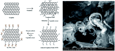
We have developed a facile and green route to prepare reduced graphene oxide (rGO) from graphene oxide (GO) and decorated its surface edges with self assembled Ag Nanowires (Ag NWs). This was carried out by a novel approach using vegetable extract (Abelmoschus esculentus) as a reducing and stabilizing agent. Currently seed mediated and polyol methods are most common in the preparation of any metallic nanowires. These methods would generally be carried out in two steps, whereas in the present study we have developed Ag NWs on a rGO surface (Ag NWs–rGO) in a one step process. Here, the vegetable extract acts as a reductant, functionalizing and capping agent in the reduction of GO followed by the effective functionalization of the rGO surface which in turn in the presence of Ag+ ions, permits the growth of self assembled Ag NWs on the edges of the rGO. The as-prepared Ag NWs–rGO was characterized by UV-visible spectroscopy, XRD, FE-SEM, FT-IR, Raman spectroscopy and elemental analyses. The resultant Ag NWs–rGO exhibits excellent catalytic activity towards the reduction of toxic nitroarene substrates. Importantly, this study could open an avenue for environmentally friendly, simple and cost effective methods in the surface functionalization and generation of self assembled Ag NWs on the edges of rGO nanosheets. Eventually, the unique green reductant used in this work has been proved to be the better reductant and surface functionalizing agent in synthesizing Ag NWs–rGO nanosheets compared to any hazardous chemical reductants.
1. Introduction
Graphene is a two dimensional (2D) single layered, honey comb, crystal lattice structure of sp2 hybridized carbon atoms with high theoretical surface area, superior electrical and thermal conductivity, and possesses excellent optical properties to use in solar cells, energy storage devices, and sensors.1–4 Synthesis of graphene has received much attention and it would be carried out by different methods.5,6 In order to get high purity and low defect graphene from graphite, exfoliation is necessary, i.e., the separation of single layer during the course of graphene synthesis.6,7 It is a simple and effective step carried out by different pathways such as micro mechanical exfoliation using scotch tape,8 ultrasonic exfoliation,7 laser exfoliation9 and intercalation exfoliation10,11 etc., where graphite is directly converted into graphene. In micro mechanical exfoliation by using scotch tape, where scalable is difficult in bulk production.12 On the other hand, ultrasonic exfoliation of graphite is generally used for bulk scale production.6,7 Instead of ultrasonic wave, an ultrafast single shot femtosecond laser is also used to detach the graphite flakes into single layered graphene.8 In addition, other approaches like solvothermal,13 arc discharge13 and CVD14 were also reported in graphene synthesis.15–17 Amid direct exfoliation process, graphene possess high conductivity whereas the effective functionalization on its surface is very difficult.6,7
Despite GO does not reduce completely into rGO, the chemical reduction method is currently considered as the most impressive method due to its economic and ease in bulk scale productivity.18,19 It means that in chemical reduction methods, little amount of oxygen moieties would be present on the rGO surface which is necessary in surface functionalization followed by its wide range of applications such as thin films, coatings, and composites, etc.19–22 Generally, in chemical reduction methods, reducing agents such as hydrazine, dimethylhydrazine, and pyrogallol and capping agents such as polyvinylpyrrolidone would be used in the reduction of GO into rGO.23 These reducing agents are highly harmful, toxic to mankind and to the environment even in trace quantity.23,24 Therefore, an alternative green reducing agent for graphene reduction is being explored, for instance, ascorbic acid, reducing sugar, and amino acid, etc.25–28 In addition, radiation induced reduction, using sunlight and UV light,29,30 microbial reduction using E. coli bacteria, microorganism extracted from river bank, and reduction using bacterial respiration are also reported.31–33 Similarly, using green leaf extracts such as tea leaf extract and spinach leaf extract are also studied in the green syntheses of rGO.34,35 Usually, in the synthesis of rGO, a major setback could arise due to graphene layer's π electron clouds exist in above and below the plane of sheets which tend to possess strong affinity between the two adjacent layers of graphene that lead to form an irreversible aggregates.36,37 This drawback could be easily overcome by the decoration of metals on the surface of rGO which avert the direct π–π interaction between the layers and the aggregation got minimized.3,38 Thereby, unique outstanding properties of graphene like conductivity, optical properties and catalytic efficiency are enhanced.3,38 In addition, one dimensional metal nanoparticles like nanorods and nanowires have been extensively studied due to their impressive potential applications rather than normal spherical nanoparticles.39
In the present study, Abelmoschus esculentus, vegetable extract is used for the first time as reductant and surface functionalizing agent which consecutively reduces GO to rGO and Ag+ ion into Ag NWs on the surface edges of rGO. The importance of our work is highlighted as: (1) in the absence of any toxic reducing agent using this vegetable extract, GO got reduced to rGO (2) secondly, in the absence of any external templates, using the same extract, the surface functionalization of rGO followed by self assembled growth of Ag NWs are achieved on its edges with ease (3) thirdly, the as synthesized Ag NWs–rGO shows better catalytic activity in different nitroarenes reduction. Thus, it ensures that the protocol followed in this work is completely eco-friendly and has no hazardous wastes neither used nor formed in the one pot synthesis. Usually, traditional NWs preparation are achieved in two steps.40 In addition, the preparations of NWs decorated rGO would be carried out in another two steps.40 On the other hand, in this work rGO–Ag NWs formation has been achieved in one step reaction. To the best of our knowledge, there is no report related to the direct growth of Ag NWs on the surface of rGO using a reductant like green extract or any other chemical methods.
Subsequently, choosing suitable nitroarene substrates, catalytic efficiency of the self assembled Ag NWs–rGO were investigated. Using this catalyst (Ag NWs–rGO), the different nitroarene substrates such as 4-nitrophenol, 2-nitrophenol, 4-nitroaniline, 2-nitroaniline got converted into aminoarenes in short interval time. The resultant products were analyzed against respective nitroarenes. In addition, rate constant and Turnover Frequency (TOF) for the reduction reaction were calculated.41–44
2. Materials and methods
2.1. Chemicals
Graphite flakes (300 mesh) were purchased from Alfa Aesar, sulphuric acid, potassium permanganate, hydrogen peroxide, hydrochloric acid, sodium borohydride, sodium nitrate and 2-nitroaniline (2-NA) were purchased from Merck. Silver nitrate, 4-nitroaniline (4-NA), 4-nitrophenol (4-NP), 2-nitrophenol (2-NP) were obtained from Aldrich. All chemicals were of analytical grade and used without any further purification.
2.2. Synthesis of graphene oxide (GO)
GO was prepared from graphite flakes using modified hummer method.25–28 Briefly 1 g graphite flakes was mixed with 0.5 g NaNO3 and the mixture was added to 25 mL concentrated H2SO4 followed by 30 min stirring. Then, 3 g of KMnO4 was added slowly while keeping the round bottom (RB) flask under ice cooling with vigorous stirring. The ice bath was removed and the RB flask was kept in water bath at 35 ± 3 °C for at least 2 h. 50 mL of H2O was slowly added to RB flask which turns into brownish solution. After 15 min, 10 mL of 30% H2O2 and 100 mL of warm water were added to RB flask. Afterwards, yellowish brown color resultant solution was filtered by using Whatman no. 1 filter paper and subsequently washed with 5% HCl and double distilled water. The filtered product was dried in an oven at 50 °C for overnight to attain brown graphite oxide powder. 100 mg graphite oxide was dispersed in 100 mL double distilled water using ultrasonicator (sonication power: 250 W, PCI India) for 3 h, where the graphite oxide allowed to exfoliated and it turns into a stable brown color graphene oxide (GO) dispersion.
2.3. Synthesis of reduced graphene oxide (rGO)
Fresh Abelmoschus esculentus (ladies finger) vegetable was purchased from local market. In order to collect the vegetable extract, we developed a protocol. First, 20 g chopped vegetable was added to double distilled water (100 mL) and boiled for 15 min and filtered using filter paper. To the filtrate, 100 mL of GO dispersion was added and refluxed for 12 h at 100 °C. Finally, the completion of reduction was observed from the color change of brown GO dispersion to black precipitate which indicates the formation of rGO (Fig. 1).
 |
| Fig. 1 Photographs of GO and rGO dispersion in water. | |
2.4. One pot synthesis of self assembled Ag nanowires on reduced graphene oxide nanosheets (Ag NWs–rGO)
First, rGO dispersion was carried out in double distilled water by sonication. 5 mM AgNO3 (40 mL) was added to same volume of rGO dispersion and stirred vigorously for 30 min. Then, 40 mL of green vegetable extract was added and stirring continued to 6 h and kept aside for 8 h. Finally, the colloidal solution formed as a result was centrifuged at 9000 rpm for 20 min and dried in an oven; ultimately Ag NWs–rGO would be generated.
2.5. Materials characterization
Absorption spectra for GO, rGO, Ag NWs–rGO, nitroarene substrates and time-course measurement of kinetic studies for the reduction reactions were carried out in the double beam UV-visible spectrophotometer (Jasco V-630, Japan). The X-ray powder diffraction (XRD) patterns were analyzed using Shimadzu XRD 6000 with Cu-Kα radiation (λ = 1.5418 Å) (Japan). The functional groups present in the samples were analysed using FT-IR spectrophotometer (Jasco 4000 series, Japan). The surface morphology and the formation of nanowires on the surface of reduced graphene oxide nanosheets were confirmed through FE-SEM (Quanta 250, FEG FEI Company, Czech Republic) analysis. The existence of silver was confirmed from the Elemental Analysis using Energy dispersive X-ray analysis (Brooker Nano GmbH, Germany). The presence of nitrogen was confirmed by using CHN elemental analyser (Elementar Vario EL III, Germany). The conversion of nitroarene into amino arenes was confirmed by using HPLC (Shimadzu SPD-M20A, Japan).
3. Results and discussion
GO dispersion is the starting material in the synthesis of rGO. The GO reduction is primarily confirmed using UV-visible spectra and XRD results. Initially, the reduction of GO and formation of Ag NWs are recognized using UV-visible spectra (Fig. 2A). The GO shows absorbance at 230 nm (Fig. 2A, (inset)), which corresponds to π → π* transition of aromatic C
C bonds and another hump observed at 300 nm due to n → π* transition of carbonyl groups.27,45 After GO reduction, the resultant product, rGO shows absorbance at 264 nm (Fig. 2A(a)) and the hump at 300 nm disappears. This confirms the complete removal of oxygen functional groups from GO and the formation of graphitic π conjugation network on the resultant rGO.26 The vegetable extract used in this work, consists of major composition of antioxidants like vitamin C, vitamin A, vitamin B6, carbohydrates, sugars, fibers, proteins and flavonols. These antioxidants are responsible for the reduction of GO into rGO.46–49 The reducing ability of this vegetable extract is comparable with the existing reductant like NaBH4, vitamin C and pyrogallol.26 Generally, surface functionalization and the existence of templates are the key factors, in forming nanorods or NWs on the surface of rGO.50–52 Frequently, amine or alkyl surface functionalizing agent would be used in the surface functionalization on rGO.50–52 In this work, the vegetable extract plays a vital role such as reductant to GO reduction which is followed by surface functionalizing, which in turn successively generated the direct growth of self assembled Ag NWs on the edges of rGO. Importantly, vegetable extract attributed to form a NH2 functionalization on the surface of rGO nanosheet. It was confirmed by the presence of nitrogen in the CHN analysis and FT-IR spectra (vide infra). This eco-friendly, green extract, based surface functionalizing step tends to direct the germination and nucleation of Ag atoms to form as Ag NWs on the edges of rGO in one step process (Ag NWs–rGO). Fig. 2A(b) shows a new absorption peak at 400 nm which coincide with the surface plasmon of Ag NWs on rGO (Ag NWs–rGO) surface. It means Ag NWs should grow on NH2 functionalized rGO surface with ease. The observed broad surface plasmon band at 400 nm was due to the transverse plasmon of Ag NWs.53 The peak observed at 264 nm corresponds to rGO which is slightly blue shifted in Ag NWs–rGO (Fig. 2A(b)) due to the coupling effect of rGO with Ag NWs.38
 |
| Fig. 2 (A) UV-visible absorption spectra of rGO (a) and Ag NWs–rGO (b). Inset shows the UV-visible absorption spectrum of GO. (B) XRD patterns of (a) pristine graphite, (b) GO, (c) rGO, (d) Ag NWs–rGO. | |
Scheme 1A and B proposes a schematic representation of self assembled Ag NWs grown on the edges of rGO. Scheme 1A explained the three stage synthesis of Ag NWs–rGO where stage 1 shows the oxidation of graphite to graphene oxide, stage 2 shows the reduction of graphene oxide in the presence of vegetable extract and stage 3 shows the one pot synthesis of Ag NWs on the edges of rGO. Similarly, Scheme 1B proposes the mechanism of one pot synthesis of self assembled Ag NWs to grown on the surface edges of rGO. Fig. 2B shows the XRD patterns of pristine graphite (a), GO (b), rGO (c) and Ag NWs–rGO (d). Fig. 2B(a) shows a sharp peak at 26° which is disappeared and new peak appeared at 10.6° in Fig. 2B(b). This peak shift strongly confirmed the exfoliation of graphite sheets due to the generation of oxygen moieties and the distance between the graphite layers tend to increase from 34 nm to 83.6 nm which in turn attributes the formation of GO.55,56 Fig. 2B(c) shows a broad peak due to GO reduction in the range of 19–27° which confirms the formation of few layer structures, rGO.55,56 Fig. 2B(c) clearly indicates the reduced oxygen moieties (1) and decreased interlayer distance in rGO (2). In addition, it is also due to smaller size of rGO layer thickness or relatively short domain order of the stacked sheets each of which broaden the peaks.55,56 Henceforth, the abovementioned two causes are mainly responsible for the broadness of peak. The XRD pattern of Ag NWs–rGO (Fig. 2B (d)) exhibit peaks at 38.1°, 33.5°, 44.3° indexed to (111), (311), (200), respectively which is corresponds to FCC lattice of Ag. These peaks are coinciding with JCPDS data (040783 and 011167) and correlate the presence of Ag on rGO surface. In addition, Fig. 2B(d) shows a sharp peak at 29° which corresponds to rGO.
 |
| Scheme 1 Schematic representation of two stage synthesis of graphite to rGO and third stage one pot synthesis of templated Ag NWs grown on the edges of rGO using indigenous vegetable extract (A), and the proposed mechanism of templated one pot Ag NWs grown on the edges of rGO surface (B). | |
The structural and surface morphology of rGO and Ag NWs–rGO are further examined using Field Emission-Scanning Electron Microscope (FE-SEM) (Fig. 3). This result clearly shows the formation of fewer layered two dimensional rGO nanosheets. In addition, it satisfactorily confirmed the generation of bundles of Ag NWs on its edge. The average thickness of Ag NWs is carefully measured as 92 nm and the length is in few micrometer scales. The FE-SEM results visibly depict Octopus like morphology of Ag NWs formed mostly on the edges of two dimensional rGO nanosheets. Thus, the FE-SEM images confirmed the surface functionalization on the edges of two dimensional rGO nanosheets where controlled aspects of Ag NWs did successfully grown as self assembled structures. Importantly, the one pot synthesis of self assembled Ag NWs on the edges of rGO is achieved by the addition of green reductant, vegetable extract is shown in Scheme 1A. The functional groups present in the vegetable extract, GO, rGO and Ag NWs–rGO are examined by FT-IR spectroscopy (Fig. 4). The vegetable extract (Fig. 4a) shows intense bands at 3434 cm−1, 1624 cm−1, 1041 cm−1 liable for O–H stretching, amide deformation and C–OH stretching, respectively. In addition, to it, some weak bands are noticed at 1246 cm−1, 2926 cm−1, 1410 cm−1 corresponds to stretching vibrations of N–H, C–H, COO−, respectively. The GO spectrum (Fig. 4b) shows a sharp bands at 1730 cm−1 and 1224 cm−1 corresponds to stretching vibrations of C
O and epoxy groups, respectively. A broad band at 3461 cm−1 corresponds to O–H stretching exist on the GO surface. In addition, the bands at 1048 cm−1 and 1620 cm−1 corresponds to stretching vibrations of C–O and C
C. Neither C–O nor O–H stretching exists in Fig. 4(c) and (d) i.e., the bands correspond to the oxygen functional groups in the IR spectra of rGO and Ag NWs–rGO. This attributes to the complete removal of oxygen moieties from rGO surface during the course of GO reduction using the indigenous reductant, vegetable extract. Fig. 4(c) and (d) shows a weak band at 1539 cm−1 corresponds to aromatic C–H stretching in rGO and Ag NWs–rGO. In addition, rGO and Ag NWs–rGO shows a band at 1014 cm−1 and 1019 cm−1, respectively corresponding to C–N stretching and a weak band at 1246 cm−1 indicating the presence of N–H stretching on both the surface.54,57,58 The above bands indicates that both C–N and N–H functional groups retained on the surface of rGO and Ag NWs–rGO. Further, in order to validate the IR results, CHN analysis was carried out to study the existence of nitrogen on rGO and Ag NWs–rGO surface (Table 1). Finally, based on IR spectra and CHN analysis, the existing nitrogen is confirmed the presence of NH2 functionalizaton on the surface of rGO or Ag NWs–rGO. Thus, the two key roles played by the indigenous reductant vegetable extract in the generation of Ag NWs–rGO were understood. First, it reduced GO into rGO and generating the NH2 functionalized rGO surface. Secondly and importantly, Ag+ ion got reduced to Agseed which in turn tend to grow as self assembled Ag NWs on NH2 functionalized rGO surface (Scheme 1B). As a result, Agseed grow as self assembled Ag NWs on the edges of rGO in the presence of Ag+ ions in one pot synthesis. Thus, the green vegetable extract acts as a reductant and capping agent in arranging bundles of Ag NWs on the edges of rGO.
 |
| Fig. 3 FE-SEM images of rGO (a and b) and Ag NWs–rGO (c and d) in two different magnification. | |
 |
| Fig. 4 FT-IR spectra of (a) green vegetable extract, (b) GO, (c) rGO and (d) Ag NWs–rGO. | |
Table 1 Elemental composition of N, C, and H obtained from CHN analysis
S. no |
Sample name |
N% |
C% |
H% |
O% by difference |
1 |
GO |
ND |
39.91 |
1.28 |
58.81 |
2 |
rGO |
2.69 |
78.40 |
2.23 |
16.78 |
Raman spectroscopy is the dynamic tool to differentiate the ordered and disordered nature of the graphene network. Fig. 5 shows a sharp bands at 1350 cm−1 and 1580 cm−1 that are known as D and G band, respectively.56–58 Fig. 5(a) shows Raman band at 1350 cm−1 and 1594 cm−1 with Id/Ig ratio as 0.9 corresponds to GO whereas after GO reduction (i.e., rGO) by supplying thermal energy, the intensity of D and G band did alter and the corresponding bands observed at 1361 cm−1 and 1596 cm−1, respectively (Fig. 5b) where Id/Ig ratio tend to increases to 1.09. The increasing intensity of D band (Fig. 5b) is due to thermal agitation during rGO formation and decreases its sp2 hybridization. It means that after reduction, the resulting rGO leads to increase its surface defect and disorderness. On the other hand, after self assembled Ag NWs generation on the edges of rGO (i.e., Ag NWs–rGO), the D and G bands tend to decrease and increase, respectively. It means that sharp Raman bands observed at 1356 cm−1 and 1592 cm−1 corresponding to D and G band where Id/Ig ratio got decreased to 0.87 (Fig. 5c). Thus, it states that the existence of Ag NWs on the edges of rGO would prevent the aggregation of graphene sheets, reduce the thickness of the graphene sheets, and increase its surface orderness.59–61
 |
| Fig. 5 Raman spectra of (a) GO, (b) rGO and (c) Ag NWs–rGO. | |
3.1. Catalytic reduction of nitroarenes using Ag NWs–rGO
The aromatic nitro compounds are one of the well known high toxic pollutants often present in industrial effluents of textiles, pesticides and military plants.62 The hydrogenated product of the aforementioned nitro compounds are very useful to prepare analgesic, antipyretic drugs, hair dyeing agent and photographic film developing process.63 Importantly, Ag NWs–rGO tends to be an excellent nanocatalyst whereas in the absence of Ag NWs, rGO did not exhibit any catalytic phenomena in the reduction of different nitroarene substrates. In order to investigate the reaction kinetics, time-course absorption spectra were measured at a fixed wavelength of 400 nm using the reaction mixture which consists of 50 mM tris, 0.1 mM nitroarene substrate, and excess amount of NaBH4 (100 times concentration of nitroarene substrates). The catalytic activity of Ag NWs–rGO was studied against different 0.l mM nitroarene substrates in the presence of NaBH4 (10 mM) as the reducing agent. In the absence of any catalyst, the absorption peak due to different nitroarenes such as 4-nitrophenol, 2-nitrophenol, 4-nitroaniline and 2-nitroaniline around 400 nm remains unaltered even for a couple of days in the presence of NaBH4.64 Thus, the addition of Ag NWs–rGO nanocatalyst to nitroarene substrate containing reaction mixture causes fading and bleaching of its color in short interval of time. As the amount of Ag NWs–rGO nanocatalyst is very small, it hardly interferes in the absorption spectra of nitroarene substrates. The reduction of different nitroarene substrates would be visualized by the disappearance of peak around 400 nm with the parallel appearance of a new peak at 300 nm (Fig. 6) which is attributed to any aminoarene substrate generation. The rate of nanocatalytic reduction for all the nitroarene substrates has been completed within 2–6 min. It is confirmed through time-course absorption measurement in UV-vis spectrophotometer, which is studied in 10 s time interval for 20 μl of Ag NWs–rGO nanocatalyst (10 μg), respectively (Fig. 7). Finally, the rate constants and Turnover Frequency (TOF) for the nanocatalytic reaction of different nitroarene substrates reduction are calculated. The rate constant, TOF and the time consumed for the different nitroarene substrates reduction reaction are given in the Table 2.
 |
| Fig. 6 UV-visible absorption spectra for the reduction of 0.1 mM of nitroarenes into amino arenes in 50 mM Tris buffer containing 10 mM NaBH4 with 10 μg Ag–rGO nanocatalyst. (a) 4-nitro phenol (4-NP) to 4-amino phenol (4-AP), (b) 4-nitro aniline (4-NA) to p-phenylene diamine (p-pd), (c) 2-nitro phenol (2-NP) to 2-amino phenol (2-AP) and (d) 2-nitro aniline (2-NA) to o-phenylene diamine. | |
 |
| Fig. 7 Time course measurement of UV-visible absorption spectra for the reduction of 0.1 mM of 4-nitro phenol (4-NP), 4-nitro aniline (4-NA) (a) and 2-nitro phenol (2-NP) and 2-nitro aniline (2-NA) (b) in 50 mM Tris buffer containing 10 mM NaBH4 with 10 μg Ag–rGO nanocatalysts. | |
Table 2 Determination of rate constant and TOF toward catalytic reduction of different nitroarene substrates using Ag NWs–rGO
S. no |
Substrate |
Product |
Time (Sec) |
Rate constant (S−1) 10−3 |
Turn over frequency (moles g−1 S−1) |
1 |
4-Nitrophenol |
4-Aminophenol |
360 |
9.08 |
0.0277 |
2 |
2-Nitrophenol |
2-Aminophenol |
600 |
7.01 |
0.0167 |
3 |
4-Nitroaniline |
p-Phenylene diamine |
240 |
10.22 |
0.0417 |
4 |
2-Nitroaniline |
o-Phenylene diamine |
600 |
8.35 |
0.0167 |
4. Conclusions
We have proposed a simple, low cost and effective green route for synthesizing stable Ag NWs–rGO nanosheets using Abelmoschus esculentus vegetable extract. The extract satisfactorily acts as an effective reducing agent in the reduction of GO and Ag+ ion and it functionalizes the rGO nanosheets, which motivate the self assembly of Ag+ into Ag NWs on the edges of rGO. The surface and the material characterization were confirmed the existence of self assembled Ag NWs on the edges of rGO. The catalytic activity of Ag NWs–rGO nanosheets was studied using different nitroarene reduction in the presence of NaBH4 and proved as a better nanocatalyst. The uniqueness in this work is the protocol followed is completely eco-friendly and has no hazardous wastes neither used nor formed during the course of Ag NWs–rGO synthesis.
Acknowledgements
Financial support from the DST-SERB, New Delhi (File no. SR/FT/CS-44/2011 dated 04.05.2012) are gratefully acknowledged. We would like to thank Dr. I.V. Muthuvijayan Enoch and Mr. S. Chandrasekaran, Karunya University for UV-Visible absorption measurements.
References
- K. Geim and K. S. Novoselov, Nat. Mater., 2007, 6, 183 CrossRef PubMed.
- A. H. C. Neto, F. Guinea, N. M. R. Peres, K. S. Novoselov and A. K. Geim, Rev. Mod. Phys., 2009, 81, 109 CrossRef.
- X. Huang, X. Qi, F. Boeyab and H. Zhang, Chem. Soc. Rev., 2012, 41, 666 RSC.
- D. Chen, H. Feng and J. Li, Chem. Rev., 2012, 112, 6027 CrossRef CAS PubMed.
- K. S. Subrahmanyam, L. S. Panchakarla, A. Govindaraj and C. N. R. Rao, J. Phys. Chem. C, 2009, 113, 4257 CAS.
- C. N. R. Rao, K. S. Subrahmanyam, H. S. S. Ramakrishna Matte, B. Abdulhakeem, A. Govindaraj, B. Das, P. Kumar, A. Ghosh and D. J. Late, Sci. Technol. Adv. Mater., 2010, 11, 054502 CrossRef.
- V. Nicolosi, M. Chhowalla, M. G. Kanatzidis, M. S. Strano and J. N. Coleman, Science, 2013, 340, 1226419 CrossRef.
- K. S. Novoselov, A. K. Geim, S. V. Morozov, D. Jiang, Y. Zhang, S. V. Dubonos, I. V. Grigorieva and A. A. Firsov, Science, 2004, 306, 666 CrossRef CAS PubMed.
- M. Lenner, A. Kaplan, Ch. Huchon and R. E. Palmer, Phys. Rev. B: Condens. Matter Mater. Phys., 2009, 79, 184105 CrossRef.
- L. M. Viculis, J. J. Mack, O. M. Mayer, H. T. Hahn and R. B. Kaner, J. Mater. Chem., 2005, 15, 974 RSC.
- K. H. Park, B. H. Kim, S. H. Song, J. Kwon, B. S. Kong, K. Kang and S. Jeon, Nano Lett., 2012, 12, 2871 CrossRef CAS PubMed.
- P. Kumar, RSC Adv., 2013, 3, 11987 RSC.
- H. Huang and X. Wang, J. Mater. Chem., 2012, 22, 22533 RSC.
- J. B. Park, W. Xiong, Y. Gao, M. Qian, Z. Q. Xie, M. Mitchell, Y. S. Zhou, G. H. Han, L. Jiang and Y. F. Lu, Appl. Phys. Lett., 2011, 98, 123109 CrossRef PubMed.
- D. V. Kosynkin, A. L. Higginbotham, A. Sinitskii, J. R. Lomeda, A. Dimiev, B. K. Price and J. M. Tour, Nature, 2009, 458, 872 CrossRef CAS PubMed.
- U. Maitra, H. Matte, P. Kumar and C. N. R. Rao, Chimia, 2012, 66, 941 CrossRef CAS PubMed.
- P. Kumar, B. Das, B. Chitara, K. S. Subrahmanyam, K. Gopalakrishnan, S. B. Krupanidhi and C. N. R. Rao, Macromol. Chem. Phys., 2012, 213, 1146 CrossRef CAS.
- D. Li, M. B. Muller, S. Gilje, R. B. Kaner and G. G. Wallace, Nat. Nanotechnol., 2008, 3, 101 CrossRef CAS PubMed.
- S. Park and R. S. Ruoff, Nat. Nanotechnol., 2009, 4, 217 CrossRef CAS PubMed.
- S. Stankovich, D. A. Dikin, G. H. Dommett, K. M. Kohlhaas, E. J. Zimney, E. A. Stach, R. D. Piner, S. T. Nguyen and R. S. Ruoff, Nature, 2006, 442, 282 CrossRef CAS PubMed.
- X. Wang, L. J. Zhi and K. Mecullen, Nano Lett., 2008, 8, 323 CrossRef CAS PubMed.
- L. J. Cote, F. Kim and J. Huang, J. Am. Chem. Soc., 2009, 131, 1043 CrossRef CAS PubMed.
- J. Paredes, S. Villar-Rodil, M. J. Fernandez-Merino, L. Guardia, A. Martínez-Alonso and J. M. D. Tascon, J. Mater. Chem., 2011, 21, 298 RSC.
- W. Schmidt, Hydrazine and Its Derivatives: Preparation, Properties, Applications, Wiley-Interscience, New York, 2001, vol. 1, ch. 4 Search PubMed.
- J. Gao, F. Liu, Y. Liu, N. Ma, Z. Wang and X. Zhang, Chem. Mater., 2010, 22, 2213 CrossRef CAS.
- M. J. Fernandez-Merino, L. Guardia, J. I. Paredes, S. V. Rodil, P. S. Fernandez and A. M. Alonso, J. Phys. Chem. C, 2010, 114, 6426 CAS.
- C. Zhu, S. Guo, Y. Fang and S. Dong, ACS Nano, 2010, 4, 2429 CrossRef CAS PubMed.
- S. Chen, W. Cai, R. D. Piner, J. W. Suk, Y. Wu, Y. Ren, Y. Kang and R. S. Ruoff, Nanotechnology, 2011, 22, 325601 CrossRef PubMed.
- P. Kumar, K. S. Subrahmanyam and C. N. R. Rao, Int. J. Nanosci. Ser., 2011, 10, 559 CAS.
- P. Kumar, K. S. Subrahmanyam and C. N. R. Rao, Mater. Express, 2011, 1, 252 CrossRef CAS PubMed.
- S. Gurunathan, J. W. Han, V. Eppakayala and J. H. Kim, Nanoscale Res. Lett., 2013, 8, 393 CrossRef PubMed.
- Y. Tanizawa, Y. Okamoto, K. Tsuzuki, Y. Nagao, N. Yoshida, R. Tero, S. Iwasa, A. Hiraishi, Y. Suda, H. Takikawa, R. Numano, H. Okada, R. Ishikawa and A. Sandhu, J. Phys.: Conf. Ser., 2010, 352, 012011 CrossRef.
- C. Salas, Z. Sun, A. Lu and J. M. Tour, ACS Nano, 2010, 4, 4852 CrossRef PubMed.
- Y. Wang, Z. Shi and J. Yin, ACS Appl. Mater. Interfaces, 2011, 3, 1127 CAS.
- D. Mhamane, W. Ramadan, M. Fawzy, A. Rana, M. Dubey, C. Rode, B. Lefez, B. Hannoyerd and S. Ogale, Green Chem., 2011, 13, 1990 RSC.
- C. Nethravathi, E. A. Anumol, M. Rajamathi and N. Ravishankar, Nanoscale, 2011, 3, 569 RSC.
- J. B. Liu, S. H. Fu, B. Yuan, Y. L. Li and Z. X. Deng, J. Am. Chem. Soc., 2010, 132, 7279 CrossRef CAS PubMed.
- P. V. Kamat, J. Phys. Chem. Lett., 2010, 1, 520 CrossRef CAS.
- Y. Cui, Q. Wei, H. Park and C. M. Lieber, Science, 2001, 293, 1289 CrossRef CAS PubMed.
- Y. Sun and Y. Xia, Adv. Mater., 2002, 14, 833 CrossRef CAS.
- B. Baruah, G. J. Gabriel, M. J. Akbashev and M. E. Booher, Langmuir, 2013, 29, 4225 CrossRef CAS PubMed.
- M. Liang, Z. L. Wang, Z. R. Su, W. Q. M. Wang, Y. Yu and Z. He, Catal. Sci. Technol., 2013, 3, 1910 CAS.
- S. K. Ghosh, M. Mandal, S. Kundu, S. Nath and T. Pal, Appl. Catal., A, 2004, 268, 61 CrossRef CAS PubMed.
- A. Gangula, R. Podila, M. Ramakrishna, L. Karanam, C. Janardhana and A. M. Rao, Langmuir, 2011, 27, 15268 CrossRef PubMed.
- Z. Zhang, H. Chen, C. Xing, M. Guo, F. Xu, X. Wang, H. Gruber, B. Zhang and J. Tang, Nano Res., 2011, 4, 599 CrossRef CAS.
- P. A. Savello, F. W. Martin and J. M. Hill, J. Agric. Food Chem., 1980, 28, 1163 CrossRef CAS.
- P. Arapitsas, Food Chem., 2008, 110, 1041 CrossRef CAS PubMed.
- S. Benchasri, Ratar. Povrt., 2012, 49, 105 CrossRef PubMed.
- O. E. Adelakun, O. J. Oyelade, B. I. O. Ade-Omowaye, I. A. Adeyem and M. V. Venter, Food Chem. Toxicol., 2009, 47, 1123 CrossRef CAS PubMed.
- Y. K. Kim, H. K. Na, Y. W. Lee, H. Jang, S. W. Han and D. H. Min, Chem. Commun., 2010, 46, 3185 RSC.
- Y. K. Kim, H. K. Na, Y. W. Lee, S. W. Han and D. H. Min, Langmuir, 2010, 26, 13065 CrossRef CAS PubMed.
- Z. Wei, A. J. Mieszawska and F. P. Zamborini, Langmuir, 2004, 20, 4322 CrossRef CAS.
- Y. Sun, B. Gates, B. Mayers and Y. Xia, Nano Lett., 2002, 2, 165–168 CrossRef CAS.
- X. Shen, Y. Liu, Y. Pang and W. Yao, Electrochem. Commun., 2013, 30, 13 CrossRef CAS PubMed.
- H. L. Guo, X. F. Wang, Q. Y. Qian, F. B. Wang and X. H. Xia, ACS Nano, 2009, 3, 2653 CrossRef CAS PubMed.
- V. Eswaraiah, S. S. J. Aravind and S. Ramaprabhu, J. Mater. Chem., 2011, 21, 6800 RSC.
- M. L. Woolfe, M. F. Chaplid and G. Otchere, J. Sci. Food Agric., 1917, 28, 519 CrossRef.
- L. K. Tamm and S. Tatulian, Q. Rev. Biophys., 1997, 30, 365 CrossRef CAS.
- B. Das, R. Voggu, C. S. Rout and C. N. R. Rao, Chem. Commun., 2008, 5155 RSC.
- M. Wall, Adv. Mater. Process., 2012, 170, 35 CAS.
- J. Li and C. Y. Liu, Eur. J. Inorg. Chem., 2010, 1244 CrossRef CAS.
- H. S. Toh, A. Ambrosi and M. Pumera, Catal. Technol., 2013, 3, 123 RSC.
- Z. Jiang, J. Xie, D. Jiang, X. Wei and M. Chen, Cryst. Eng. Commun., 2013, 15, 560 RSC.
- J. Das, Md. A. Aziz and H. Yang, J. Am. Chem. Soc., 2006, 128, 16022 CrossRef CAS PubMed.
|
This journal is © The Royal Society of Chemistry 2014 |
Click here to see how this site uses Cookies. View our privacy policy here.