Effect of solvent on the uncatalyzed synthesis of aminosilane-functionalized graphene

Muhammad Z. Iqbalab, Marios S. Katsiotisa, Saeed M. Alhassana, Matthew W. Liberatoreb and Ahmed A. Abdala*ac
aDepartment of Chemical Engineering, The Petroleum Institute, PO Box 2533, Abu Dhabi, United Arab Emirates. E-mail: aabdala@pi.ac.ae; Fax: +971 2 6075200; Tel: +971 2 607 5584
bDepartment of Chemical and Biological Engineering, Colorado School of Mines, Golden, CO 80401, USA
cDepartment of Chemical Engineering and Petroleum Refining, Faculty of Petroleum and Mining Engineering, Suez University, Suez, Egypt

Received 11th November 2013 , Accepted 24th December 2013

First published on 3rd January 2014


Abstract

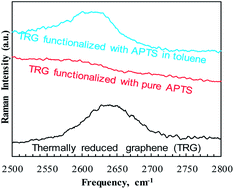
Uncatalyzed functionalization of thermally reduced graphene (TRG) with 3-aminopropyltriethoxy silane (APTS) is reported and the effect of the solvent on selective functionalization is discussed. The chemical, morphological and thermal properties of the functionalized TRG (f-TRG) have been studied using FTIR, XPS, EELS, Raman spectroscopy, TEM, and TGA. Our results indicate that the use of organic solvent during the silylation reaction not only increases grafting yield from 7 to 8 atomic% of Si attachment but also directs APTS groups to the edges of TRG as revealed using energy filtered TEM elemental mapping. A reaction mechanism based on attachment of the silane groups on the TRG surface through residual, surface bound phenolic and carbonyl groups is proposed and discussed. The present approach provides an economical route for mass production of APTS-f-TRG and sheds light on the role of organic solvents in silane functionalization of graphene.


Introduction

Since its discovery in 2004, graphene has attracted the attention of researchers owing to its extraordinary electronic, thermal and mechanical properties.1–5 Due to this combination of extraordinary properties in addition to the very high surface area, graphene is widely used as a nano-filler in manufacturing of polymer nanocomposites with improved mechanical, thermal and electrical properties.6–10 Nevertheless, homogenous dispersion of graphene and strong interfacial interactions between graphene and the polymer matrix are required to obtain enhanced properties. The chemical functionalization of graphene alters the Van der Waals interactions among the nanofiller aggregates, making them easier to disperse in a polymer matrix and can also enhance the interface between graphene and the polymer matrix. On the other hand, the extremely high surface area of graphene makes it an ideal candidate as an adsorbent for H2 storage,11 removal of pollutants from water,12 oil,13 and gases.14 In order to enhance the compatibility of graphene with polymer matrices and increasing its adsorption affinity functionalization of graphene may be required.

The use of residual oxygen pendent groups on thermally or chemically reduced graphene for further attachment of the organic moieties is an attractive route exploited for covalent functionalization of graphene.15 Silane coupling agents are historically applied to surface modification of nanofillers.16–20 Graphene produced from reduction of graphite oxide contains sufficient amounts of hydroxyl, epoxy and carboxyl groups on their basal planes and edges.21 These groups can be dehydrated with silane coupling agents under appropriate conditions. Also, the functional groups of silane coupling agents can be chemically attached to the polymer. Amongst, 3-aminopropyltriethoxysilane (APTS) has been widely reported as a reactive coupling functionalization for CNTs17,22 and graphene.18,19,23 Specifically, Wang et al. reported attachment of APTS on graphene surface and used the functionalized graphene in graphene–epoxy nanocomposites.18 The grafting of APTS was proposed to follow dehydration of carboxylic and hydroxyl groups. In another report by Yang et al., APTS reaction with chemically converted graphene resulted in 4% Si grafting on atomic basis.19 However, the proposed mechanism of APTS grafting was through the epoxide groups on the graphene surface. In addition, both reports employed DCC catalyst to accelerate reaction kinetics and enhance the grafting yield. A 3.4 atomic% Si yield has also been reported for graphene by Ganguli et al.23 Gaspar et al. reported detailed reaction mechanism for grafting various types of silanes on CNT.22 Therefore, these studies outline the importance of silane functionalized graphene in various applications. For the mass production of APTS-f-graphene, a method that does not utilize a catalyst is needed. Catalyst can increase the production cost by many fold. Hence, a catalyst-free method in functionalizing graphene by silane based coupling agents is highly desirable. In addition, in order to use the APTS-f-graphene in different applications, a better understanding of APTS chemistry is needed. This study focuses on the above mentioned question using various characterization techniques.

In the present work, we present a catalyst-free method for APTS attachment on TRG with higher yield of silane grafting. Thermally reduced graphene (TRG) is produced via simultaneous thermal exfoliation and reduction of graphite oxide (GO). The effect of solvent on the localization of functionalization on graphene surface is also discussed. Two simple routes for reacting APTS with TRG are presented; (1) using toluene as a solvent, and (2) using pure silane in absence of solvent. A mechanism for APTS attachment onto graphene that reflects the effect of organic solvent on functionalization chemistry is proposed and supported by the results of various characterization tools. The resulting functionalized graphene was thoroughly characterized using physicochemical methods to understand the nature of functionalization reaction. The success of the simple reaction routes is expected to promote mass production of APTS-functionalized graphene.

Experimental details

Natural flake graphite (−10 mesh, 99.9%, Alfa Aesar), Sulfuric Acid (95–97%, J.T. Bakers), hydrochloric acid (37%, Reidel-deHaen), hydrogen peroxide (30% solution, BDH), potassium permanganate, sodium nitrate (Fisher Scientific), 3-aminopropyltriethoxysilane (98%, Merck) and toluene (99.5%, Panreac) were used as received.

Preparation of TRG

TRG was produced via the thermal exfoliation of graphite oxide (GO).24 In this method, graphite is oxidized using Staudenmaier method25 as follows: graphite (5 g) was placed in ice-cooled flask containing a mixture of H2SO4 (90 mL) and HNO3 (45 mL) then potassium chlorate (55 g) was slowly added to the pre-cooled (0–5 °C) reaction mixture under stirring. After the reaction proceeds for 96 h, it was stopped by pouring the reaction mixture into water (4 L). GO was filter and washed with HCl solution (5%) until no sulfite ions were detected. Finally, the resulting deep brown colored mixture was repeatedly washed with copious amount of water until no chloride ions was detected. The wet GO was dried under vacuum overnight. TRG was made by rapid heating of dry GO powder at 1050 °C in a tube furnace (Model 21100, Barnstead Thermolyne) under flow of nitrogen for 30 s.

Silylation reaction

Two simple routes were used to examine the solvent effect on the mechanism of APTS attachment.
Method 1. 50 mg of TRG was dispersed in 30 mL of pure APTS in a 50 mL reaction flask. The mixture was refluxed at 100 °C for 3 hours under stirring then cooled to room temperature and the functionalized TRG (f-TRGP) was recovered by vacuum filtration.
Method 2. Same as Method 1 except the 30 mL of pure APTS was replaced by 50 mL of 30 vol% APTS in toluene. TRG functionalized using this method is denoted (f-TRGS).

To remove any physically adsorbed APTS from the surface of TRG, f-TRG was dispersed in toluene under stirring for 15–20 minutes followed by tip-sonication for additional 5 minutes and filtering under vacuum. To ensure the complete removal of any unreacted APTS, this washing procedure was repeated twice. The samples were then dried in air at 120 °C for 2 h.

Characterization

Transmission Electron Microscopy (TEM) analyses were performed using FEI Tecnai G20 with 0.11 nm point resolution and operated at 200 kV. Electron Energy Loss Spectroscopy (EELS) was performed using post column energy filtered camera (Gatan GIF 963). Energy filtered TEM (EFTEM) mapping was applied to map the location of the elements on the surface of f-TRG samples by measuring core, post-edge and pre-edge losses of the respective elements using the “three-window method” technique.26 X-ray Photoelectron Spectroscopy (XPS) measurements were performed using SSX-100 system (Surface Science Laboratories, Inc.) equipped with a monochromated Al Kα X-ray source, a hemispherical sector analyzer (HSA) and a resistive anode detector. For the high-resolution spectra, the lowest binding energy C1s was set at 285.0 eV which was used as the reference for all of the other elements. Fourier Transformed Infrared (FTIR) spectra of TRG, f-TRGP, and f-TRGS in the range of 400–4000 cm−1 were collected using Thermo Nicole FTIR at a resolution of 4 cm−1 and 32 scans. The spectra of the dried samples were obtained in KBr pellets (Merck, spectroscopic grade) containing 0.2 wt% of TRG. A LabRAM HR (Horiba Scientific) was used to obtain Raman spectra. Typically, a 50× objective was used with 633 nm excitation line. Thermo-gravimetric analysis was carried out by STA coupled with a mass spectrometer (STA-QMS, 409 PC Netzsch). The temperature range was from 35 °C to 450 °C at a ramp rate of 10 °C in an inert atmosphere of nitrogen (30 mL min−1). The electrical conductivity was measured by a custom-made conductivity cell at the National Renewable Energy Laboratory (Golden, CO). The samples were bath-sonicated in 10 mL of water–isopropanol solution (3/1) followed by 2 minutes of tip-sonication. The dispersed samples were dried on copper stubs for the conductivity measurements. The resistivity was measured using a multimeter (Waveteck 28XT, accuracy: ±1% + 0.1 Ohm). Further details on experimental procedures are provided in the ESI.

Results and discussion

The simultaneous thermal exfoliation and reduction of GO to TRG was successfully confirmed by XRD and TEM (see ESI Fig. s1).

The APTS-functionalized TRG was systematically characterized to investigate the effect of the solvent on the chemical, thermal, electrical and morphological properties of APTS-f-graphene. The chemical nature of the f-TRG is studied by FTIR, XPS, Raman, and electron energy loss (EEL) spectroscopy to elucidate the mechanism of APTS grafting on TRG. TEM micro-images and elemental mapping were used to study the morphology of f-TRG. The electrical conductivity and thermal stability of f-TRG are also discussed.

Chemical analysis

FTIR spectroscopy (Fig. 1) confirms the successful attachment of APTS on TRG by the appearance of additional functional group stretches associated with APTS–TRG linkages. The intensity of peak D (1189 cm−1), which corresponds to –COOH groups has been reduced. This suggests that the carboxylic groups are among the active moieties for APTS attachment. Peak H (1544 cm−1), which corresponds to C[double bond, length as m-dash]C aromatic carbons stretching in non-graphitic domains, remains the same for f-TRGP but shifts to a higher frequency (∼1558 cm−1) for f-TRGS. The shift to higher frequency for TRGs suggests the possibility of APTS reacting with C-hexagons of the TRG sheets in addition to the reaction with TRG's functional groups (also confirmed by XPS analysis). The carbonyl group [double bond splayed left]C[double bond, length as m-dash]O at ∼1740 cm−1 (peak J) also remains the same for pure and f-TRG. The appearance of new peaks in f-TRGP and f-TRGS is attributed to –Si–O stretching [peak A (–Si–OH at 914 cm−1), peak B (Si–O–Si at 1015 cm−1) and peak C (–Si–O–R at 1110 cm−1)], indicating the covalent functionalization of graphene. The effect of the solvent is demonstrated by the broadening of Si bonding peaks and the appearance of –Si–O–Si– peaks, which suggests session of the alkyl group and crosslinking of multiple APTS molecules. This may be attributed to the ability of toluene to transfer free radical to APTS in a similar manner to its chain-transfer effect during polymerization of monomers such as styrene. Also, the NH– amine vibration, which is expected around 3300–3400 cm−1 in f-TRGS (peak M), is not prominent, likely due to overlapping with the characteristic bands of adsorbed water in the same region.19 Peak F (1380 cm−1) corresponds to in-plane –OH deformation and peak G (1485 cm−1) is assigned to phenyl groups. The –NH bending vibration mode27 (peak I at 1629 cm−1) also confirms the direct attachment of amine groups to graphene and its very low intensity indicates the presence of a very small amount of this sequence. Moreover, the appearance of two new peaks at ∼2853 and 2925 cm−1 for the f-TRG samples is attributed to CH2–CH2 symmetric and asymmetric vibrations from APTS.22
image file: c3ra46586a-f1.tif
Fig. 1 FTIR spectra of the pure TRG, f-TRGP and f-TRGS.

While FTIR confirmed the attachment of APTS to graphene, a quantitative estimate of the level of functionalization is provided by XPS analysis. The results shown in Table 1 reveal that the functionalized graphene samples contain 7.4% Si and 6.9% N for f-TRGP and 8.0% Si and 6.5% N for f-TRGS. Not only we are able to achieve such high yield without the use of any catalyst but also the observed yield is higher than all previously reported for nanocarbons, e.g., graphene oxide19 and CNT.22 The Si/N ratio for f-TRGP is close to the stoichiometric APTS ratio of 1. However, The Si/N ratio for f-TRGS is 1.2. A Si/N atomic ratio greater than unity suggests the session of the amino branch from APTS induced possibly by solvent induced radical transfer to carbon atoms in the amino branch of APTS, which was also suggested by the FTIR analysis.

Table 1 Atomic composition of pure and f-TRG as obtained from XPS
  Atomic%
C O1s N1s Si2p
TRG 89.1 10.9
f-TRGP 66.8 20.0 6.9 7.4
f-TRGS 67.4 18.2 6.4 8.0


In order to further explore the structure of f-TRG, high resolution C1s, O1s, N1s and Si2p spectra were collected (Fig. 2 and Table 2). In C1s spectra, all the samples exhibit the same graphitic C[double bond, length as m-dash]C peak whose area percentage decreases in the f-TRG due to the appearance of new bands. The C–O peak19 in TRG decreased in f-TRGP and f-TRGS samples. Due to the smaller electronegativity difference between C and N compared to between C and O, the peak for C–N is observed at a lower binding energy (BE). The relative percentage of ∼285 eV peak has increased significantly in f-TRGP and f-TRGS. The peak for C[double bond, length as m-dash]O carboxylic groups for f-TRGS has a reduced intensity compared to the C[double bond, length as m-dash]O peak in TRG, it vanishes in f-TRGP. Thus, C[double bond, length as m-dash]O is a potential attachment site for APTS on TRG (i.e., the carboxylic group on TRG). The disappearance of C[double bond, length as m-dash]O in f-TRGP suggests selective attachment of APTS onto the carboxyl group. Meanwhile, two additional peaks are observed in C1s spectra of f-TRGP. The first peak at 286.16 eV is assigned to aliphatic C–N group19,28 whereas the second peak at 288.85 eV is attributed to either N–C[double bond, length as m-dash]O or O–C[double bond, length as m-dash]O.22 Since APTS is a bi-functional silane, its grafting to TRG can also take place through a linkage between the amine group in APTS and the carbonyl or carboxylic functionalities on TRG. The small peak at 288.85 eV BE is attributed to such linkage.22 However, the relative area of this peak is very small indicating that fraction of APTS that attaches to TRG through the amine linkage, is relatively small. Therefore, we conclude that APTS reacts bi-functionally with TRG through attacking the phenolic and carbonyl groups on the surface. Moreover, the π–π* interactions18 are observed in TRG and f-TRGS whereas no π–π* interactions are observed in f-TRGP possibly due to the disappearance of C[double bond, length as m-dash]O peak in f-TRGP.


image file: c3ra46586a-f2.tif
Fig. 2 High resolution XPS spectra of pure and f-TRG samples. Y-axis scale for all C1s spectra is the same and Y-axis scale for all O1s spectra is the same.
Table 2 High resolution fitting of C1s and O1s groups appearing in the XPS spectra of TRG, f-TRGP, and f-TRGS
  C1s O1s
C[double bond, length as m-dash]C (sp2) C–O/C–N C–N C[double bond, length as m-dash]O N–C[double bond, length as m-dash]O/O–C[double bond, length as m-dash]O π–π* C[double bond, length as m-dash]O Si–O C–O–C/O–C[double bond, length as m-dash]O O–C[double bond, length as m-dash]O
TRG 284.57 285.86 287.26 290.54 531.56 533.55 534.84
f-TRGP 284.57 285.37 286.16 288.85   531.26 532.75
f-TRGS 284.67 285.67 287.26 290.14 531.66 532.65 533.74


Unlike C1s core level, where the assignment of the peaks to single and multiple C–O bonds is quite straight forward, the assignment of O1s peaks is more difficult. The carbonyl groups C[double bond, length as m-dash]O are expected between 531 and 532 eV, C–O bonds in ethers and hydroxyls between 532 and 533.5 eV, whereas ether oxygen atoms in esters and anhydrides appear at 533.8–534.6 eV, or higher, and the carboxilates should originate from a single component at BE similar to that of C[double bond, length as m-dash]O groups in the case of oxidized carbon surfaces. Although C[double bond, length as m-dash]O is observed for TRG, f-TRGP and f-TRGS, the relative intensity of C[double bond, length as m-dash]O is lower in the f-TRG samples as a result of APTS attachment in agreement with the analysis of the C1s spectra. The C–O–C/O–C[double bond, length as m-dash]O band in TRG appears at higher BE for TRGS showing the bonding with a more electronegative structure at this point. The peak at ∼533.5 eV originates from Oδ contribution of TRG due to the presence of O−2 ions.

The appearance of new Si–O bands in f-TRGP and f-TRGS is a sign of successful APTS functionalization and its intensity and relative contribution depend on the degree of APTS functionalization.29 Moreover, the carboxylic band observed for TRG at ∼534 eV disappears in the f-TRG samples in excellent agreement with the results from C1s XPS spectra and FTIR.

The prominent changes in O1s spectra of the pure and functionalized samples clearly indicate that the silanization has changed the structure of the graphene sheets. Table 2 summarizes the results of the high resolution C1s and O1s spectra. Furthermore, the details of high resolution N1s and Si2p XPS spectra of f-TRG are provided in the ESI.

Considering the XPS data and the structure of f-TRG, the APTS may attach to graphene through the phenolic as well as the carbonyl surface groups where a similar mechanism has been reported by Gaspar et al.22 for CNTs. However, this multiple functionality may differ from the previously reported mechanisms for silylation where the aminosilanes are proposed to attack the epoxy groups19 on the graphene surface. In addition, the second possibility of enhanced grafting efficiency of APTS is NH2–silicon polymerization22 on the surface of TRG.

Previous reports demonstrated APTS grafting efficiency on MWCNT22 up to 4.5 Si atomic%, and 3.4% (ref. 23) and 4.1% for graphene.19 The current method is able to generate much higher yield of ∼7.4% Si attachment in f-TRGP and ∼8% Si in f-TRGS. The increase in Si attachment can be attributed to the reaction of APTS with not only the surface functionalities of graphene but also with the surface C-atoms. Schematics of possible mechanisms are also discussed later.

The effect of silane attachment on the electronic structure of graphene is also confirmed by EELS. The core-level energy losses of C, O, N and Si (Fig. 3 and 4) are measured at the areas of minimum overlapping between the graphene sheets. All collected EELS spectra are background corrected.


image file: c3ra46586a-f3.tif
Fig. 3 EELS of carbon K-edge (a) and oxygen K-edge (b).

image file: c3ra46586a-f4.tif
Fig. 4 EELS of nitrogen K-edge (a) and silicon L2–3-edge (b).

The carbon K-edge (Fig. 3a) is the characteristic of graphene.30 The sp2 hybridization of TRG is clearly observed as sharp peaks corresponding to the 1s ≅ → π* and 1s → σ* transitions at 289 and 294 eV, respectively. Interestingly, the hybridization state of graphene is altered in the functionalized samples. In specific, the sp2 structure becomes less dominant in the case of f-TRGP and f-TRGS in excellent agreements with FTIR, XPS and Raman results. The ratio sp2/(sp2 + sp3) was calculated using the peak-ratio method31 and found to be 98 ± 1.5%, 94 ± 1.1%, and 87 ± 1.9% for TRG, TRGP, and TRGS, respectively. A plausible explanation for this change in hybridization is attributed to the sp3 structure of carbon atoms in silane. Based on the current information obtained from FTIR and XPS on the successful grafting of APTS on graphene, the apparent change in hybridization most possibly represents the combination between the sp2 structure of graphene and the sp3 structure of silane. The oxygen K-edge region is presented in Fig. 3b and exhibits an overall shape equivalent to that of the N K-edge. Two π* peaks can be observed at 530 and 535 eV which are attributed to the C–O bonds. The dominating structure however, is the absorption beyond 541 eV with a particular triangular shape. Similar findings were observed in literature32,33 and the aforementioned structure was attributed to the increased presence of Si–O bonds originating from the APTS-attachment on graphene.

Fig. 4a displays the nitrogen K-edge evolution, where a strong π* absorption is observed at ∼399 eV and a structure-less σ* absorption of triangular shape is observed at ∼410 eV.

The π*-transition is usually attributed to unsaturated C–N bonds.34 Since this type of bonds is not present in the silane molecule, the peak at ∼399 eV can be attributed to a possible loose attachment of the amine part of silane on the graphene surface in agreement with FTIR and XPS spectra. Regarding the σ* absorption, this can be attributed in the Si–N bond present in APTS. In the case of silicon L-edge (Fig. 4b), the EELS spectra from the functionalized samples are very similar to the spectrum of silicon oxide.35 Peak at 99 eV corresponds to elemental silicon (Si), at 105 eV to Si–N bond (Si1+) and peaks at 108 and 116 eV to Si–O (Si2+) and Si[double bond, length as m-dash]O (Si4+) bonds respectively.36,37 Regarding Si L1 edge at ∼155 eV, strong overlapping with the threefold L2–3 edge prevents the acquisition of any useful information. Regardless, silicon EELS strongly supports the presence of silane grafted on graphene.

The effect of APTS functionalization and the degree of exfoliation in TRG was also examined by the Raman spectroscopy (Fig. 5), which is known for its unambiguous nondestructive identification.38 For graphitic materials, the typical Raman bands are: a defect-induced D band at 1350 cm−1, an in-plane vibration of sp2 carbon at 1580 cm−1 (G band), and a two-phonon double-resonance process at ca. 2700 cm−1 (2D band). The G-band position increases with decreasing number of layers in graphene.39 Herein, the G band centred at 1770 cm−1 for TRG has been upshifted to 1778 cm−1 on functionalization (Fig. 5a), thus revealing the decrease in the layer stacking,40 mainly due to the presence of APTS. Moreover, compared to TRG (Fig. 5b), the characteristic 2D band for f-TRGS and f-TRGP is observed to be blue shifted by about 28 cm−1 and accompanied with peak broadening, confirming that the functionalization lead to better exfoliation of TRG and formation of few-layer graphene flakes.41


image file: c3ra46586a-f5.tif
Fig. 5 Raman spectra of TRG samples (a) D and G band and (b) 2G (G′) band.

Additionally, the appearance of D peak at 1339 cm−1 has been attributed to the presence of structural disorder.42 The D band arises from the activation in the first order scattering process of sp3 carbons in graphene, and the intensity ratio of D and G bands (ID/IG) expresses the sp2/sp3 carbon ratio, a measure of the extent of disorder.43 The ID/IG for f-TRGS and f-TRGP has increased from 0.677 for TRG to 0.903 and 1.026, respectively. The increased ID/IG ratio is consistent with the functionalization of graphene through covalent bonding44,45 and also in agreement with the EELS results. In addition, the appearance of a new peak at lower frequencies between 500 and 600 cm−1 is ascribed to the Si grafting on TRG.46 Hence, overall the Raman findings confirm the successful functionalization of TRG and the exfoliation state of TRG remains unaffected by APTS attachment.

Morphological analysis

TEM and EELS analyses are performed in order to understand the effect of functional moieties on the morphology of graphene and to define the bonding between APTS and TRG which can arise due the presence or absence of organic solvent. Pure TRG is composed of very thin sheets with lateral dimension from 0.5 to 1.5 μm (Fig. 6). These thin sheets are composed of overlapping graphene layers; the sheets are well defined and separated. The inset in Fig. 6a shows a selected area electron diffraction (SAED) pattern collected from the center of the image, which verifies the crystallographic identity of graphene.
image file: c3ra46586a-f6.tif
Fig. 6 TEM images of (a) pure TRG particle composed of overlapping graphene sheets (inset: SAED pattern from the center of the graphene particle), (b) HRTEM image of two overlapping graphene sheets.

The high resolution TEM imaging (HRTEM) performed in areas where graphene was “folded”, showed the presence of multi-layer structure with number of layers between 5 and 10. It worth noting that TEM does not provide any statistically significant estimate of the thickness or number of layer of graphene but is used here as a to provide a qualitative comparison between TRG and f-TRG. Finally, EDS results indicate the presence of a small amount of chlorine impurity (<0.5%), probably originating from the HCl washing (see ESI).

Although f-TRGP exhibits a structure similar to that of pure TRG, however, the sheets do not appear as well separated as were in the pure TRG (Fig. 7). The silane creates defects in the graphene sheets (tears on the graphene surface) (see Fig. 7a for f-TRGP). On the other hand, no considerable change in the morphology of graphene sheets was observed for f-TRGS. Instead, the sheets are very well defined and separated, (Fig. 7b). The solvent appears to soften the severe attack of APTS on graphene and hinders breaking down of sheets to smaller ones. We infer that using organic solvent during the course of silane functionalization reaction helps in maintaining the intrinsic morphology of graphene. The EDS analyses (see ESI, Table s2) from the two samples were in good agreement with the XPS elemental analysis: on average Si/N ratio is found to be equal to 1.05 for f-TRGP, and 1.25 for f-TRGS (average of EDS spectra collected at various points for each sample).


image file: c3ra46586a-f7.tif
Fig. 7 TEM images of functionalized graphene; (a) f-TRGP, (b) f-TRGS.

Furthermore, in order to understand the preferable attachment sites for silane on TRG, EFTEM elemental mapping was performed on both f-TRGP and f-TRGS. To the best of our knowledge, there is currently no publication on EFTEM mapping of the functionalized graphene to determine the functionalization selectivity. However, the EFTEM elemental mapping, completed here (Fig. 8), can be a simple and versatile method for studying the local functionalization. Bright spots in Fig. 8b and d represent Si from EFTEM Si-elemental mapping of Fig. 8a and c. It is interesting to note that, in f-TRGP, Si (and N which is not shown here) is distributed over the entire graphene surface (Fig. 8b) representing a homogeneous distribution of the grafted silane on f-TRGP surface. Thus, we infer that pure silane reacted homogeneously on the graphene surface. A small amount of localized Si is also observed on the sheet edge. Thus, the mode of silane grafting, when TRG is reacted with pure APTS, is with the surface functional groups as well as the functional groups on the sheet edges. On the other hand, in f-TRGS, the Si is observed to localize on the sheet edges. The homogeneous surface distribution of Si was not observed as was seen in f-TRGP. Thus, it appears that in the presence of organic solvents like toluene, the edge-functionalization of TRG can be the preferable mode of grafting. The selectivity of edge-functionalization might further be increased by choosing appropriate solvents and reaction conditions. In summary, solvent can play a vital role in the selective functionalization of TRG. Further studies will evolve better control on its chemistry for selective applications of silane functionalized TRG.


image file: c3ra46586a-f8.tif
Fig. 8 TEM bright field image (a) and corresponding EFTEM maps of silicon (b) for f-TRGP; TEM bright field image (c) and corresponding EFTEM maps of silicon (d) for f-TRGS.

Based on the observations presented above, proposed reaction schemes for APTS with TRG are elucidated (Scheme 1). The TRG oxy-functionalities are distributed over the surface and sides of the sheets where the carboxylic groups are mainly located near the edges and most of hydroxyl and epoxy groups are distributed over the surface. In f-TRGP, APTS reacts with most of the surface groups and there is less reaction with the side carboxylic groups.


image file: c3ra46586a-s1.tif
Scheme 1 Reaction of APTS with TRG. TRG contains carboxylic functionalities on the sides whereas hydroxyl and epoxy groups are mainly distributed over the surface.

In the case of f-TRGS, the APTS molecules form covalent bonds with the carboxylic groups present on the sides of TRG sheets.

The use of various solvents assisting silane functionalization is not uncommon. Toluene being a non-polar solvent with a dipole moment of 0.36 D has induced significant effects on the functionalization selectivity. Owing to the non-polarity of toluene and TRG being polar due to the presence of oxy-functional groups on its surface, the solvent helps assisting preferable distribution of silane on TRG surface; leading silane molecules to more reactive areas such as the edges. On the other hand, in the absence of solvent, the silane (being polar) is chemically adsorbed throughout the surface and edges. Increasing solvent polarity in silylation reaction been reported to decrease the rate of silylation of inorganic surfaces.47,48 Thus, using non-polar solvents such as cyclohexane, benzene, diethyl ether, chloroform, etc. can lead to interesting aspects for selective silane functionalization. Nevertheless, the probability and further quantification of the attached functional groups depending on other solvents is beyond the scope of this article.

Thermal stability and conductivity

The thermal characterization of the pure and f-TRG by TGA provides a reliable quantification of the relative amount of the introduced functionalities. As shown in Fig. 9, both TRGP and TRGS have lower thermal stability compared to the pure TRG. The lower thermal stability of f-TRG samples compared to pure TRG is attributed to the full or partial detachment of the APTS groups from the graphene surface. The observed weight losses in the temperature range of 50–500 °C are 16, 22, and 25% for TRG, f-TRGS, and f-TRGP, respectively.
image file: c3ra46586a-f9.tif
Fig. 9 Thermograms of TRG, f-TRGP, and f-TRGS. Samples were heated from 50–500 °C at 10° min−1 under nitrogen.

As discussed in the previous section, the XPS results indicated that f-TRGP contains 7.4 atomic% Si, which corresponds to 15 wt% APTS and f-TRGS contains 8 atomic% Si corresponding to 16 weight% APTS.

Therefore, about 40% of APTS in f-TRGP and 55% of APTS in f-TRGS is lost between 50 and 500 °C, respectively. Moreover, the higher weight loss for f-TRGS compared to f-TRGP might be due to the most of the attached silane groups on the edges (as observed from the elemental mapping), and in f-TRGP, the silane moieties are distributed over the surface and on the edges of the sheets. Thus, it can be inferred that the end-functional groups are thermally less stable as compared to the surface distributed labile groups.

The electrical resistance of pure and functionalized TRG samples has been investigated using a custom made conductivity setup. The functionalization, as expected, has appeared to increase the resistance of graphene. The measured room temperature resistance of TRG of 42 ohms slightly increases to 50.8 ohms for f-TRGP but increased significantly to 230 ohms for f-TRGS. The increased resistance (decrease in conductivity) is expected due to the increase in sp3-contents in the f-TRGS as indicated by ID/IG ratio in the Raman spectra (see Fig. 5). The edge distributed APTS in f-TRGS seems to have a more pronounced effect on the conductivity compared to surface distributed groups in f-TRGP. This may be due to the effect of the group distribution on the dispersion of sp2-electronic cloud of TRG.

Conclusion

A simple catalyst-free method for synthesizing APTS-functionalized graphene was successfully demonstrated along with the effective role of the organic solvent in directing the functionalities over the graphene surface and to the edges. The amount of APTS attached onto graphene was determined by XPS and TGA and the covalent linkages were confirmed by FTIR, XPS, EELS and Raman spectroscopy. TEM and EELS analyses show that no substantial change in surface morphology of graphene was observed upon APTS functionalization in solution whereas pure APTS appears to attack the sheets adversely and changed the morphology considerably.

The quantification of the f-TRG showed more than 7–8 atomic % attachment of Si, which is higher than any previously reported functionalization. The method of APTS intercalation/incorporation has a significant effect on the structure and properties of the resulting functional material. Based on the EFTEM elemental mapping, it can be proposed that the selective end-functionalization can be achieved using a suitable solvent during the course of the silylation reaction. The f-TRG has shown good thermal stability over the temperature range of 50–500 °C where only 16% weight loss is observed for pure TRG compared to 22% for TRGp and ∼25% for TRGs. The decrease in electrical conductivity (increase in electrical resistance) of TRG with functionalization has been attributed to the increase in sp3-contents. Further studies can evolve a better mechanism for selective functionalization of TRG with APTS.

Acknowledgements

The authors would like to thank Mr. Issam Ismail at the Department of Chemical Engineering and Materials Science, University of Minnesota for the XPS measurements, Kelly Mason at National Renewable Energy Laboratory (Golden, CO) for helping in the conductivity measurements, and Dr Sunil Lonkar at the Department of Chemical Engineering, The Petroleum Institute for his valuable comments. This project has been supported by The Petroleum Institute in Abu Dhabi through the Cooperative Research Partnership (CRP) with Colorado School of Mines and by the ADNOC Gas Subcommittee under research Grant number 13701.

Notes and references

  1. K. Novoselov, A. Geim, S. Morozov, D. Jiang, Y. Zhang, S. Dubonos, I. Grigorieva and A. Firsov, Science, 2004, 306, 666–669 CrossRef CAS PubMed.
  2. S. L. Cheekati, MS thesis, Wright State University Dayton, Ohio, 2011.
  3. A. Balandin, S. Ghosh, W. Bao, I. Calizo, D. Teweldebrhan, F. Miao and C. Lau, Nano Lett., 2008, 8, 902–907 CrossRef CAS PubMed.
  4. D. A. Dikin, S. Stankovich, E. J. Zimney, R. D. Piner, G. H. B. Dommett, G. Evmenenko, S. B. T. Nguyen and R. S. Ruoff, Nature, 2007, 448, 457–460 CrossRef CAS PubMed.
  5. A. Geim and K. Novoselov, Nat. Mater., 2007, 6, 183–191 CrossRef CAS PubMed.
  6. T. Ramanathan, A. Abdala, S. Stankovich, D. Dikin, M. Herrera-Alonso, R. Piner, D. Adamson, H. Schniepp, X. Chen and R. Ruoff, Nat. Nanotechnol., 2008, 3, 327–331 CrossRef CAS PubMed.
  7. H. Pang, T. Chen, G. Zhang, B. Zeng and Z. Li, Mater. Lett., 2010, 64, 2226–2229 CrossRef CAS PubMed.
  8. T. Kuilla, S. Bhadra, D. Yao, N. Kim, S. Bose and J. Lee, Prog. Polym. Sci., 2010, 35, 1350–1375 CrossRef CAS PubMed.
  9. H. Kim, PhD thesis, University of Minnesota, Minnesota, 2009.
  10. H. Kim, A. Abdala and C. Macosko, Macromolecules, 2010, 43, 6515–6530 CrossRef CAS.
  11. L. Schlapbach and A. Züttel, Nature, 2001, 414, 353–358 CrossRef CAS PubMed.
  12. M. Iqbal and A. A. Abdala, RSC Adv., 2013, 3, 24455–24464 RSC.
  13. M. Z. Iqbal and A. A. Abdala, Environ. Sci. Pollut. Res., 2012, 20, 3271–3279 CrossRef PubMed.
  14. F. Schedin, A. Geim, S. Morozov, E. Hill, P. Blake, M. Katsnelson and K. Novoselov, Nat. Mater., 2007, 6, 652–655 CrossRef CAS PubMed.
  15. V. Georgakilas, M. Otyepka, A. B. Bourlinos, V. Chandra, N. Kim, K. C. Kemp, P. Hobza, R. Zboril and K. S. Kim, Chem. Rev., 2012, 112, 6156–6214 CrossRef CAS PubMed.
  16. P. Ma, J. Kim and B. Tang, Carbon, 2006, 44, 3232–3238 CrossRef CAS PubMed.
  17. A. Shanmugharaj, J. Bae, K. Lee, W. Noh, S. Lee and S. Ryu, Compos. Sci. Technol., 2007, 67, 1813–1822 CrossRef CAS PubMed.
  18. X. Wang, W. Xing, P. Zhang, L. Song, H. Yang and Y. Hu, Compos. Sci. Technol., 2012, 72, 737–743 CrossRef CAS PubMed.
  19. H. Yang, F. Li, C. Shan, D. Han, Q. Zhang, L. Niu and A. Ivaska, J. Mater. Chem., 2009, 19, 4632–4638 RSC.
  20. K. Yang and M. Gu, Composites, Part A, 2010, 41, 215–221 CrossRef PubMed.
  21. W. Gao, L. Alemany, L. Ci and P. Ajayan, Nat. Chem., 2009, 1, 403–408 CrossRef CAS PubMed.
  22. H. Gaspar, C. Pereira, S. Rebelo, M. Pereira, J. Figueiredo and C. Freire, Carbon, 2011, 49, 3441–3453 CrossRef CAS PubMed.
  23. S. Ganguli, A. K. Roy and D. P. Anderson, Carbon, 2008, 46, 806–817 CrossRef CAS PubMed.
  24. R. Prud'Homme, I. Aksay, D. Adamson and A. Abdala, US Pat., 7,658,901, 2010.
  25. L. Staudenmaier, Ber. Dtsch. Chem. Ges., 1898, 31, 1481–1487 CrossRef CAS.
  26. M. Worch, H. J. Engelmann, W. Blum and E. Zschech, Thin Solid Films, 2002, 405, 198–204 CrossRef CAS.
  27. A. Shanmugharaj, J. Bae, K. Y. Lee, W. H. Noh, S. H. Lee and S. H. Ryu, Compos. Sci. Technol., 2007, 67, 1813–1822 CrossRef CAS PubMed.
  28. R. Waltman, J. Pacansky and C. Bates Jr, Chem. Mater., 1993, 5, 1799–1804 CrossRef CAS.
  29. S. Y. Jing, H. J. Lee and C. K. Choi, J. Korean Phys. Soc., 2002, 41, 769–773 CAS.
  30. S. Wakeland, R. Martinez, J. K. Grey and C. C. Luhrs, Carbon, 48, 3463–3470 CrossRef CAS PubMed.
  31. S. Urbonaite, S. Wachtmeister, C. Mirguet, E. Coronel, W. Y. Zou, S. Csillag and G. Svensson, Carbon, 2007, 45, 2047–2053 CrossRef CAS PubMed.
  32. A. Falqui, V. Serin, L. Calmels, E. Snoeck, A. Corrias and G. Ennas, J. Microsc., 2003, 210, 80–88 CrossRef CAS.
  33. K. Giannakopoulos, N. Boukos and A. Travlos, Superlattices Microstruct., 2006, 39, 115–123 CrossRef CAS PubMed.
  34. L. Laffont, M. Monthioux, V. Serin, R. B. Mathur, C. Guimon and M. F. Guimon, Carbon, 2004, 42, 2485–2494 CrossRef CAS PubMed.
  35. W. M. Skiff, R. W. Carpenter and S. H. Lin, J. Appl. Phys., 1987, 62, 2439–2449 CrossRef CAS PubMed.
  36. G. A. Botton and M. W. Phaneuf, Micron, 1999, 30, 109–119 CrossRef.
  37. G. J. Auchterlonie, D. R. McKenzie and D. J. H. Cockayne, Ultramicroscopy, 1989, 31, 217–222 CrossRef CAS.
  38. S. BittoloáBon, J. Mater. Chem., 2011, 21, 3428–3431 RSC.
  39. M. Pimenta, G. Dresselhaus, M. S. Dresselhaus, L. Cancado, A. Jorio and R. Saito, Phys. Chem. Chem. Phys., 2007, 9, 1276–1290 RSC.
  40. Y.-T. Liu, J.-M. Yang, X.-M. Xie and X.-Y. Ye, Mater. Chem. Phys., 2011, 130, 794–799 CrossRef CAS PubMed.
  41. D. Graf, F. Molitor, K. Ensslin, C. Stampfer, A. Jungen, C. Hierold and L. Wirtz, Nano Lett., 2007, 7, 238–242 CrossRef CAS PubMed.
  42. A. Ferrari, J. Meyer, V. Scardaci, C. Casiraghi, M. Lazzeri, F. Mauri, S. Piscanec, D. Jiang, K. Novoselov and S. Roth, Phys. Rev. Lett., 2006, 97, 187401 CrossRef CAS.
  43. K. N. Kudin, B. Ozbas, H. C. Schniepp, R. K. Prud'Homme, I. A. Aksay and R. Car, Nano Lett., 2008, 8, 36–41 CrossRef CAS PubMed.
  44. M. Fang, K. Wang, H. Lu, Y. Yang and S. Nutt, J. Mater. Chem., 2010, 20, 1982–1992 RSC.
  45. M. Quintana, K. Spyrou, M. Grzelczak, W. R. Browne, P. Rudolf and M. Prato, ACS Nano, 2010, 4, 3527–3533 CrossRef CAS PubMed.
  46. Y.-S. He, P. Gao, J. Chen, X. Yang, X.-Z. Liao, J. Yang and Z.-F. Ma, RSC Adv., 2011, 1, 958–960 RSC.
  47. J. Clark and D. Macquarrie, Chem. Commun., 1998, 853–860 RSC.
  48. A. Wight and M. Davis, Chem. Rev., 2002, 102, 3589–3614 CrossRef CAS PubMed.

Footnote

Electronic supplementary information (ESI) available: Experimental details, XRD, TEM of TRG and EDS results are included. See DOI: 10.1039/c3ra46586a

This journal is © The Royal Society of Chemistry 2014
Click here to see how this site uses Cookies. View our privacy policy here.