DOI:
10.1039/C3RA46775A
(Paper)
RSC Adv., 2014,
4, 6840-6844
Synthesis of meso/macroporous aluminum nitrides via aluminum alloy ceramization
Received
18th November 2013
, Accepted 2nd January 2014
First published on 6th January 2014
Abstract
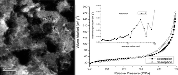
A novel and simple route, nitridization/ceramization of aluminum alloys, was introduced to produce meso/macroporous AlN. The meso/macroporous AlN were characterized by X-ray powder diffraction (XRD), scanning electron microscopy (SEM), transmission electron microscopy (TEM), and scanning transmission electron microscopy (STEM) and N2 adsorption/desorption isotherm at −195.7 °C. Results show that pores with diameters of 2 nm to hundreds of nanometers are distributed inside the materials. The surface areas of the materials can be as large as 142.9 m2 g−1 and the cumulative volume of pores with average radii between 1 nm and 100 nm is 0.40 cm3 g−1. The formation mechanism of pores was suggested to be that the pores would be left when the Mg3N2 was removed in nitridization products of Al and Mg alloy particles.
1 Introduction
Inorganic mesoporous materials have been widely used in many fields, for example catalysis, filters, fuel cells, sensors, biotechnology, energy storage and photovoltaics, and have been fabricated by different template routes1–7 and/or free-template processes.8–13 Porous wide band-gap IIIA nitrides exhibit a lot of new properties14,15 with a large potential for application in bioengineering, environmental engineering, catalysis, interfacial structures and sensor systems, due to their high surface-to-volume ratio and size selective adsorption. Because the nitrogen anion prefers to combine with the hydrogen cation in an aqueous solution and to be transformed into an ammonium cation or ammonia, it is difficult to obtain a nitride in an aqueous solution via assembling directly the nitrogen anion with a metal cation. Thus there are few reports on the synthesis of porous nitrides directly by such methods. It has long been recognized as a difficulty of preparing mesoporous IIIA-nitrides materials16,17 especially high-surface-area porous IIIA-nitrides.18–21 Till now, there are only a few reports on the successful synthesis of porous IIIA-nitrides. By a template method, Lin, Colin and Gérald obtained porous GaN with a high specific surface area (up to 136–156 m2 g−1) through the nitridization of porous Ga2O3 or Ga/C composite19,21,22 or a careful thermal treatment of metal-organic precursor containing the elements of Ga and N.18 Because of the high cost, tedious synthetic steps and possible shrinkage of the porous frameworks causing the loss of the desired porosity during the process of removing the templates, the method is not suitable for scale-up production of mesoporous materials.23–25 According to Carvajal, CVD is not suitable for the fabrication of the meso/macroporous GaN particles with a high specific surface area.26 Through sintering AlCl3·xNH3 cores into hollow AlN nanospheres, it is unknown how to control the thickness of the shell of hollow AlN nanosphere and how to control the percentage of void volume in a hollow nanosphere.27 By directly nitridizing an Al melt in the presence of Mg, it is difficult to obtain porous AlN with a high value of specific surface.28
In this paper, a novel and simple method was introduced to produce mesoporous/macroporous AlN, through Al–Mg alloy microparticles nitriding and post washing process. The AlN materials were characterized by XRD, TEM, SEM, STEM and N2 adsorption/desorption isotherm at −195.7 °C. Results show that the diameters of pores in the AlN samples are in the range of 2 nm to hundreds of nanometers, the BET surface area of AlN samples is up to 142.9 m2 g−1 and the cumulative volume of pores with average radii between 1 nm and 100 nm is 0.40 cm3 g−1.
2 Experimental
An Al ingot with purity of 99.9% was melted together with another metal X ingot with purity of 99.9% (X can be Mg, Li or Ca, here Mg was used as an example) in an electric furnace. The melt was then quenched to room temperature to obtain an Al–X alloy ingot. The contents of Al in the alloy are 30% to 70% (wt). The alloy ingots were processed into particles through high-energy ball milling and then sieving processes. The sieving process was carried out in a 400 mesh sieve. After the balling and sieving processes, the size distribution of particles, which was obtained on BT-2003 Laser Particle Size analyzer, was shown in Fig. 1. It can be seen that the diameter of particles is less than 50 μm. These particles were then put into a stainless steel crucible and were introduced into a vacuum furnace filled with pure nitrogen afterwards. The temperature of the furnace was raised to 800 °C at a speed of 150 °C per hour and maintained at the temperature for 10 hours. At the same time, the pressure of nitrogen in the furnace was kept at 1.05 atmospheres in the heating process. Because the alloy will be nitridized into ceramics during this process of heating, it will be called as a nitridization process or ceramization process later on, and the temperature is called as a nitridization temperature or ceramization temperature, respectively. Finally, the samples were cooled to room temperature in the furnace filled with pure nitrogen. The obtained products were washed by 0.2 M hydrochloric acid and filtered to remove the Mg2N3 and the residual Al and Mg. The final product was dried at 120 °C for 4 hours in a vacuum drying chamber and was collected for characterization.
 |
| Fig. 1 The size distribution of 50%Mg–50%Al (Al, Mg) alloy particles after balling and sieving processes. | |
The samples were characterized by XRD, TEM/STEM, SEM and BET method of nitrogen adsorption at a low temperature. The XRD analysis was carried out with a Rigaku Damx X-ray diffractometer with a CuKα1 radiation with a scanning speed of 10° per min. TEM bright-field (BF) and high resolution transmission electron microscopy (HRTEM) images were obtained on a JEM-3010 transmission electron microscope. SEM images were recorded on a JSM-7000F field emission microscope. High angle annular dark-field (HAADF) images in STEM and element mapping were taken using a JEOL 2010F analytical electron microscope. N2 adsorption isotherms were taken on an ASAP 2020 V3.04H surface area and pore size analyzer at −195.7 °C.
3 Results and discussion
Fig. 2 displays the XRD patterns collected from the unwashed and hydrochloric acid washed samples, respectively, which were produced from 60%Mg–40%Al alloy particles. By indexing the peaks in the patterns, conclusion can be made that there are only two phases AlN and Mg3N2 in the samples. No peaks from Al, Mg and intermediate phases show up in the patterns. This means that the content of these phases in the nitridization products is either less than the detection limit (1% wt) of the X-ray spectrometer or the alloy has almost completely turned into nitrides.
 |
| Fig. 2 XRD patterns of samples produced from 60%Mg–40%Al alloy (curve “a” corresponds to the XRD patterns of the sample unwashed by hydrochloric; curve “b” corresponds to that of sample washed by hydrochloric). | |
A SEM image taken from washed samples is shown in Fig. 3. It shows that many pores are distributed in the samples. The diameters of the pores observed vary from tens of nanometer to hundreds of nanometer. Fig. 4 is a HAADF image of washed AlN samples showing many pores (as shown by the arrows in the figure) with a diameter less than 50 nm. This means that there are many mesopores in the sample. Fig. 5 shows the N2 adsorption/desorption isotherm for washed AlN samples at −195.7 °C and the corresponding pore-size distribution from the adsorption branch using the Barrett–Joyner–Halenda (BJH) method (inset). The samples were made from the 50%Mg–50%Al alloy particles. From the N2 adsorption/desorption isotherm, it can be seen that there is a small convex surface facing towards the top near the origin of coordinate and a loop at the high P/P0, which is a characteristic of type IV, so the isotherm can be identified as type IV, which is characteristic of mesoporous materials.29–31 The inset in Fig. 5 indicated that there are many pores in the samples with an average radius from as small as 1 nm to a hundred nanometers. Through summing the incremental pore volume of every average radius, the BJH adsorption cumulative volume of pores with average radii between 1 nm and 50 nm and the volume of pores with average radii between 50 nm and 100 nm are 0.23 cm3 g−1 and 0.17 cm3 g−1 respectively (the total volume of pores with average radii between 1 nm and 100 nm is 0.40 cm3 g−1). The BET surface area of sample calculated from N2 isotherms at −195.7 °C is 142.9 m2 g−1. These large incremental pore volumes and the high BET surface area mean that there are lots of mesopores and macropores in the samples.
 |
| Fig. 3 A SEM image of washed samples. | |
 |
| Fig. 4 A HAADF image of washed sample (the pores shown by arrows with a diameter less than 50 nm). | |
 |
| Fig. 5 N2-adsorption/desorption isotherms and Barrett–Joyne–Halenda (BJH) pore size distribution plot (inset) for a washed sample made from the 50%Mg–50%Al alloy particles. | |
Fig. 6a shows a TEM image of unwashed samples fabricated from 50%Mg–50%Al alloy particles nitridized completely. Fig. 6b is the selected area electron diffraction (SAED) patterns of the “E” particles in the Fig. 6a. Because the d(321) (0.266 nm) and d(400) (0.249 nm) in Mg3N2 are very close to d(100) (0.271 nm) and d(002) (0.249 nm) in AlN, it is difficult to distinguish AlN from Mg3N2 only by the interplanar distances. In order to identify the two phases, HRTEM was taken as shown in Fig. 6c from a region in the “E” particle. The interplanar spacings marked as 0.27 nm and 0.24 nm and the interplanar angle of 90° in the image are in good agreement with the interplanar spacings 0.271 nm and 0.249 nm of the planes (100) and (002) (also with an angle of 90°) in hexagonal AlN. Even the interplanar spacing 0.27 nm is also close to the values of d{123} (0.266 nm) and the 0.24 nm is close to d{400} (0.249 nm) or d{322} (0.241 nm) of the Mg3N2 phase, but the angles between them are not 90°. So the phase in the region was identified as AlN, as shown in Fig. 6c. In the same way, the interplanar spacings 0.26 nm and 0.24 nm with an angle 71° were identified as the interplanar distances of planes (123) and (3
2) of the Mg3N2 phase, respectively, and the interplanar spacings 0.26 nm and 0.26 nm with an angle 60° as the interplanar distances of planes (123) and (3
2) of the Mg3N2, respectively. From Fig. 6c, it can be seen clearly that the Mg3N2 particle is enclosed in the AlN.
 |
| Fig. 6 The TEM and HRTEM images of an unwashed sample manufactured from 50%Mg–50%Al alloy particles ((a): a TEM image; (b): the SEAD of the particle “E” shown in (a); (c): a HRTEM image for a region in the particle “E”). | |
Fig. 7a is a HAADF image of unwashed sample obtained from 30%Mg–70%Al alloy particles, from which it can be seen that there are many pores in the samples (the dark regions), possibly coming from the sublimation of Mg3N2 during the ceramization process. Fig. 7b is the element maps showing the distributions of the elements Al and Mg in the unwashed sample. There are alternative distributions of Al and Mg with the enrichment of the Mg element nearby the border of pores. When the regions with the Mg3N2 phase are removed by an acid, pores will be formed at such sites. Thus, the formation mechanism of the pores in the AlN particles is suggested as follow: During the nitridization process at 800 °C, the Al and Mg atoms in the surface of particles are nitridized into AlN and Mg3N2, forming a thin film of AlN and Mg3N2 covering the whole surface of every particle. It keeps the particles separately from being melted into a body.32 With the increase of time, because the nitrogen atom is small, and then will have a larger diffusion coefficient than that of atom Al and Mg, it is easy for the atom to diffuse by the interstitial diffuse and/or interfacial diffuse, thus further nitridization will continue through the interaction of N that diffuses through the formed AlN and/or Mg3N2 layer with the Al and Mg atoms inside the alloy particles. In order to decrease the nucleation energy, the newly formed AlN and Mg3N2 tend attach to the surfaces of the previous formed AlN and Mg3N2 nucleation, respectively. At the same time, the rate of Mg changed into Mg3N2 is much faster than that of Al into AlN, which can be confirmed by the XRD patterns (shown in Fig. 8) of the products corresponding to the 50%Al–50%Mg alloy particles nitridized at 800 °C for 5 hours (in this condition, the alloy is not nitridized completely). From the patterns, it can been seen clearly that there is residual Al in the products, but Mg is almost turned into Mg3N2, and the content of residual Mg and/or its intermediate phases, for example Al3Mg2 and Al12Mg17, are all below the detection limit of the X-ray spectrometer (1% wt). According to Ye,33 the formation of Mg3N2 will be helpful greatly to the formation of AlN through the substitutional reaction:
 |
| Fig. 7 The images of elements mapping for an unwashed sample obtained from 30%Mg–70%Al alloy particles ((A) a HAADF image of the AlN sample; (B) the Al (shown by yellow dots) and the Mg (shown by blue dots) distribution in the sample shown in (A)). | |
 |
| Fig. 8 The XRD patterns of the unwashed sample acquired by nitridizing 50% (wt) Al–50%Mg alloy at temperature of 800 °C for 5 hours. | |
In which the Mg3N2 particles acts as a heterogeneous nucleation for the AlN formation. Thus the resultant AlN attaches to the Mg3N2 particles. When the Mg3N2 is washed by acid, pores will be left. So the formation processes of pores in AlN particles can be illustrated in Fig. 9.
 |
| Fig. 9 IIlustration of pores formation in AlN particles. | |
4 Conclusions
In conclusion, we reported a novel and simple route to produce meso/macroporous AlN with diameters of pores in the range of 2 nm to hundreds of nanometers, BET surface area up to 142.9 m2 g−1 and a cumulative volume of pores with average radii between 1 nm and 100 nm up to 0.40 cm3 g−1. We also illustrated the formation mechanism of porous AlN particles. According to the mechanism of pores formation, it may be expected that many other porous ceramic/compounds particles can be made in a similar way by choosing appropriate binary alloys and ceramization conditions.
Acknowledgements
We thank Liqun Wang and Lixia Sun (Department of Materials Physics and Chemistry, Xi'an Jiaotong University) for the SEM and TEM measurements and Dr Lijing Ma (School of Energy and Power Engineering, Xi'an Jiaotong University) for the N2 gas absorption/desorption measurements, Prof. Zhengxin Lu (Advanced Material Analysis and Test Center, Xi'an University of Technology) and Dr Ruihong Wang (School of Material Science and Engineering, Xi'an University of Technology) for the analysis of TEM images. This works were supported by the State Scholarship Fund of China (Grant no. 2010861527), the National Natural Science Foundation of China (no. 51172179), the Projects of Natural Science Foundation of Shaanxi Province (Grant no. SJ08-ZT04), the Natural Science Foundation of Shaanxi Provincial Department of Education (Grant no. 2010JK744 and 2010JK753), the Projects of Xi'an University of Technology (Grant no. 101-210909) and Specialized Research Fund for the Doctoral Program of Higher Education of China (Grant no. 20116118120003).
References
- P. Yang and T. Deng, Science, 1998, 282, 2244 CrossRef CAS.
- F. Caruso and R. A. Caruso, Science, 1998, 282(6), 1111 CrossRef CAS.
- Y. Piao and J. Kim, Nat. Mater., 2008, 7(3), 242 CrossRef CAS PubMed.
- A. Imhof and D. J. Pine, Nature, 1997, 389, 948 CrossRef CAS PubMed.
- S. Schacht and Q. Huo, Science, 1996, 273, 768 CAS.
- Y. D. Yin and R. M. Rioux, Science, 2004, 304, 711 CrossRef CAS PubMed.
- D. Zhao and J. Feng, Science, 1998, 279, 548 CrossRef CAS.
- H. X. Li and Z. F. Bian, J. Am. Chem. Soc., 2007, 129, 8406 CrossRef CAS PubMed.
- J. Li and H. C. Zeng, J. Am. Chem. Soc., 2007, 129, 15839 CrossRef CAS PubMed.
- X. W. Lou and Y. Wang, Adv. Mater., 2006, 18(17), 2325 CrossRef CAS.
- B. Liu and H. C. Zeng, J. Am. Chem. Soc., 2004, 126, 8124 CrossRef CAS PubMed.
- L. J. Mao and C. Y. Liu, J. Mater. Chem., 2008, 18, 1640 RSC.
- E. S. Toberer and R. Seshadri, Adv. Mater., 2005, 17(18), 2244 CrossRef CAS.
- X. Li and Y. W. Kim, Appl. Phys. Lett., 2002, 80, 980 CrossRef CAS PubMed.
- P. Schmuki and D. J. Lockwood, Pits and Pores: Formation, Properties and Significance for Luminescent Materials, The electrochemical Society, Montreal, 1997, pp. 97–99 Search PubMed.
- D. Zhao and H. Yang, J. Mater. Chem., 2005, 15, 1217 Search PubMed.
- J. Roggenbuck and T. Waitz, Microporous Mesoporous Mater., 2008, 113, 575 CrossRef CAS PubMed.
- G. Chaplais and S. Kaskel, J. Mater. Chem., 2004, 14, 1017 RSC.
- C. N. Lin and M. H. Huang, J. Phys. Chem. C, 2009, 113(3), 925 CAS.
- A. Fischer and J. O. Muller, ACS Nano, 2008, 2(12), 2489 CrossRef CAS PubMed.
- C. West and R. Mokaya, Chem. Mater., 2009, 21, 4080 CrossRef CAS.
- G. Chaplais and K. Schlichte, Chem. Commun., 2003, 730 RSC.
- H. C. Zeng, J. Mater. Chem., 2006, 16, 649 RSC.
- J. Liu and F. Liu, J. Mater. Chem., 2009, 19, 6073 RSC.
- S. A. Corr and D. P. Shoemaker, J. Mater. Chem., 2010, 20, 1413 RSC.
- J. J. Carvajal and J. Carlos Rojo, Cryst. Growth Des., 2009, 9(1), 320 CAS.
- F. Zhang and Q. Wu, J. Phys. Chem. C, 2008, 112, 11331 CAS.
- G. Yan and G. Chen, Mater. Lett., 2009, 63, 2205 CrossRef CAS PubMed.
- K. S. W. Sing and D. H. Everett, Pure Appl. Chem., 1985, 57, 603 CrossRef CAS.
- K. Sing, Colloids Surf., A, 2001, 187/188, 3 CrossRef.
- J. S. Hu, L. L. Ren, L. J. Wan and C. L. Bai, Angew. Chem., Int. Ed., 2005, 44, 1269 CrossRef CAS PubMed.
- T. B. Sercombe and G. B. Schaffer, Science, 2003, 301, 1225 CrossRef CAS PubMed.
- H. Z. Ye and X. Y. Liu, Mater. Lett., 2004, 58, 2361 CrossRef CAS PubMed.
|
This journal is © The Royal Society of Chemistry 2014 |
Click here to see how this site uses Cookies. View our privacy policy here.