Hoon Kim*a and
John Ralphab
aDepartment of Biochemistry and the DOE Great Lakes Bioenergy Research Center, Wisconsin Energy Institute, University of Wisconsin, 1552 University Ave., Madison, Wisconsin 53726 WI 53726, USA. E-mail: hoonkim@wisc.edu; Fax: +1 608-890-2270; Tel: +1 608 263-0166
bDepartment of Biological Systems Engineering, Wisconsin Energy Institute, Department of Biochemistry, University of Wisconsin, 1552 University Ave., Madison, Wisconsin 53706 WI 53726, USA. E-mail: jralph@wisc.edu; Fax: +1 608-890-2270; Tel: +1 608 890-2429
First published on 8th January 2014
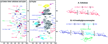
A recently developed “gel-state NMR method” that simply swells plant cell walls in a DMSO-d6/pyridine-d5 (4:
1) solvent system and uses a high-resolution solution state 2D-NMR (HSQC) technique has been successfully applied to whole plant cell wall 2D-NMR profiling studies. However, there was limited information to assign many polysaccharide peaks unlike lignin structures. Here we collected NMR data from various cellulose and xylan models using the same solvent system to assign the unknown peaks. Furthermore, DMSO-soluble cellulose and xylan fractions were prepared from ball-milled cotton linter cellulose, and the detailed chemical structures were analyzed. The major component of cotton is cellulose (95–99%), but it typically contains ∼2% hemicelluloses. Xylan in particular was isolated and identified along with the amorphous cellulose in this study. The fully assigned spectra of cellulose and xylan provided invaluable database information for peak assignment and authentication that will be directly used to screen and identify the two main polysaccharide components from various whole cell wall NMR spectra in the same solvent system.
Cellulose is the major component of secondary walls in higher plants. It has a polymer chain of linear β-(1 → 4)-linked D-glucosyl (β-1,4-glucan) units, with a degree of polymerization (DP) of ∼7000–15000.17 It forms 15–30% of the primary cell wall and 35–50% of the secondary cell wall.18 Cellulose also exists in the animal kingdom – in the tunicin from tunicates, for example.19 Cellulose has different crystalline allomorphs. Cellulose I is natural cellulose with a crystalline structure.20–22 Cellulose II refers to a thermodynamically stable structure with an antiparallel arrangement of the strands, characteristic of regenerated cellulose.4,17 Amorphous cellulose (paracrystalline cellulose) is not as regular in structure compared to crystalline forms because the neighboring strands do not align as well, resulting in a less rigid structure, and making it more accessible to water.4 It can be prepared by chemical or mechanical treatments; dimethylsulfoxide–paraformaldehyde,23 cadmium ethylenediamine solution (Cadoxen),24 cuprammonium hydroxide (Cuam, Cuoxam),25 or ball-milling followed by dissolution in sodium hydroxide,26 can all be used to generate amorphous cellulose.
NMR has long been an important tool for cellulose structural studies. Chemical shifts of highly crystalline cellulose II samples using solid-state CP/MAS 13C-NMR were measured, and amorphous cellulose in DMSO was measured with solution-state 13C-NMR.27–29 However, unmodified cellulose structures, and hemicelluloses such as xylan, have not been as well characterized with solution-state 2D NMR techniques.
Xylans are the most common of the hemicellulosic polymers. They have a backbone of xylopyranosyl residues that can be linked either via β-(1,3)- or β-(1,4)-linkages. However, all higher plants have a β-(1,4)-linked xylan as the backbone.4,30 There are several types of xylans in the plants. Arabinoxylans (AXs), which exist in most grasses, including in cereal grains, have a xylan chain that is substituted with α-L-arabinofuranosyl units at the O-2 or O-3 positions.30,31 Glucuronoarabinoxylans (GAXs) are structurally related to arabinoxylans, but they additionally have α-D-glucuronic acid (α-D-GlcA) substitutions on the xylan backbone. GAXs are the major cross-linking hemicellulosic polysaccharides in commelinoid monocots.32 The α-L-arabinosyl residues are consistently linked to the xylosyl residue at the O-3 position. The α-D-GlcA residue is linked to the O-2 position of the xylan backbone. The arabinosyl unit can be further substituted with ferulate, which acylates the O-5 position of the arabinosyl unit. Such ferulates are involved in cross-linking of polysaccharide, forming diferulate bridges, and/or cross-linking of polysaccharides with lignin via oxidative radical coupling, and may act as nucleation sites for lignin polymerization in the cell walls of grasses.33–36 Noncommelinoid monocots and all dicots also have GAX in which the α-L-arabinosyl residues are attached at either the O-2 or O-3 positions of xylan backbone.4,37 4-O-Methylglucuronoxylans (MGXs) are the most abundant type of hemicellulose in hardwood species, such as beech, birch and aspen.38,39 These polysaccharides share the same β-(1,4)-linked D-xylan backbone as arabinoxylans (AXs) and glucuronoarabinoxylans (GAXs), but are substituted with 4-O-methyl-α-D-glucuronic acid (4-O-methyl-α-D-GlcA; 4-O-MeGlcA) residues instead of arabinose. Major xylan backbone structures have been characterized in NMR studies with a variety of solvent systems.13,40–44
For the study described here, we isolated the DMSO-soluble cellulose and xylan mixture from cotton linters. Cotton's major component is cellulose (95–99%), with an average DP (degree of polymerization) of close to 15300.17 Unlike cotton lint, cotton linters are short residual fibers that are attached to the cottonseed and they are produced by the delinting process before the seed is crushed to generate cottonseed oil.45 Cotton linters typically contain ∼2% hemicelluloses that may come from seed hulls.45 On the other hand, cottonseed contains large amounts of xylan (40–45%), and its structure has been characterized as a glucuronoxylan, having both 4-O-MeGlcA and glucuronic acid (GlcA) substitutions.46,47
This paper reports the NMR data from various cellulose and xylan models and the complete peak assignments for the 2D NMR (HSQC) spectra of DMSO-soluble amorphous cellulose and xylan from cotton linter using the DMSO-d6/pyridine-d5 (4:
1) solvent system, providing an assignment database for cell wall samples.
Cellulose model compound residue | 1H & 13C Chemical shifts (ppm) | |||||||||
---|---|---|---|---|---|---|---|---|---|---|
1 | 2 | 3 | 4 | 5a | 5b | 6a | 6b | OMe | ||
a R; Reducing end, NR; non-reducing end. | ||||||||||
Glucose (α) | 1H | 5.13 | 3.35 | 3.71 | 3.31 | 3.80 | — | 3.65 | 3.78 | — |
13C | 92.61 | 72.70 | 73.49 | 70.80 | 72.22 | — | 61.50 | 61.50 | — | |
Glucose (β) | 1H | 4.50 | 3.16 | 3.37 | 3.28 | 3.28 | — | 3.61 | 3.84 | — |
13C | 97.18 | 75.14 | 77.01 | 70.53 | 76.96 | — | 61.45 | 61.45 | — | |
Cellobiose (Rα) | 1H | 5.05 | 3.34 | 3.75 | 3.45 | 3.86 | — | 3.74 | 3.85 | — |
13C | 92.08 | 72.14 | 71.46 | 80.66 | 69.81 | — | 60.52 | 60.52 | — | |
Cellobiose (Rβ) | 1H | 4.48 | 3.14 | 3.45 | 3.45 | 3.36 | — | 3.74 | 3.85 | — |
13C | 96.69 | 74.51 | 75.00 | 80.66 | 74.76 | — | 60.52 | 60.52 | — | |
Cellobiose (NR) | 1H | 4.40 | 3.17 | 3.33 | 3.23 | 3.33 | — | 3.54 | 3.83 | — |
13C | 103.24 | 73.23 | 76.63 | 69.94 | 76.63 | — | 60.91 | 60.91 | — | |
Cellotriose (Rα) | 1H | 5.05 | 3.33 | 3.75 | 3.46 | 3.85 | — | 3.80 | 3.84 | — |
13C | 92.10 | 72.13 | 71.51 | 80.77 | 69.88 | — | 60.37 | 60.37 | — | |
Cellotriose (Rβ) | 1H | 4.47 | 3.14 | 3.45 | 3.46 | 3.35 | — | 3.80 | 3.84 | — |
13C | 96.74 | 74.51 | 75.10 | 80.77 | 74.79 | — | 60.37 | 60.37 | — | |
Cellotriose (internal) | 1H | 4.46 | 3.23 | 3.48 | 3.48 | 3.32 | — | 3.74 | 3.90 | — |
13C | 102.86 | 73.04 | 74.81 | 80.28 | 76.72 | — | 60.29 | 60.29 | — | |
Cellotriose (NR) | 1H | 4.39 | 3.16 | 3.32 | 3.22 | 3.32 | — | 3.54 | 3.82 | — |
13C | 103.32 | 73.25 | 76.72 | 69.95 | 76.72 | — | 60.94 | 60.94 | — | |
Cellotetraose (Rα) | 1H | 5.07 | 3.36 | 3.77 | 3.49 | 3.88 | — | 3.78 | 3.86 | — |
13C | 92.10 | 72.13 | 71.46 | 80.82 | 69.88 | — | 60.46 | 60.46 | — | |
Cellotetraose (Rβ) | 1H | 4.50 | 3.16 | 3.48 | 3.49 | 3.37 | — | 3.78 | 3.86 | — |
13C | 96.74 | 74.54 | 75.15 | 80.82 | 74.77 | — | 60.46 | 60.46 | — | |
Cellotetraose (internal 1 & 2) | 1H | 4.48 | 3.24 | 3.50 | 3.50 | 3.34 | — | 3.74 | 3.92 | — |
13C | 102.89 | 72.99 | 74.78 | 80.23 | 76.68 | — | 60.22 | 60.22 | — | |
Cellotetraose (NR) | 1H | 4.40 | 3.18 | 3.34 | 3.24 | 3.34 | — | 3.55 | 3.85 | — |
13C | 103.31 | 73.22 | 76.68 | 69.99 | 76.68 | — | 60.92 | 60.92 | — |
Xylan model compound residue | 1H & 13C Chemical shifts (ppm) | |||||||||
---|---|---|---|---|---|---|---|---|---|---|
1 | 2 | 3 | 4 | 5a | 5b | 6a | 6b | OMe | ||
a R; reducing end, NR; non-reducing end, MGA; 4-O-Me-α-D-GlcpA (MeGlcA), XMGA; 4-O-Me-α-D-GlcpA linked O-2 to (1 → 4)-β-D-Xylp (α-(1 → 2)-linkage), MGA (NR) and XMGA (internal) assignment was based on previous xylan 2D NMR study (Jensen et al., Plant J., 2011, 66, 387–400). | ||||||||||
Xylose (α) | 1H | 5.00 | 3.28 | 3.57 | 3.39 | 3.47 | 3.62 | — | — | — |
13C | 92.68 | 72.46 | 73.43 | 70.28 | 61.62 | 61.62 | — | — | — | |
Xylose (β) | 1H | 4.36 | 3.06 | 3.23 | 3.40 | 3.10 | 3.72 | — | — | — |
13C | 97.87 | 74.86 | 76.92 | 69.92 | 65.82 | 65.82 | — | — | — | |
Xylobiose (Rα) | 1H | 5.01 | 3.32 | 3.68 | 3.59 | 3.62 | 3.71 | — | — | — |
13C | 92.36 | 72.33 | 71.09 | 75.81 | 58.87 | 58.87 | — | — | — | |
Xylobiose (Rβ) | 1H | 4.38 | 3.10 | 3.37 | 3.61 | 3.18 | 3.90 | — | — | — |
13C | 97.64 | 74.74 | 74.49 | 75.54 | 63.20 | 63.20 | — | — | — | |
Xylobiose (NR) | 1H | 4.33 | 3.17 | 3.26 | 3.42 | 3.16 | 3.80 | — | — | — |
13C | 102.08 | 72.49 | 76.35 | 69.47 | 65.78 | 65.78 | — | — | — | |
Xylotriose (Rα) | 1H | 5.00 | 3.31 | 3.68 | 3.60 | 3.62 | 3.70 | — | — | — |
13C | 92.39 | 72.32 | 71.14 | 75.75 | 58.85 | 58.85 | — | — | — | |
Xylotriose (Rβ) | 1H | 4.37 | 3.10 | 3.36 | 3.62 | 3.18 | 3.90 | — | — | — |
13C | 97.69 | 74.76 | 74.44 | 75.41 | 63.16 | 63.16 | — | — | — | |
Xylotriose (internal) | 1H | 4.35 | 3.20 | 3.40 | 3.62 | 3.25 | 3.96 | — | — | — |
13C | 101.98 | 72.59 | 74.07 | 75.41 | 63.31 | 63.31 | — | — | — | |
Xylotriose (NR) | 1H | 4.35 | 3.16 | 3.26 | 3.42 | 3.16 | 3.80 | — | — | — |
13C | 101.98 | 72.59 | 76.38 | 69.53 | 65.86 | 65.86 | — | — | — | |
Xylan (Birchwood) | 1H | 4.36 | 3.19 | 3.40 | 3.62 | 3.24 | 3.96 | — | — | — |
13C | 101.83 | 72.67 | 74.09 | 75.42 | 63.30 | 63.30 | — | — | — | |
MGA (NR) | 1H | 5.23 | 3.36 | 3.74 | 3.23 | 4.65 | — | — | — | 3.43 |
13C | 97.61 | 71.99 | 72.08 | 81.90 | 70.02 | — | — | — | 59.25 | |
XMGA (internal) | 1H | 4.57 | 3.35 | 3.48 | 3.61 | 3.34 | 4.11 | — | — | — |
13C | 101.28 | 76.70 | 72.53 | 76.51 | 62.76 | 62.76 | — | — | — |
![]() | ||
Fig. 2 Anomeric region of 2D 13C–1H correlation (HSQC) spectra of cellulose and xylan from cotton linter polysaccharides and other polysaccharide models in DMSO-d6/pyridine-d5 (4![]() ![]() |
Birchwood xylan is pure 4-O-methylglucuronoxylan (MGX).57,58 The xylan backbone NMR data was successfully obtained from this sample and helped to distinguish the xylan peaks in the polysaccharide mixtures (Fig. 2d & 3d).
Typical gel-state 2D HSQC NMR experimental parameters were used for the DMSO-extracted cellulose and xylan as described above.5,6 Such NMR experimental parameters had been optimized for gel samples of ball-milled whole plant cell walls, and the advantage is the short acquisition time based on the rapid relaxation as a result of the high viscosity of the polymer. We optimized the single-scan acquisition times to as short as 100 ms in F2 (1H), and used interscan relaxation delays (D1) of 500 ms, as explained in the NMR experimental section. The typical 5 h acquisition for the whole plant cell wall samples was sufficient for this cellulose and xylan sample to obtain all necessary correlations.
Adiabatic 2D HSQC NMR experiments run under typical conditions normally provide well-resolved resonances, but Non-Uniform Sampling (NUS) 2D HSQC NMR experiments were used here to provide significantly improved 13C-dimension resolution without requiring additional NMR time. Non-Uniform Sampling has been long suggested as one method of improving both the resolution and the signal-to-noise ratio.61,62 This newly-implemented Bruker standard NMR method allows high resolution along the indirect (13C) dimension by sampling more data points in that dimension (equating to longer evolution times) without sampling the FID at each dwell-time-interval during the acquisition.63 The NUS 2D HSQC NMR experiment provided clearly better resolution for the cotton linter polysaccharide sample.
Glycosyl residue | 1H & 13C Chemical shifts (ppm) | |||||||||
---|---|---|---|---|---|---|---|---|---|---|
1 | 2 | 3 | 4 | 5a | 5b | 6a | 6b | OMe | ||
a Cellulose; (1 → 4)-β-D-Glcp, xylan; (1 → 4)-β-D-xylp, NR; non-reducing end, R; reducing end, MGA; 4-O-Me-α-D-GlcpA (MeGlcA), XMGA; 4-O-Me-α-D-GlcpA linked O-2 to (1 → 4)-β-D-Xylp (α-(1 → 2)-linkage). | ||||||||||
Cellulose (internal) | 1H | 4.45 | 3.21 | 3.48 | 3.47 | 3.32 | — | 3.71 | 3.90 | — |
13C | 102.79 | 72.95 | 74.71 | 80.16 | 76.56 | — | 60.07 | 60.07 | — | |
Cellulose (NR) | 1H | 4.38 | 3.15 | 3.32 | 3.21 | 3.32 | — | 3.53 | 3.82 | — |
13C | 103.18 | 73.15 | 76.56 | 69.88 | 76.56 | — | 60.85 | 60.85 | — | |
Cellulose (Rα) | 1H | 5.05 | 3.31 | 3.74 | 3.46 | 3.85 | — | 3.77 | 3.84 | — |
13C | 91.99 | 72.16 | 71.38 | 80.57 | 69.82 | — | 60.33 | 60.33 | — | |
Cellulose (Rβ) | 1H | 4.47 | 3.13 | 3.44 | 3.46 | 3.35 | — | 3.77 | 3.84 | — |
13C | 96.56 | 74.40 | 74.91 | 80.57 | 74.66 | — | 60.33 | 60.33 | — | |
Xylan (internal) | 1H | 4.37 | 3.19 | 3.39 | 3.62 | 3.25 | 3.96 | — | — | — |
13C | 101.63 | 72.55 | 73.92 | 75.29 | 63.19 | 63.19 | — | — | — | |
Xylan (NR) | 1H | 4.37 | 3.19 | 3.26 | 3.41 | 3.16 | 3.80 | — | — | — |
13C | 101.63 | 72.55 | 76.26 | 69.43 | 65.72 | 65.72 | — | — | — | |
Xylan (Rα) | 1H | 5.00 | 3.31 | 3.67 | 3.62 | 3.62 | 3.70 | — | — | — |
13C | 92.26 | 72.16 | 70.99 | 75.29 | 58.70 | 58.70 | — | — | — | |
Xylan (Rβ) | 1H | 4.37 | 3.09 | 3.35 | 3.62 | 3.18 | 3.89 | — | — | — |
13C | 97.53 | 74.69 | 74.66 | 75.29 | 63.12 | 63.12 | — | — | — | |
MGA (NR) | 1H | 5.24 | 3.37 | 3.75 | 3.23 | 4.68 | — | — | — | 3.43 |
13C | 97.34 | 71.77 | 71.95 | 81.70 | 69.69 | — | — | — | 59.09 | |
XMGA (internal) | 1H | 4.57 | 3.37 | 3.40 | 3.62 | 3.34 | 4.12 | — | — | — |
13C | 101.16 | 76.52 | 72.58 | 76.35 | 62.60 | 62.60 | — | — | — |
In the whole cell wall samples, most of the polysaccharide anomeric correlation peaks appear in the range of 90–106 ppm (13C) and 3.7–6.0 ppm (1H) in HSQC spectra. The structural changes and compositional distribution of polysaccharide backbone, branches between different species and samples can always be easily recognized.6
From the cotton linter sample, we were able to clearly identify the cellulose peaks, such as the internal peak, non-reducing-end peak, and both α and β reducing-end peaks (Fig. 2a).
The internal cellulose [C–I1; (1 → 4)-β-D-Glcp] unit is the most important plant cell wall component; its anomeric (C1/H1) correlation peak appeared in spectra at 102.79/4.45 ppm. The same correlation appears, unsurprisingly, in the spectra from all plants we have surveyed.5,6 The anomeric peak from the non-reducing-end terminal cellulose residues (C-NR1) appeared at 103.18/4.38 ppm (C1/H1), and was extremely close to the internal cellulose peak (C-I1). The cellulose DP (degree of polymer) was reasonably estimated as ∼6 by integrating the non-reducing and internal anomeric peak; however, we are aware that the more mobile end-units may be over-quantitated by the NMR technique, so we assume that the actual DP is considerably higher.
The α- and β-anomer form reducing-end correlations of cellulose were clearly separated from those of the internal backbone units. A higher frequency of reducing ends will be present if there are shorter polymers that are mostly produced from the ball-milling process. The C1/H1 correlation from the reducing-terminal-end of α-D-Glcp (C-Rα1) was at 91.99/5.05 ppm (C1/H1); the analogous β-D-Glcp (C-Rβ1) correlation was at 96.56/4.47 ppm (Fig. 2a, Table 3). The cellulose reducing-end peaks and xylan reducing-end peaks are quite close together, but they were readily differentiated and assigned.
The non-anomeric polysaccharide region (56–85/2.5–5.0 ppm) overlaps with the lignin side-chain region when whole cell wall samples are examined.6 There is, however, no lignin was discernible in the tested sample (Fig. 3a).
It is very difficult to resolve the polysaccharide peaks from whole cell wall samples in many cases because of the complexity and the many overlapping signals. However, most internal cellulose peaks in this region were clearly identified in the DMSO-d6/pyridine-d5 spectrum from the amorphous cellulose sample as major assigned peaks at 72.95/3.21 [C-I2 (C2/H2)], 74.71/3.48 [C-I3 (C3/H3)], 80.16/3.47 [C-I4 (C4/H4)], 76.56/3.32 [C-I5 (C5/H5)] ppm (Table 3); two internal C-I6 (C6/H6) peaks were also distinctively located at 60.07/3.71 and 60.07/3.90 ppm. Most end-group correlations were well resolved, but some peaks were superimposed with other peaks. Non-reducing-end C-NR6 (C6/H6) peaks were well separated from the internal C-I6 (C6/H6) peaks and appeared at 60.85/3.53 and 60.85/3.82 ppm. C-NR4 (C4/H4) was also clearly evident at 69.88/3.21 ppm, and C-NR2 (C2/H2) was located close to the internal C-I2 (C2/H2) at 73.15/3.15 ppm, but was reasonably resolved. However, C-NR3 (C3/H3) and C-NR5 (C5/H5) had coincident chemical shifts, and also overlapped with the internal C-I5 (C5/H5) correlation at 76.56/3.32 ppm. The peak assignments for reducing ends were more complicated because these ends had α- and β-anomeric forms, unlike the non-reducing end. There were different chemical shift distribution patterns of the α and β anomeric peaks that will be discussed in the model study section later. Despite the conformational complexity, however, all peaks were evidently assigned: 72.16/3.31 [C-Rα2 (C2/H2)], 71.38/3.74 [C-Rα3 (C3/H3)], and 69.82/3.85 [C-Rα5 (C5/H5)] ppm. C-Rβ2 (C2/H2) and C-Rβ5 (C5/H5) were also well resolved at 74.40/3.13, 74.66/3.35 ppm. However, C-Rβ3 (C3/H3) was coincident with C-I3 at 74.91/3.44 ppm, C-Rα4 (C4/H4) and C-Rβ4 (C4/H4) were coincident at 80.57/3.46 ppm, and C-Rα6 (C6/H6) and C-Rβ6 (C6/H6) also were close together at 60.33/3.77 and 60.33/3.84 ppm, and were buried between the internal C-I6 peaks.
All xylan peaks were present in the same chemical shift range where the cellulose peaks appeared in the 2D HSQC NMR spectrum. Like cellulose, there were internal, non-reducing-end and both α and β reducing-end peaks (Fig. 2a).
The internal xylan correlation peak [X-I1; (1 → 4)-β-D-Xylp] from the backbone was at 101.63/4.37 ppm (C1/H1). Non-reducing-end xylan residues (X-NR1), unfortunately, shared the same chemical shifts (Table 3). However, the xylan α- and β-anomer reducing-end correlations appeared next to the cellulose reducing-end peaks and were clearly resolved. The α reducing-terminal-end of D-Xylp (X-Rα1) was at 92.26/5.00 ppm (C1/H1), and the correlation from the other reducing-terminal-end, β-D-Xylp (C-Rβ1), was present at 97.53/4.37 ppm (C1/H1). The xylan from the cotton linter was identified as a 4-O-methylglucuronoxylan (MGX) as is usually found in hardwoods.30 The 2D NMR data of previously studied 4-O-methyl-α-D-glucuronic acid (4-O-MeGlcA; MGA) and the MGA branched β-D-Xylp (X-MGA) were well matched with this current NMR data.13,58 There were no traces of arabinofuranose (Araf) peaks, which can usually be found in 4-O-methylglucuronoarabinoxylans (GAXs) or arabinoxylans (AXs).64 in the range of 105–110 ppm (13C) and 4.5–5.8 ppm (1H). This result corroborated the previous studies of cotton-seed xylan and simple glucuronoxylan, with 4-O-methylglucuronic acid (and glucuronic acid) structures found from acid and enzyme hydrolysis experiments.46,47
The xylan non-anomeric peaks also share the same chemical shift range (56–85/2.5–5.0 ppm) with cellulose non-anomerics and lignin side-chains. Despite the congestion of the correlations, most peaks were well resolved from the cellulose components and were readily assigned (Fig. 3a).
Two internal X-I5 (C5/H5) peaks were located at 63.19/3.25 and 63.19/3.96 ppm. The internal residue of xylan backbone, X-I2 (C2/H2), X-I3 (C3/H3), and X-I4 (C4/H4), appeared as major peaks at 72.55/3.19, 73.92/3.39, and 75.29/3.62 ppm, but X-I2 shared its chemical shifts with other correlations. X-NR2 (C2/H2), a non-reducing-end, appeared at ∼72.55/3.19 ppm where the X-I2 peak is, and C-I2 (C2/H2), an internal cellulose peak, also appeared nearby. Other non-reducing-end peaks were clearly resolved from other correlations. X-NR3 (C3/H3) and X-NR4 (C4/H4) appeared at 76.26/3.26 and 69.43/3.41 ppm, and two X-NR5 (C5/H5) peaks were located at 65.72/3.16 and 65.72/3.80 ppm. Peak assignment of reducing ends was also as complicated as for cellulose because of the α- and β-anomers. X-Rα2 (C2/H2) appeared at 72.16/3.31 ppm and is therefore in the same location as a cellulose reducing-end (C-Rα2) peak. X-Rα3 (C3/H3) was at 70.99/3.67 ppm, but the peak intensity was weak. Two X-Rα5 (C5/H5) peaks appeared at 58.70/3.62 and 58.70/3.70 ppm; however, they also appeared near the noise level. X-Rβ2 (C2/H2) appeared very close to C-Rβ2 at 74.69/3.09 ppm, and the two X-Rβ5 (C5/H5) also appeared right next to two X-I5 peaks at 63.12/3.18 and 63.12/3.89 ppm. X-Rβ3 (C3/H3) was fairly well resolved at 74.66/3.35 ppm, but it shared its chemical shifts with a cellulose reducing-end peak (C-Rβ5). X-Rα4 (C4/H4) and X-Rβ4 (C4/H4) appeared with an internal xylan peak (X-I4) at 75.29/3.62 ppm.
When MeGlcA (or GlcA) is attached to an O-2 position of a xylan residue, the chemical shift of the xylan unit noticeably changes.39,65,68,69,73,74 The X-MGA1 correlation peak appeared next to the internal anomeric xylan (X-I1) peak at 101.16/4.57 ppm (C1/H1). X-MGA3 (C3/H3) and X-MGA4 (C4/H4) also appeared close to the corresponding internal xylan peaks (X-I3 and X-I4) at 72.78/3.48 and 76.35/3.62 ppm. Two X-MGA5 (C5/H5) peaks were at 62.60/3.34 and 62.60/4.12 ppm, right next to the internal xylan peaks (X-I5). X-MGA2 (C2/H2) was, logically, the peak most affected by the O-2 substitution. It moved far away from the original internal X-I2 and appeared at 76.52/3.37 ppm.55,75 These X-MGA structures were also examined in our previous research on isolated Arabidopsis xylans as described earlier.13
Using the DMSO-d6/pyridine-d5 (4:
1) solvent system for polysaccharide research is a huge advantage for structural studies because the same NMR solvent has been successfully used for cell wall profiling research.5,6,14 In other words, polysaccharide components can be easily profiled without complicated sample preparation or isolation processes, and without any of the chemical modifications that may accompany extraction methods. NMR profiling provides a simple but powerful method to obtain detailed information on complex cell wall composition and structure. Using DMSO-d6/pyridine-d5 (4
:
1) as the common solvent system would be beneficial for examining not only lignins but also polysaccharides in whole cell wall profiling studies.
NMR experiments for polysaccharides from cotton were performed as previously described for the gel-state samples from ball-milled cell walls.5,6 NMR spectra were acquired on a Bruker Biospin (Billerica, MA) Avance 500 MHz spectrometer fitted with a 5 mm TCI (triple resonance; 1H, 13C, 15N) gradient cryoprobe with inverse geometry (proton coils closest to the sample). The central DMSO solvent peak was used as internal reference (δC 39.5, δH 2.49 ppm). The 13C–1H correlation experiment was an adiabatic HSQC experiment (Bruker standard pulse sequence ‘hsqcetgpsisp.2’; phase-sensitive gradient-edited-2D HSQC using adiabatic pulses for inversion and refocusing).76 HSQC experiments were carried out using the following parameters: acquired from 10 to 0 ppm in F2 (1H) with 1000 data points (acquisition time 100 ms), 200 to 0 ppm in F1 (13C) with 400 increments (F1 acquisition time 8 ms) of 72 scans with 500 ms interscan delay; the d24 delay was set to 0.89 ms (1/8J, J = 145 Hz). The total acquisition time was 5 h. Processing used typical matched Gaussian apodization (GB = 0.001, LB = −0.1) in F2 and squared cosine-bell and one level of linear prediction (32 coefficients) in F1.
For the structural elucidation and assignment authentication of the model compounds, the number of scans can be adjusted as usual depending on the signal-to-noise required from a sample. The standard Bruker implementations of the traditional suite of 1D and 2D (gradient-selected, 1H-detected, e.g., DEPT-135, COSY, HSQC, HSQC-TOCSY, HMBC) NMR experiments were used for model compounds. Adiabatic HSQC experiments (Bruker standard pulse sequence ‘hsqcetgpsisp.2’ or ‘hsqcetgpsisp2.2’) were also used for both model compounds and the cotton linter polysaccharide mixture sample and had the following parameters: spectra were acquired from 10 to 0 ppm in F2 (1H) using 1998 data points for an acquisition time of ≤200 ms, 200 to 0 ppm in F1 (13C) using 512 increments (F1 acquisition time 10.1 ms) of 8 scans with a 1 s interscan delay. The d24 delay was set to 0.89 ms (1/8J, J = 145 Hz). The total acquisition time was 1 h 24 min.
NUS (Non-Uniform Sampling) 2D NMR experiments were performed to obtain highly resolved data. The HSQC experiment was carried out using the following parameters: acquired from 10 to 0 ppm in F2 (1H) with 1998 data points (acquisition time 200 ms), 200 to 0 ppm in F1 (13C) with 1024 increments (F1 acquisition time 20.4 ms) of 64 scans with 1 s interscan delay; NUS, sampling 25% of the FID. NUS allowed a 2.6-fold TD1 (the number of points sampled in the second dimension) increase to improve resolution while reducing the total acquisition time from the normal HSQC experiment. The total acquisition time was 5 h 34 m for the higher S/N spectrum with two and a half times the F1 resolution.
Volume integration of contours in HSQC plots used the Bruker's TopSpin 3.1 (Mac version) software (on data upon which no linear prediction was applied).
Footnote |
† Electronic supplementary information (ESI) available. See DOI: 10.1039/c3ra46338a |
This journal is © The Royal Society of Chemistry 2014 |