DOI:
10.1039/C3RA45940C
(Paper)
RSC Adv., 2014,
4, 11130-11135
Polarization dependent diffraction from anisotropic Ag nanorods grown on DVD grating templates by oblique angle deposition†
Received
19th October 2013
, Accepted 11th February 2014
First published on 11th February 2014
Abstract
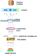
In this work, we demonstrate surface plasmon (SP) excitations by white light irradiation on Ag nanorod covered diffraction grating substrates. Recordable digital versatile discs (DVD) were used as the diffraction grating substrates on which aligned Ag nanorods arrays were deposited by oblique angle deposition. A simple experimental method based on normal incidence optical transmission was used to monitor the first order diffraction spectra from these Ag nanorod arrays on DVD gratings. The SP peak positions were observed to have dependence on polarization of the incident light and get shifted according to the aspect ratio of nanorods. The results illustrate that the radiative properties of Ag nanorod arrays on the DVD grating can be tailored just by controlling the geometric dimensions of the Ag nanorods. These Ag nanorod arrays on DVD grating templates may be used for cost efficient and sensitive surface plasmon based applications such as refractive index measurement and biosensing.
Introduction
The great variety of nanostructured object applications leads to an increased interest in investigating the light scattered from the surface of thin films.1–5 Coupling of light with nanostructured materials leads to a unique and potentially useful optical phenomenon.6 Some of the interesting examples involve the interaction of light with the sculptured metal surfaces which results in an extraordinary optical transmission.7 Metal nanorods with high aspect ratios represent an anisotropic medium and hence, the optical properties were found to be strongly dependent upon incident angle as well as the polarization of the incident light.8,9 The optical properties of metallic nanostructures, especially Ag and Au have attracted recent attention due to their potential applications in plasmonics.10,11 Guo et al.12 reported a nanoplasmonic circuit with less loss and subwavelength confinement. Byun et al.13 demonstrated enhancement of SPR biosensors with a gold nanowire grating. When excited with visible light, the free electron metals such as Ag, Au and Cu fulfill the necessary surface plasmon (SP) requirements by having dielectric constant with large negative real and small imaginary components. It has already been demonstrated through experiments that plasmon peaks strongly depend on the shape,14 structure,15 dimensions16 and dielectric constant of the host medium.17–19 A variety of fabrication strategies such as electron beam lithography20 and codeposition of Ag and polymer21 have been utilized in order to tune the surface plasmon peaks. Beyond these specialized methods one can utilize cheap and commercially available diffraction gratings (Compact Disc (CD), Digital Versatile Disc (DVD) and blue-ray disc) as templates. The optical properties of the metal nanostructure grown over these grating templates can be used to tune the position of surface plasmon peaks. The metal coated DVD grating shows highly sensitive surface plasmon based potential applications such as cheap SERS substrate,22 diffraction-based tracking of enhanced transmission of light23 and refractive index measurement.24
So far, most of the studies have been focused on determining the transmission and reflection of light from thin films, whereas studies on radiative properties such as diffraction from Ag nanorod arrays grown on grating templates are still lacking. Recently, Zhao et al.25 have used a laser scatterometer with an integrating sphere to observe anisotropic diffraction from inclined silver nanorods arrays on CD grating templates. They have shown that the orientation of the tilted nanorods with respect to the direction of grooves has a significant effect on the diffracted rays and the angular distribution of the scattered light. In the present study, we report a simple optical microscope-based method for monitoring the diffraction peaks from Ag nanorods arrays on DVD grating templates. The peaks associated with SP excitation were observed by tracking the light transmitted through ±1 diffraction orders.
Experimental section
In this work, we have utilized the grooved polycarbonate layer of the DVD-R as an inexpensive source of high quality diffraction grating as substrate. The preparation of DVD grating substrates is given as the ESI in Fig. S1.† The dried DVD grating is then placed into vacuum chamber for deposition of silver nanostructures. Silver films were grown over DVD grating substrates by thermal evaporation of silver powder (99.9%) using oblique angle deposition (OAD) method.26–30 For the growth of silver films the substrates were inclined in such a way that the substrate normal made a very high angle (α = 85°) with respect to the incident vapor flux direction. The random nucleation centers are formed initially and later on due to the geometric shadowing effect, the preferential growth of nanorods towards the direction of deposition takes place. The OAD chamber was evacuated to a pressure of about 2 × 10⁻⁶ Torr, and the substrates were kept at room temperature (∼30 °C).
Characterizations
The morphology and structural analysis of the as-prepared Ag nanostructures were characterized by scanning electron microscope (SEM) (Zeiss, EVO 50). The topographic images of the bare DVD grating were recorder by atomic force microscopy (AFM) (Dimension Icon Model, Bruker). Optical transmission measurements were performed using an optical microscope (Nikon Eclipse LV 100) in transmission mode with white light source. Fig. 1 shows schematic of the optical setup used for the diffraction measurements. The light source consisted of a 50 W halogen light source. Control of incident light polarization was done by placing a linear polarizer (Thorlabs Inc.) in the path of the incident beam and the sample. Light was focused onto the sample using a condenser lens. The sample was mounted on the vibration resistant rotating stage for xyz movement. Diffraction spectra on the screen were captured by Nikon camera (Coolpix 810). Subsequent data analysis was done by using ImageJ software (National Institute of Health, USA).31 The sensitivity of the three colors on a color camera is different, but we have mentioned the transmittance in arbitrary unit and discussed about the relative transmittance of the Ag nanorods grown DVD templates having different film thickness. For determination of film thickness (d) acetone cleaned glass slide with a step made by scotch tape is placed at α = 0° with respect to the incident vapor flux prior each deposition. After removing the scotch tape, the thickness of the metal deposited was determined by measuring the step height by line profiler (KLA Tencor).
 |
| Fig. 1 Schematic of optical configuration used to record transmission diffraction spectra induced surface plasmon resonance. | |
Results and discussion
Fig. 2(a) presents the image of a bare grooved polycarbonate diffraction grating constructed from DVD-R. It is observed that the ridges form a regular periodic pattern throughout the surface.
 |
| Fig. 2 (a) AFM image (5.0 μm × 5.0 μm) of the topography of the commercial DVD-R, and (b) line profile of the AFM image of DVD-R. | |
From the line profile (Fig. 2(b)), it was observed the periodicity of the ridges reflects grating period (Λ) of 700 nm and height (a) of 140 nm. A colored diffraction image of bare DVD as shown in Fig. 3 exhibits three dominant features; a white circular spot at the center which represents the light transmitted directly through the sample referred as 0th order and two elliptical spots which were observed on right and left sides of the central spot referred as +1 (right) and −1 (left) diffracted order. Due to the curvature of the DVD grating the diffraction spots were elongated in one direction. The 0th order peak appears white whereas in ±1 diffraction order, the dispersion of light takes place resulting into the blue light nearest to the center and the red light at the edges. The relation between the position (L) of the diffracted spot and grating period (Λ) can be expressed as32
|
 | (1) |
where
L is the distance measured from the 0th order spot,
D is the constant associated with the magnification level,
m is the diffracted order,
λ is the wavelength, and
Λ is the grating period. The
eqn (1) clearly indicates that the dispersion of colors in the diffracted spot depends on the wavelength, with longer wavelength appearing at the farther distance from the center spot. The diffraction image of the bare DVD grating is independent of the polarization (
s or
p) under direct illumination as shown in the
Fig. 3.
 |
| Fig. 3 The diffraction spectra of the bare DVD and the corresponding line profile for unpolarized, s-polarized and p-polarized light. | |
Fig. 4(a–f) shows the top-view SEM images of the tilted nanorod growth of Ag on DVD grating templates for different film deposition thickness (d) values. At the initial stage of the deposition, random nucleation centers are formed as shown in Fig. 4(a–c). With increase in the film deposition thickness, the larger islands will act as shadowing centers and will receive more vapor flux resulting in the nanorods formation (Fig. 4(d–f)). The surface morphologies indicate the periodic nanorods formation at different radial locations due to the shadowing effect. The X-ray diffraction spectrum in Fig. S2 in the ESI† shows no apparent oxidation of Ag nanorods samples upon exposure to the atmospheric conditions.
 |
| Fig. 4 SEM image presenting the top-view of the Ag nanorods on DVD with varying thickness of (a) d = 55 nm, (b) d = 100 nm, (c) d = 185 nm, (d) d = 305 nm, (e) d = 405 nm, and (f) d = 485 nm, respectively. | |
Fig. 5 presents the schematic for the nanorods formation on the DVD grating template. The side of the DVD facing the incident vapor flux results into the nanorods formation on that side whereas the other side gets shadowed.
 |
| Fig. 5 The schematic for columnar growth during oblique angle deposition. | |
According to Karabacak et al.,33 the height (h) of the nanorods increases almost linearly with film thickness and diameter (D) of the nanorods vary with h according to the power law D ∝ hγ with a fitting parameter of γ = 0.14 ± 0.1 for obliquely deposited silver nanorods.8 It is well known for oblique angle deposited nanorods that the tilt angle (β) is not same as the vapor incident angle (α).34 The alignment of the Ag nanorods makes the medium optically anisotropic with its optical axis parallel to the major long axis of the nanorods. Due to the anisotropic nature of the Ag nanorods, the diffraction spectra show a strong polarization dependence.9 Fig. 6 schematically shows the electric field component for obliquely aligned nanorods in both s- and p-polarization directions. Inset in Fig. 6 shows the schematic of the orientations and coordinates used in the diffraction measurements. For the light incident normally, the incident beam with wave vector k point towards the negative y axis and the x–y plane represents the plane of incidence. The grooves of DVD grating were parallel to the z-axis. The s-polarized and p-polarized light corresponds to an incidence plane wave whose E and H vector lies in a plane perpendicular to the plane of incidence. For s-polarization, propagation along y-direction has only electric field component Ez, tangential to the top surface of the Ag nanorods. Since this component of electric field is perpendicular to the major axis of the nanorods, therefore only transverse mode (TM) appears in the spectra. Although most of the Ag nanorods are aligned in one direction still there is a small distribution of the alignment angle as shown in Fig. 4(f). For the nanorods which are not in the main alignment direction of the incident vapor flux, the E-field will no longer remain perpendicular to the long axis of nanorods and a small component will lie along the long axis of the nanorods as shown in Fig. 6(a). This field produces longitudinal mode (LM) in s-polarization spectra. In case of s-polarization, LM appears for larger thickness of the film. For p-polarization, propagation in the same y-direction have Ex and Ey components, in the plane of incidence. Since, the nanorods have an angle β with respect to the surface normal, the E-field can be decomposed into two components: parallel Ep∥ and perpendicular Ep⊥ fields to the major axis of the nanorods (Fig. 6(b)). Thus, Ep∥ generates LM, while Ep⊥ excites the TM. The intensity of these modes further depends upon the aspect ratio of the nanorods.
 |
| Fig. 6 The cartoon sketch of the E-field component of (a) s-polarized, and (b) p-polarized light for obliquely aligned nanorods. Inset shows the coordinates used in diffraction measurement. | |
The diffraction spectra from Ag nanorods films on DVD gratings having different thickness values (d) were obtained for s- and p-polarization of the incident light. Fig. 7 shows only −1 diffracted order of the diffraction spectra. Through ImageJ software, using narrow bandpass filters of 475 nm, 510 nm and 650 nm, the intensity spectrum corresponding to each wavelength was obtained from the diffraction spectra. The line profile (intensity versus pixel location) of the diffraction images were converted into spectral data (transmittance versus wavelength). The details of the calibration are given as the ESI in Fig. S3.† Using only −1 order diffraction peak, the resulting spectral data for Ag nanorods of different thickness values deposited on DVD grating are shown in Fig. 8. However, the UV-visible transmittance spectra of the samples having different film thickness are given as the ESI in Fig. S4.† The evolution of the plasmon peaks position as a function of film thickness d was found to be different for the two different polarizations (s- and p-) of the incident light. For s-polarization, the broad surface plasmon peak centered at λp = 592 nm for d = 55 nm is the main TM of the Ag nanostructured film since it dominates the s-polarization transmission spectra. Initially, the surface plasmon peak shows a blue shift from λp = 592 nm to 534 nm for the variation in thickness value from d = 55 nm to 185 nm in case of s-polarization. As OAD progress, more and more atoms will add onto the structure that has already been deposited on the substrate. Thus the shift in the plasmon wavelength directly indicates that the relative aspect ratio (D/h) of the Ag nanostructure in s-polarization direction becomes smaller. However, in case of p-polarization, for the same film thickness two plasmon peaks (LM mode (∼490 nm) and TM mode (∼592 nm)) were observed. The intensity of both the peaks were found almost the same as Ep⊥ was weak in this case to generate TM and the nanorods height h was not sufficient for the generation of LM. With increase in the film thickness d upto the value of 185 nm, the intensity of both the peaks increases in a similar way. As d increases from 305 nm to 485 nm, both the s- and p-polarized spectral data show a very similar trend: there is one broad peak nearby λp = 545 nm for all the cases. The broadening of the peak is due to the inter-rods coupling effect. Since, the diameter of the nanorods D increases slowly as compare to the height h so the relative aspect ratio of the nanostructure decreases in s-polarization direction, as compare to p-polarization direction. It can also be observed from Fig. 4. So, for the film thickness value d = 305 nm and above, it is only LM which dominates for both the cases. The plasmon resonance is closely related to the topographic structure of the metallic nanostructure. Thus, the changes in the plasmon resonant mode as a function of the film thickness can be used to observe the change of nanostructure topology during growth at initial stages.
 |
| Fig. 7 Diffraction images of DVD grating using s-polarized (left) and p-polarized (right) light showing −1 and 0 order diffracted peaks with samples having Ag nanorods of varying thickness of (a) d = 55 nm, (b) d = 100 nm, (c) d = 185 nm, (d) d = 305 nm, (e) d = 405 nm, and (f) d = 485 nm, respectively. | |
 |
| Fig. 8 Transmission spectrum from −1-order diffracted peak with s-polarised (left), and p-polarized (right) light, for Ag nanorods on DVD with varying thickness of (a) d = 55 nm, (b) d = 100 nm, (c) d = 185 nm, (d) d = 305 nm, (e) d = 405 nm, and (f) d = 485 nm, respectively. | |
Conclusion
In conclusion, Ag nanorod structures were deposited on a DVD grating template using oblique angle deposition (OAD) techniques. The resulting Ag nanorod films grown over DVD grating templates were found to be anisotropic. This anisotropy was demonstrated by recording the polarization dependent diffraction spectra using simple optical microscopy. Diffraction induced surface plasmon peak positions appear to be dependent on the nanorods height. Thus, we have shown that the radiative properties of the sample can be tailored just by controlling the deposition parameters and hence the geometric dimensions, such as nanorod length, diameter and volume filling fraction. Ag nanorods grown on DVD templates may offer a potential application such as sensitive surface plasmon based refractive index measurement and biosensing.
Acknowledgements
The author PG kindly acknowledges Council of Scientific and Industrial Research (CSIR), India for the senior research fellowship. This research was funded by DST, India grant number SR/S2/CMP-13/2010 and Nanoscale Research Facility, IIT Delhi.
References
- S. A. Maier and H. A. Atwater, J. Appl. Phys., 2005, 98, 011101 CrossRef PubMed.
- P. Avouris, M. Freitag and V. Perebeinos, Nat. Photonics, 2008, 2, 341 CrossRef CAS.
- Z. Li, Y. Chen, X. Li, T. I. Kamins, K. Nauka and R. S. Williams, Nano Lett., 2004, 4, 245 CrossRef CAS.
- J. Zhu, Z. Yu, G. F. Burkhard, C. M. Hsu, S. T. Connor, Y. Xu, Q. Wang, M. McGehee, S. Fan and Y. Cui, Nano Lett., 2009, 9, 279 CrossRef CAS PubMed.
- Y. Cui, Q. Wei, H. Park and C. M. Lieber, Science, 2001, 293, 1289 CrossRef CAS PubMed.
- W. L. Barnes, A. Dereux and T. W. Ebbesen, Nature, 2003, 424, 824 CrossRef CAS PubMed.
- T. W. Ebbesen, H. J. Lezec, H. F. Ghaemi, T. Thio and P. A. Wolff, Nature, 1998, 391, 667 CrossRef CAS PubMed.
- Y. P. Zhao, S. B. Chaney and Z. Y. Zhang, J. Appl. Phys., 2006, 100, 063527 CrossRef PubMed.
- M. J. Bloemer, T. L. Ferrell, M. C. Buncick and R. J. Warmack, Phys. Rev. B: Condens. Matter Mater. Phys., 1988, 37, 8015 CrossRef CAS.
- P. C. Andersen and K. L. Rowlen, Appl. Spectrosc., 2002, 56, 124A CrossRef CAS.
- A. P. Alivisatos, Science, 1996, 271, 933 CAS.
- X. Guo, M. Qiu, J. Bao, B. J. Wiley, Q. Yang, X. Zhang, Y. Ma, H. Yu and L. Tong, Nano Lett., 2009, 9, 4515 CrossRef CAS PubMed.
- K. M. Byun and S. J. Kim, Opt. Express, 2005, 13, 3737 CrossRef.
- S. Link and M. A. El-Sayed, J. Phys. Chem. B, 1999, 103, 8410 CrossRef CAS.
- J. J. Mock, M. Barbic, D. R. Smith, D. A. Schultz and S. Schultz, J. Chem. Phys., 2002, 116, 6755 CrossRef CAS PubMed.
- S. Chen, S. Webster, R. Czerw, J. Xu and D. L. Carroll, J. Nanosci. Nanotechnol., 2004, 4, 254 CrossRef CAS PubMed.
- G. Xu, M. Tazawa, P. Jin, S. Nakao and K. Yoshimura, Appl. Phys. Lett., 2003, 82, 3811 CrossRef CAS PubMed.
- P. Orfanides, T. F. Buckner, M. C. Buncick, F. Meriaudeau and T. L. Ferrell, Am. J. Phys., 2000, 68, 936 CrossRef.
- J. J. Mock, D. R. Smith and S. Schultz, Nano Lett., 2003, 3, 485 CrossRef CAS.
- H. J. Lezec, A. Degiron, E. Devaux, R. A. Linke, L. Martin-Moreno, F. J. Garcia-Vidal and T. W. Ebbesen, Science, 2002, 297, 820 CrossRef CAS PubMed.
- A. Biswas, O. C. Aktas, U. Schurmann, U. Saeed, V. Zaporojtchenko, F. Faupel and T. Strunskus, Appl. Phys. Lett., 2004, 84, 2655 CrossRef CAS PubMed.
- G. Giallongo, R. Pilot, C. Durante, G. A. Rizzi, R. Signorini, R. Bozio, A. Gennaro and G. Granozzi, Plasmonics, 2011, 6, 725 CrossRef CAS.
- W. H. Yeh, J. W. Petefish and A. C. Hillier, Anal. Chem., 2011, 83, 6047 CrossRef CAS PubMed.
- B. Kaplan, H. Guner, O. Senlik, K. Gurel, M. Bayindir and A. Dana, Plasmonics, 2009, 4, 237 CrossRef.
- X. J. Wang, A. M. Haider, J. L. Abell, Y. P. Zhao and Z. M. Zhang, Nanoscale Microscale Thermophys. Eng., 2012, 16, 18 CrossRef CAS.
- D. P. Singh, P. Goel and J. P. Singh, J. Appl. Phys., 2012, 112, 104324 CrossRef PubMed.
- J. P. Singh, H. Y. Chu, J. Abell, R. A. Tripp and Y. P. Zhao, Nanoscale, 2012, 4, 3410 RSC.
- Y. P. Zhao, D. X. Ye, G. C. Wang and T. M. Lu, Nano Lett., 2002, 2, 351 CrossRef CAS.
- T. Karabacak, G. C. Wang and T. M. Lu, J. Vac. Sci. Technol., A, 2004, 22, 1778 CAS.
- D. P. Singh and J. P. Singh, J. Phys. Chem. C, 2011, 115, 11914 CAS.
- M. D. Abramoff, P. J. Magalhaes and S. J. Ram, Biophotonics International, 2004, 11, 36 Search PubMed.
- J. Meyer-Arendt, Introduction to Classical and Modern Optics, Prentice Hall, Englewood Cliffs, NJ, USA, 1995 Search PubMed.
- T. Karabacak, J. P. Singh, Y. P. Zhao, G. C. Wang and T. M. Lu, Phys. Rev. B: Condens. Matter Mater. Phys., 2003, 68, 125408 CrossRef.
- R. N. Tait, T. Smy and M. J. Brett, Thin Solid Films, 1993, 226, 196 CrossRef CAS.
Footnote |
† Electronic supplementary information (ESI) available. See DOI: 10.1039/c3ra45940c |
|
This journal is © The Royal Society of Chemistry 2014 |
Click here to see how this site uses Cookies. View our privacy policy here.