Nivika R. Guptaa,
B. L. V. Prasadb,
Chinnakonda S. Gopinath*cd and
Manohar V. Badiger*ad
aPolymer Science and Engineering Division, CSIR-National Chemical Laboratory, Dr Homi Bhabha Road, Pune-411 008, India. E-mail: mv.badiger@ncl.res.in; Fax: +91-20-2590-2612; Tel: +91-20-2590-2187
bPhysical and Materials Chemistry Division, CSIR-National Chemical Laboratory, Dr Homi Bhabha Road, Pune-411 008, India
cCatalysis Division, CSIR-National Chemical Laboratory, Dr Homi Bhabha Road, Pune-411008, India. E-mail: cs.gopinath@ncl.res.in; Web: http://www.nclwebapps.ncl.res.in/csgopinath/ Fax: +91-20-2590-2633; Tel: +91-20-2590-2043
dCenter of Excellence on Surface Science, CSIR-National Chemical Laboratory, Dr Homi Bhabha Road, Pune-411 008, India
First published on 17th December 2013
Major research efforts are continuously being made to look for alternative, environment friendly green chemicals for the synthesis of nanoparticles in place of conventional and hazardous reducing agents such as sodium borohydride and hydrazine. We report here on the synthesis and characterization of AgNPs using a thermo-associating polymer namely, carboxymethyl guar grafted poly(ethylene oxide-co-propylene oxide) [CMG-g-PEPO]. The polymer acts as both reducing agent as well as stabilizing/capping agent. The formation of AgNPs with polymer was confirmed by UV/Vis spectroscopy and the TEM images indicated the size of nanoparticles to be in the range of 10–20 nm. We also demonstrated the use of these nanoparticles in the controlled release of doxorubicin hydrochloride (Dox), an anticancer drug. The binding of Dox onto the polymer and AgNPs was investigated by XPS and Raman spectroscopy which indicates that a charge-transfer mechanism is operative between the Dox and polymer holding both the entities together. The first synthesis of AgNPs using non-toxic thermo-associating polymer and subsequent release of Dox with body temperature (37 °C) as a trigger is the highlight of the present work.
Amongst the metal nanoparticles, AgNPs exhibit antibacterial properties being used in personal care products, food products, textiles and paints and pharmaceutical formulations. The current global production of AgNPs is estimated to be ∼500 tonnes per year. Despite the large/wide applications of AgNPs due to its antimicrobial property, AgNPs have been found to show some toxicity as reported in the literature. For example, AgNPs were found to be toxic to both human lung fibroblast (e.g. IMR-90) and the human glioma (e.g. U251) cell lines.1 ATP assays have been utilized to determine the toxicity of AgNPs. Enrique Navarro et al., have reported on the toxic effects of AgNPs in aquatic organism algae, Chlamydomonas reinhardtii.2
Soohee Kim et al., reported on the oxidative stress dependent toxicity of AgNPs in human hepatoma cells.3 It was indicated that the AgNP cytotoxicity is primarily the result of oxidative stress and is independent of the toxicity of Ag+ ions which could be due to a negligible amount of free Ag+ ions in the AgNP solution. The toxic effects of Ag+ ions and AgNPs on bacteria and human cells were also reported by Greulich et al.4 Ronny van Aerle et al. have examined the mechanism of toxicity in AgNPs (∼10 nm), bulk Ag (∼1.0 μm) and Ag+ ions using high-throughput (HT-1 SuperSAGE approach).
In most cases, the toxicity of AgNPs is reported to arise from Ag+ ions associated with AgNPs. The surface oxidation of AgNPs can liberate Ag+ ions that can enhance the toxicity of AgNPs. Ag+ ion toxicity is 10 times higher than that of the AgNPs. However, the toxicity of AgNPs can be prevented by the use of antioxidant such as N-acetylcysteine. It is also reported that AgNPs at an optimal concentration have shown no toxicity towards human cells.5 Furthermore, capping agents such as glutathione (GSH) and cysteine or citrate can be used to prevent the toxicity of AgNPs.6
A variety of techniques have been reported in the literature for the preparation of metallic nanoparticles and notable examples include salt reduction,7–9 reverse micelle process,10–13 thermal process,14–16 irradiation,17,18 laser ablation,19–22 and electrochemical synthesis.23–27
In the commonly used salt reduction method, reducing agents such as sodium borohydride, hydrazine, sodium citrate and phosphines are used. However, the stability of nanoparticles obtained using these reagents is not very good and there seems to be an aggregation of nanoparticles even with small changes in pH and electrolyte environment. Furthermore, the use of toxic reducing agents such as sodium borohydride and hydrazine can pose an environmental hazard. Therefore, alternative, environment friendly green chemicals are being continuously investigated for the synthesis of nanoparticles.
Polymers play an important role in controlling the formation and dispersion stability of nanoparticles, while avoiding their aggregation. Synthetic polymers like poly(vinyl alcohol) [PVA],28–31 poly(acrylic acid) [PAA],32–34 poly(methacrylic acid) [PMAA],35,36 poly(vinylpyrrolidone) [PVP],37–40 poly(allylamine hydrochloride) [PAH],41,42 poly(ethyleneimine) [PEI],43–45 PEGs46,47 and PVP39,40,48 have been used for the synthesis of nanoparticles. Patakfalvi et al.48 reported the synthesis of silver nanoparticles using hydroquinone and sodium citrate as reducing agents with neutral polymers such as PVP and PVA as stabilizing agents. Besides in situ reduction of silver ions in aqueous solution, a polymer matrix mediated reduction of silver ions has been found to be more suitable for the synthesis of polymer–silver nanocomposite particles for various biomedical applications.49–54 Kim et al.55 used various silver salts as starting materials and examined the effect of the initial precursor content on the rate of nanoparticle formation. They found that in the presence of AgBF4, AgPF6, and AgClO4 the initial fast rate was reduced after some time, whereas in the case of silver nitrate (AgNO3) the reaction rate was slower but constant. Park et al.56 obtained gold nanoparticles by rapid addition of an aqueous solution of HAuCl4 to an aqueous solution of ascorbic acid. At the same time, the use of natural polymers like starch, gellan gum,57 dextran, gum arabic, heparin and hyaluronic acid have also been evaluated for the synthesis of nanoparticles.57–63 Particularly, some of the polysaccharides have become prominent reducing agents in the preparation of metal nanoparticles. Because of their biocompatible and biodegradable nature, polysaccharides are becoming increasingly important in the preparation of metal nanoparticles for their applications in drug delivery. Further, since polysaccharides contain carbohydrate groups, the capping of Ag or Au NPs with polysaccharides makes the surface of nanoparticles carbohydrate rich which enable easy loading of drugs into nanoparticles.
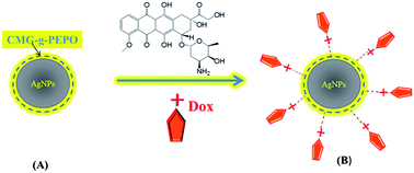
In this paper, we report on the synthesis and characterization of silver nanoparticles (AgNPs) using thermoassociating polymer namely, carboxymethylguar-g-poly(ethyleneoxide-co-propylene oxide) [CMG-g-PEPO] as reducing agent. The advantage with CMG-g-PEPO is that, it plays a dual role of capping as well as reducing agent during the synthesis of AgNPs. Further, the polymer layer on the nanoparticles being thermosensitive can be made hydrophilic or hydrophobic with a small change in the temperature which will help in drug delivery applications. The AgNPs formation was confirmed using UV/Vis spectroscopy. Further, physico-chemical and textural properties of the AgNPs were studied using X-ray diffraction (XRD), X-ray photoelectron spectroscopy (XPS), transmission electron microscopy (TEM) and Raman spectroscopy. Finally, we demonstrated the use of these nanoparticles in controlled drug delivery applications. Doxorubicin hydrochloride (Dox), a well-known anticancer drug was loaded onto AgNPs and the release profiles of Dox from AgNPs were studied.
Two different concentrations of CMG-g-PEPO, [(i) 4 mg of CMG-g-PEPO in 20 mL of deionized water (0.02 wt%) and (ii) 60 mg of CMG-g-PEPO in 20 mL of deionized water (0.3 wt%)] were used to prepare AgNPs and the samples were coded as 0.02 wt% and 0.3 wt% polymer treated AgNPs, respectively. The flow diagram for the synthesis of AgNPs is shown in Scheme 1.
The SLS measurements were performed using a Brookhaven Instruments corporation UK 90 Plus particle size analyzer. The scattering intensity was measured at a 90° angle. All the AgNPs solutions were prepared in doubly distilled water at 2 mg mL−1 concentration. Before the measurements, the scattering cell was rinsed thrice with the filtered solution. All the experiments were performed at room temperature in triplicate. The surface charge of AgNPs before and after loading of Dox was determined by measuring zeta potential. The electrode probe was dipped in a dilute AgNPs solution. All the AgNPs solutions were prepared in doubly distilled water at 2 mg mL−1 concentration. Electric field of 7.0 V cm−1 was applied across two electrodes. Zeta potential was determined with an input of pH. For each measurement, 5 runs were averaged with each run employing 10 cycles for 3 minutes. The data was analyzed using zeta pals software of Brookhaven instruments. The morphology of nanoparticles was investigated using a high-resolution transmission electron microscope (HR-TEM) at 300 kV (Cs = 0.6 mm, resolution 1.7 Å), Technai-FEI 3010. A drop of dilute AgNPs solution (2 mg mL−1) was placed on a polymer coated copper grid. The grid was then allowed to dry in air and stored in a desiccator before microscopic analysis.
![]() | (1) |
The release of Dox from Dox-loaded AgNPs was studied using the following procedure: Dox loaded AgNPs were placed in a sealed dialysis bag (MWCO 3.5 kDa) immersed in 25 mL of release buffer in a capped centrifuge tube. The centrifuge tubes were placed in an incubator shaker with gentle shaking at either 25 °C or 37 °C. At predetermined time intervals of every 1 h for day 1 and continued for every 12 h for 14 days, 2 mL aliquot of release medium was withdrawn from the tube and replaced with the same amount of fresh buffer. The amount of doxorubicin present in the collected buffer sample was determined by UV-Visible spectral analysis. The release experiments were conducted in duplicate and average values were taken for calculations. The percentage of Dox released was calculated using the equation below,
![]() | (2) |
The formation of AgNPs (A) was established by the UV-Vis spectra. AgNPs exhibit an intense absorption peak at 410 nm, due to the surface plasmon excitation, which represents the collective excitation of conduction electrons in the metal. The evolution of UV-Vis spectra of AgNPs using two different concentrations of CMG-g-PEPO solutions is shown in Fig. 2a and b. The observation of yellow color in the reaction mixture clearly indicates the formation of AgNPs. The UV-Vis spectra show strong peaks with maxima around 410–420 nm corresponding to the typical surface plasmon resonance (SPR) of conduction band electrons from the surface of silver nanoparticles. It can be readily seen from Fig. 2 that the intensity of absorption increased with reaction time; no significant difference in the peak position or shift in SPR (at 410–420 nm), signifying a continuous reduction of the Ag+ by polymer. It is also observed that increasing polymer concentration enhanced the synthesis of nanoparticles especially at short times (∼30 min). The in situ reduction of silver salts to AgNPs can be expressed as:
>R–OH + Ag+ → >R–O–Ag + H+ | (3) |
>R–O–Ag → –R![]() | (4) |
>R–OH + Ag+ → –R![]() | (5) |
![]() | ||
Fig. 2 The surface-plasmon resonance absorption spectrum of colloidal AgNPs formed with CMG-g-PEPO polymer concentrations of (a) 0.02 wt% (b) 0.3 wt%. |
![]() | ||
Fig. 4 TEM images of AgNPs capped by functionalized CMG-g-PEPO (a) 0.02 wt%, (b) 0.3 wt%, (c) Dox loaded 0.02 wt% and (d) SAED pattern of AgNPs. |
We also show in Fig. 4d the selected area electron diffraction (SAED) pattern of AgNPs obtained with 0.02 wt% CMG-g-PEPO. The SAED pattern shown in Fig. 4d exhibits concentric rings with intermittent bright dots which indicate the crystalline nature of AgNPs obtained. These rings arise due to the diffraction from the (111), (200), (220) and (311) planes of face-centered cubic (fcc) silver. The crystalline nature of AgNPs was further confirmed by X-ray diffraction studies (data not shown).
The zeta potential of AgNPs obtained treating with 0.02 wt% and 0.3 wt% of CMG-g-PEPO were found to be −18.34 mV and −24.34 mV, respectively. This clearly indicates that the AgNPs are surrounded with CMG-g-PEPO, which is an anionic polymer and helps nanoparticles to attain stability by means of electrostatic repulsion. The higher the polymer content on the nanoparticles, the higher is the zeta potential which is evident from the above results for the zeta potentials. Upon loading of the cationic drug molecule, Dox onto nanoparticles, the zeta potential decreased to −13.32 mV and −11.98 mV, for AgNPs treated with 0.02 wt% and 0.3 wt% CMG-g-PEPO, respectively. This decrease is attributed to the presence of positively charged Dox onto the AgNPs. Although the Z-potential values of Dox-loaded AgNPs were low as compared to the threshold value of ±30 mV for aggregation, we observed no inter particle aggregation. The AgNPs were formed to be stable, which was evidenced by UV-Vis spectroscopy of samples recorded over 15 days. In order to study the influence of drug loading and temperature on the size of nanoparticles, we undertook SLS studies of the AgNPs as a function of (a) temperature and (b) with and without drug loading (Fig. 5). It can be seen from Fig. 5a that, the AgNPs wrapped with CMG-g-PEPO exhibit thermo-associating behavior when the temperature was raised from 25 to 50 °C. The particle size increased from 10–15 to 20–25 nm upon increasing the temperature above the LCST of polymer. This could be attributed to the swelling of polymer coils and increase in viscosity as a result of the association of PEPO chains in the polymer above the LCST. We observe a slight increase in the size of nanoparticles upon loading of the drug, Dox onto AgNPs. We show in Fig. 5b the size of AgNPs with and without Dox at 25 °C. It can be seen that, the size of AgNPs increased from 10–15 to 25–35 nm upon incorporation of Dox. About 20% of AgNPs are in the size range of 40–60 nm. Increase in particle size is attributed mainly to the Dox loading. A slight broadening of the size distribution in AgNPs upon loading of Dox can be due to small change in the drug loading which may not be totally homogeneous. This can affect the diffusion of particles which in turn changes the distribution of particles.
![]() | ||
Fig. 5 Particle size determination as function (a) temperature, and (b) AgNPs with and without Dox loading. Later two measurements were with 0.02 wt% polymer. |
O1s core level spectra of AgNPs recorded before and after Dox loading as well as with low and high concentration of polymer. Before Dox loading, O 1s peak displays very broad spectra at binding energies around 531 eV with a full-width at half maximum of 4 eV (Fig. 6c and d). It possibly implies that two or more oxygen atoms from different groups68 are involved in the polymer–silver interactions, irrespective of polymer concentration. Upon Dox loading, a large reduction in peak width was observed with only one peak appearing at 532.9 eV, irrespective of the polymer content. Above observation underscores the interaction between Dox and AgNP which is likely through certain types of functional groups. High BE of O 1s peak observed around 533 eV, after Dox loading; suggest that functional groups, such as carboxyl or carbonyl, should be preferentially interacting with the silver surface. Indeed these being strong electron donating groups which are present at the interface, acts as a bridge between Dox molecules and silver surfaces69 and facilitates charge transfer from Dox to silver.
Charge transfer suggested from the above observations is further confirmed by C 1s core level spectral data (Fig. 6e and f). C 1s feature that appears at binding energy below 286 eV is due to alkyl, aryl groups and single hydroxyl group containing carbons. Upon Dox loading, a peak at high BE (287.5 eV) appears in both cases indicating that the –COOH and >CO are the likely binding sites coming from Dox and the polymer. In fact, the above species are not prominently observed before Dox loading suggesting that they are mostly from Dox. These studies clearly indicate the presence of Dox onto AgNPs upon loading with drug molecule.
Upon incorporating Dox molecule onto AgNPs, one can see the characteristic peaks of the Dox molecule which appear at 444 cm−1 and between 1200 and 1600 cm−1. All the characteristic peaks are observed with neat Dox and Dox loaded Ag–NPs. These studies confirm the capping and stabilization of AgNPs by CMG-g-PEPO and further the incorporation of Dox onto AgNPs.
![]() | ||
Fig. 8 Release profiles for doxorubicin loaded onto AgNPs at two temperatures 25 °C and 37 °C for 0.02 wt%. |
Dox release from AgNPs was plotted in terms of cumulative drug released versus time. It can be seen from Fig. 8 that a burst release of Dox (∼5–10% of the total loaded Dox) occurred in the first 2–3 h which is attributed to the quick release of loosely bound/unbound Dox molecules onto the AgNPs. After the burst release in the initial period, the Dox release was almost negligible at 25 °C for the next 13–14 days; indeed it is a good indication that the binding between Dox and AgNPs is significant and requires some trigger. However, at 37 °C the release of Dox was much faster and more drug was released with respect to time. This could be due to the expansion of polymer coils and loosening of the polymer structure on the AgNPs above LCST as a result of the thermo-associating behavior of the polymer. This can enhance the diffusion of higher amounts of Dox molecules at a faster rate. This is indeed, observed in the release profiles of Dox at 37 °C suggesting the body temperature acts as a trigger. The above results clearly indicate that the release of drug, Dox is influenced by the thermo-associating nature of the polymer surrounding the AgNPs. In metal nanoparticle, the surface of the ‘naked’ particle is imparted with charge due to the lower coordination experienced by metal ions on the surface. In our case, such a surface is covered by anionic polymer, CMG-g-PEPO which over compensates the charges of metal and imparts an overall negative charge. The cationic drug, Dox is bound to the anionic polymer. The release of the drug depends on the ionic strength, environment around the nanoparticles, temperature and also on the equilibrium that exists between the drug molecules bound to the nanoparticles and outside the solution. Although the Dox release in the first 10 h is high, by controlling the LCST of the polymer, there is good scope to devise drug incorporated nanoparticles with tailored drug release profiles. It is likely possible to control the drug release rate by fine tuning the charge transfer interactions between AgNPs and Dox.
Footnote |
† Electronic supplementary information (ESI) available. See DOI: 10.1039/c3ra45022h |
This journal is © The Royal Society of Chemistry 2014 |