Delivery of doxorubicin in vitro and in vivo using bio-reductive cellulose nanogels†
Hanqing
Qian
a,
Xin
Wang
a,
Kangjun
Yuan
a,
Chen
Xie
a,
Wei
Wu
a,
Xiqun
Jiang
*a and
Lijiang
Hu
b
aDepartment of Polymer Science & Engineering, College of Chemistry & Chemical Engineering, Nanjing University, Nanjing 210093, P. R. China. E-mail: jiangx@nju.edu.cn
bDepartment of General Surgery, Drum Tower Hospital affiliated to Medical School of Nanjing University, P. R. China
First published on 7th October 2013
Abstract
A methacrylation strategy was used to functionalize carboxymethyl cellulose and prepare redox-sensitive cellulose nanogels which contained disulfide bonds. Dynamic light scattering, zeta potential and electron microscopy were utilized to characterize these nanogels. It was found that these nanogels had a spherical morphology with a diameter of about 192 nm, and negative surface potential. These redox-sensitive nanogels were stable against high salt concentration but de-integrated in the reducing environment containing glutathione. When doxorubicin (DOX) was loaded into the nanogels, a high drug loading content (36%) and a high encapsulation efficiency (83%) were achieved. Confocal laser scanning microscopy and co-localization images showed that DOX-loaded nanogels were internalized by the cancer cells through endocytosis and the DOX could be delivered into the nucleus. Near-infrared fluorescence imaging biodistribution examination indicated that the nanogels could passively target to the tumor area by the EPR effect and had a significantly prolonged circulation time. In vivo antitumor evaluation found that DOX-loaded nanogels exhibited a significantly superior antitumor effect than the free DOX by combining the tumor volume measurement and the examination of cell apoptosis and proliferation in tumor tissues.
Introduction
Polymer nanoparticles have recently been attracting increasing interest in the area of controlled and long-term delivery of chemotherapeutic drugs in vitro and in vivo, of which potential advantages include their ability to improve the pharmacological profiles of drugs as well as the enhancement of therapeutic efficacy.1,2 Due to their biocompatibility and biodegradability, natural polymers, such as polysaccharides, proteins and cellulose, are considered good candidates for constituting drug delivery systems. Among these, carboxymethyl cellulose (CMC) is a commercially available derivative of cellulose which has been approved by FDA for biomedical applications.3 Commonly, the preparation of natural polymer nanoparticles is based on the self-assembly of amphiphilic molecules in aqueous solution.4–16 For instance, cholesterol group-modified glycogen,4 dextran–poly(ε-caprolactone),6 maltoheptaose–poly(N-isopropyl acryl-amide),7 cellulose–poly(ε-caprolactone),10 hyaluronic acid–polyethyleneimine,12 heparin–cisplatin,14 and docetaxel–CMC16 are reported to fabricate natural polymer based nanoparticles.However, these nanoparticles are usually in a dynamic state and present only temporal stability.17 The inadequate stability of these particles is one of the most important practical challenges for clinical use. When injected into the body, nanoparticles are extensively diluted and massively interact with the biological components in the blood stream. Thus, the dissociation or aggregation of polymer nanoparticles may occur upon intravenous administration, leading to premature drug release, indefinite biodistribution and low drug accumulation in tumor sites.18 So far, a variety of chemically cross-linking approaches to stabilize the structures of the nanoparticles have been proposed.19 Traditionally, divinyl sulfone, epichlorohydrin, epoxides (1,2,3,4-diepoxybutane), and diisocyanate were used to cross-link cellulose,20–22 but most of these syntheses for producing cross-linked cellulose often involve multiple steps and the chemicals required are not environmentally friendly, and can even be cytotoxic. Moreover, one preparation protocol can hardly be adapted to another system, lacking generality. Recently, some improved methods to cross-link polysaccharides and cellulose have been developed. For example, hyaluronic acid was functionalized with a vinyl sulfone group through a simple “click” reaction and was subsequently cross-linked with a thiol counterpart to produce hydrogels.23 Heparin was chemically modified with thiol groups and then cross-linked with disulfide linkages to produce reducible heparin nanoparticles.24 Allyl isocyanate modified hydroxypropyl cellulose was cross-linked by γ-ray irradiation to prepare hydrogels.25 A cellulose network was also generated by glyoxalization.26 Nevertheless, there is still a lack of effective and mild strategies to prepare cellulose nanogels for biomedical application. On the other hand, the cross-linking of nanoparticles may cause a diffusion barrier in nanoparticles for the efficient release of their payloads and alter the stealthiness of the nanoparticles. Thus, the development of natural polymer-based drug delivery vehicles, which are stable against interactions with substrates in blood during circulation and accomplish efficient intracellular drug release following arrival at the pathological site, remains a considerable technological challenge.
It has been found that there exists a large difference in the concentration of glutathione (GSH) between the relatively oxidizing extracellular milieu and the reducing intracellular fluids.27 For example, glutathione is present at 4-fold higher concentrations in some tumor tissues than that in normal tissues.28,29 Furthermore, subcellular organelles such as endosomes and lysosomes also have reductive activity due to the presence of the reducing enzyme gamma-interferon-inducible lysosomal thiol reductase (GILT) as well as the reductive cysteine.30 Considering this biological feature in the tumor microenvironment and the prominent stability of the nanogel structure, in the present work we report a methacrylation strategy to functionalize CMC with vinyl groups, which can be easily adapted to other polysaccharides. Subsequently, the methacrylated cellulose was reacted with disulfide-containing cystamine bisacrylamide (CBA) to prepare redox-sensitive CMC nanogels in completely aqueous medium. These CMC nanogels were utilized to efficiently carry doxorubicin (DOX) for intracellular delivery. Additionally, due to the presence of disulfide linkages, the CMC nanogels are sensitive to reductive environments such as tumor tissues with high GSH concentration and readily de-cross-link, allowing tumor- and intracellular-specific release of the entrapped drugs. The morphology, colloidal stability and GSH sensitivity of CMC nanogels were characterized by dynamic light scattering and electron microscopy. The in vitro intracellular fate and in vivo biodistribution of these cellulose-forming nanogels were examined. Finally, the in vivo antitumor activity of DOX-loaded CMC nanogels in H22 tumor-bearing mouse was assessed.
Materials and methods
Materials
Sodium carboxymethyl cellulose (Mw 90 kDa, substitution degree = 0.7) was purchased from Sigma-Aldrich Co. LLC. Acryloyl chloride (Shanghai Chemical Reagent Co., China) was distilled before use. Potassium persulfate (K2S2O8) was recrystallized from deionized water. Doxorubicin hydrochloride (DOX) was obtained from Shenzhen Main Luck Pharmaceuticals Inc. (Shenzhen, China). Cystamine dihydrochloride, N,N′-methylene bisacrylamide, methacrylic anhydride, GSH, fluorescein isothiocyanate (FITC), NIR-797 isothiocyanate, Lyso Tracker red and other reagents and solvents were of analytical grade and used as received. The murine hepatic H22 cell line and the human neuroblastoma SH-SY5Y cell line were purchased from Shanghai Institute of Cell Biology (Shanghai, China). Male ICR mice (6–8 weeks old) were purchased from the Animal Center of Drum Tower Hospital (Nanjing, China). All animal experiments were performed under a protocol approved by the Institutional Animal Care and Use Committee, Nanjing University.Characterization
Nuclear magnetic resonance (NMR) spectroscopy was recorded using a Bruker DQX-300 spectrometer with tetramethylsilane as an internal standard. ATR-Fourier transform infrared spectroscopy (ATR-FTIR) was carried out using a Nicolet iS10 FT-IR spectroscope (Thermo, USA). Elemental analysis was performed using a Vario EL II elemental analyzer (Elementar, Germany). Transmission electron microscope (TEM) measurements were carried out using a JEM-100S (JEOL, Japan) at an acceleration voltage of 80 kV. Scanning electron microscopy (SEM, HITACHI, Japan) was employed to observe the morphology of samples at an accelerating voltage of 15 kV. The mean diameter and size distribution of the nanogels were determined by the dynamic light scattering (DLS) method using a Brookhaven BI9000AT system (Brookhaven Instruments Co., USA), and the result is shown using the intensity mode. Zeta-potential of the nanogels was obtained using a Brookhaven 90 Plus Zeta Potential Analyzer (Brookhaven Instruments Co., USA).Synthesis of cystamine bisacrylamide (CBA)
CBA was synthesized according to the previous work.31 Briefly, cystamine dihydrochloride (5.6 g, 0.025 mol) was dissolved in 25 mL of water in a three-necked flask. An acryloyl chloride solution (0.1 mol in 10 mL of dichloromethane) and a NaOH aqueous solution (10 M, 20 mL) were added dropwise simultaneously at 0 °C. After the solution addition was completed, the reaction mixture was stirred continuously at room temperature for 16 h, and a suspended solution was observed. Then a white raw product was obtained by filtration, which was washed 3 times with water. The raw product was further purified by crystallization using acetate. Finally, a white solid, prepared CBA was obtained by removing the solvent under vacuum (4.5 g, 70%). 1H NMR (CDCl3, ppm): 6.69 (2H, –CONHCH2–), 6.30 and 6.23 (4H, CHH![[double bond, length as m-dash]](https://www.rsc.org/images/entities/char_e001.gif) CHCO–), 5.69 (2H, CHHCHCO–), 3.65 (4H, –NHCH2CH2–), 2.88 (4H, –CH2CH2S–).
CHCO–), 5.69 (2H, CHHCHCO–), 3.65 (4H, –NHCH2CH2–), 2.88 (4H, –CH2CH2S–).
Synthesis of methacrylated carboxymethyl cellulose (MACMC)
MACMC was synthesized as before with little modification.32 2 g of carboxymethyl cellulose was dissolved in 100 mL of water. 4 mL of methacrylic anhydride was added and then the pH of the mixture was adjusted to around 8 with a 3 M NaOH solution. After being left to react for 24 h at 0 °C, the mixture was precipitated and washed with ethanol to remove the remaining methacrylic acid and methacrylic anhydride. Finally, the product of MACMC was obtained after being dried under vacuum. The degree of substitution of the methacrylic molecules was determined by 1H-NMR using D2O as a solvent.Synthesis of MACMC–CBA nanogels
MACMC–CBA nanogels were prepared by radical polymerization of MACMC with CBA as a cross-linking reagent. Typically, MACMC (54 mg) and CBA (66 mg) were dissolved in 15 mL of water by heating. After the solution became clear, polymerization was initiated using K2S2O8 at 80 °C and proceeded for 30 min in a nitrogen atmosphere. Finally, dialysis was used to remove residual monomers, initiators and other impurities in a dialysis bag (MWCO = 14 kDa).For comparison, N,N′-methylene bisacrylamide (MBA) was used as a cross-linker to prepare non-bio-reductive responsive MACMC–MBA nanogels by the same method.
Preparation of DOX-loaded nanogels
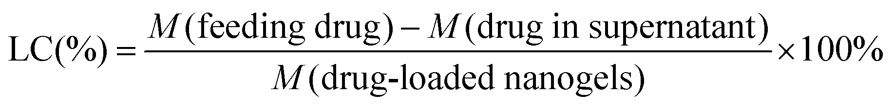
The drug-loaded nanogels were prepared by mixing 1 mL of a DOX aqueous solution at a predetermined concentration with a MACMC–CBA or MACMC–MBA nanogel suspension (3 mL, 4 mg mL−1), the pH of which was adjusted to 8 previously. The mixed solution was stirred overnight at room temperature in the dark to allow DOX to equilibrate between the aqueous phase and the nanogel matrix. To remove the unloaded DOX, the resulting mixture was centrifuged at a speed of 12![[thin space (1/6-em)]](https://www.rsc.org/images/entities/char_2009.gif) 000 r min−1 for 40 min and redispersed in PBS (10 mM, pH = 7.4), giving the DOX-loaded cellulose nanogels. The amount of free DOX in the supernatant was quantified by measuring the absorbance at 480 nm based on a calibration curve, and the mass of the DOX-loaded nanogels was weighed after being dried under vacuum overnight. The drug loading efficiency (LE) and loading capacity (LC) of the nanogels were calculated as follows:
000 r min−1 for 40 min and redispersed in PBS (10 mM, pH = 7.4), giving the DOX-loaded cellulose nanogels. The amount of free DOX in the supernatant was quantified by measuring the absorbance at 480 nm based on a calibration curve, and the mass of the DOX-loaded nanogels was weighed after being dried under vacuum overnight. The drug loading efficiency (LE) and loading capacity (LC) of the nanogels were calculated as follows: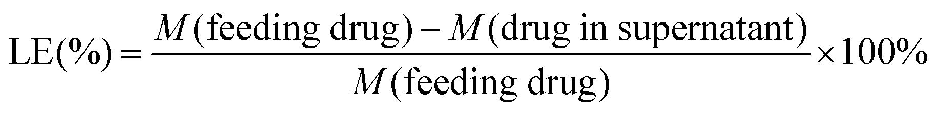 | (1) |
 | (2) |
In vitro release of DOX from the nanogels in fetal bovine serum (FBS)
DOX-loaded MACMC–CBA nanogels were suspended in 5 mL of FBS. The resulting solution was incubated at 37 °C. At a predetermined time, 0.5 mL aliquots was taken from the medium and centrifuged at a speed of 12000 r min−1 for 40 min. The amount of released DOX in the supernatant was quantified using UV/Vis spectrometry (UV-5300, Metash, China). The calibration curve was established using a set of FBS solutions containing a known concentration of DOX.
In vitro pH and reduction-triggered release of DOX from the nanogels in buffer solutions
0.5 mL of DOX-loaded nanogels with a known drug concentration (1.6 mg mL−1) was placed into a dialysis bag (MWCO = 14 kDa). Then it was immersed into 5 mL of PBS (10 mM, pH = 7.4, 6.5, 4.5, respectively) or PBS (10 mM, pH = 7.4) containing 10 mM GSH at 37 °C. At a predetermined time, all 5 mL of the release buffer solution was withdrawn completely and replaced with 5 mL of fresh PBS with corresponding pH or PBS with GSH. The amount of released DOX was determined by measuring the absorbance at 480 nm based on a calibration curve using UV/Vis spectrometry (UV-5300, Metash, China).In vitro cytotoxity
The in vitro cytotoxicity of DOX-loaded nanogels against two cancer cell lines including human neuroblastoma SH-SY5Y cell line and murine hepatic carcinoma cell line H22 was tested by MTT assays. SH-SY5Y or H22 cells (5000 cells per well) were seeded in a 96-well plate respectively, and exposed to 200 μL of medium containing free DOX, DOX-loaded nanogels (MACMC–CBA and MACMC–MBA respectively) and empty nanogels at different concentrations at 37 °C for 24 h in a humidified atmosphere with 5% CO2. After the incubation, the culture medium was removed and washed with PBS twice. Then 20 μL of 5 mg mL−1 MTT solution and 180 μL of fresh medium were added to each well. After incubating the cells for another 4 h, the medium was removed and 150 μL per well DMSO was then added to dissolve the formazan crystals, the absorbance at 570 nm was measured on a microplate reader (Huadong, DG-5031, China), and the cell viability was calculated by the following formula: | (3) |
Cellular uptake of nanogels
Initially, dye-labeled nanogels were prepared as follows: 4 mg of FITC in 1 mL of DMSO was added into an aqueous solution of MACMC (1.5 g/20 mL H2O) and the mixture was stirred overnight at room temperature. After that, the mixture was precipitated into 250 mL of ethanol and washed with ethanol twice. Finally, the fluorescence labeled MACMC was obtained after being dried under vacuum. The resultant FITC–MACMC was used to prepare fluorescence labeled FITC-nanogels and FITC–DOX-loaded nanogels with the same method described above.Subsequently, for cellular uptake examination, the SH-SY5Y cells were seeded into a 6-well plate at a density of 2.5 × 10⁵ cells per well and incubated for 12 h at 37 °C in a humidified atmosphere of 5% CO2. 200 μL of FITC–DOX-loaded nanogels (MACMC–CBA and MACMC–MBA respectively) was subsequently added into the cell culture medium. After 4 h of incubation at 37 °C, the cells were washed three times with PBS at 4 °C and 37 °C respectively. 4′,6-diamidino-2-phenylindole (DAPI) was employed to dye the nucleus zone of the cells. Then the cells were observed using CLSM (LSM 710, Zeiss, Germany) that was equipped with a 63×/NA 1.4 oil immersion objective. FITC, DOX, and DAPI excitations were achieved with a 488 nm argon laser, a 543 nm HeNe laser, and a 405 nm diode laser, respectively. The pin-hole diameter was set at 1 airy unit (AU).
Co-localization of nanogels and cells
SH-SY5Y cells were first incubated with 100 nM Lyso-Tracker Red for 30 min at 37 °C in a humidified atmosphere of 5% CO2, and washed with culture medium. 200 μL of FITC-nanogels was subsequently added into the cell culture medium. After incubation for 4 h at 37 °C, the cells were washed three times with PBS at 4 °C and 37 °C respectively. DAPI was employed to dye the nucleus zone of the cells. Then the cells were observed with CLSM. Lyso-Tracker Red excitation was achieved with a 543 nm HeNe laser.In vivo NIR fluorescence imaging
First, NIR-797 isothiocyanate was used to label MACMC–CBA nanogels. In brief, NIR-797 isothiocyanate (0.5 mg in 50 μL of DMSO) was reacted with 10 mL of MACMC–CBA nanogel suspension (4 mg mL−1) for 12 h in the dark. The resulting mixture was centrifuged at 12000 r min−1 for 40 minutes twice in order to remove the unconjugated NIR-797. Then the NIR-797-labeled nanogels were redispersed in PBS.
All the animal experiments were reviewed and approved by Animal Care and Use Committee, Nanjing University. H22 cells (4–6 × 10⁶ cells per mouse) were subcutaneously inoculated into the left flank of ICR male mice. The mice were kept 7 days with free access to food and water. After that, 0.2 mL of NIR-797-labeled nanogels was injected into H22 tumor bearing mouse (20–25 g) through a tail vein. After administration, real time biodistribution of MACMC–CBA nanogels in tumor-bearing mouse was investigated using a Maestro EX in vivo fluorescence imaging system (CRi, Inc., USA). Imaging was performed at 1 h, 2 h, 4 h, 8 h, 12 h, 24 h, 48 h, 72 h, 96 h, and 120 h post i.v. administration.
In vivo biodistribution of DOX in nanogels
21 H22 tumor bearing mice were intravenously injected with DOX-loaded MACMC–CBA nanogels at an equivalent DOX dose of 5 mg kg−1 body weight. Free DOX (5 mg kg−1) and DOX-loaded MACMC–MBA nanogels (5 mg kg−1 DOX eq.) were also administered as the control in 21 mice per group. 3 mice served as a blank group without treatment. The mice were sacrificed at predetermined time points after administration. The organs were washed with water and dried with filter paper carefully. After being wet-weighed, they were homogenized and extracted for 48 h with 4 mL of 60% (v/v) alcohol solution with 5% hydrochloric acid. After centrifugation at 12000 rpm for 20 min, the fluorescence of the supernatant was measured using a fluorescence spectroscope (Shimadzu, RF-5301PC, Japan) with an emission of 560 nm.33 The measured average values of each tissue from the blank group served as the background and were deducted from the corresponding sample of the administered mice.
In vivo antitumor efficacy of DOX-loaded nanogels
H22 cells (4–6 × 10⁶ cells per mouse) were inoculated subcutaneously into ICR mice at the left flank in the same way as described above. 7 days after inoculation (the tumor volume reached about 100 mm3), 40 H22 tumor bearing mice were randomly divided into 4 groups (10 mice per group). 200 μL of saline, empty MACMC–CBA nanogels, free DOX (single injection, 5 mg kg−1), and DOX-loaded MACMC–CBA nanogels (single injection, 5 mg kg−1 DOX eq.) were administered via a tail vein, respectively, and this day was designated Day 1. The tumor volume and body weight were measured on alternate days. The tumor volume was calculated by | (4) |
In the equation, D and d are the maximum and minimum diameters of the tumor, respectively. The tumor growth inhibition (TGI) was calculated by the following equation:
 | (5) |
For multiple dose treatments, 200 μL of saline, free DOX (5 mg kg−1 DOX × 3 injections), DOX-loaded MACMC–CBA nanogels (5 mg kg−1 DOX eq. × 3 injections) and DOX-loaded MACMC–MBA nanogels (5 mg kg−1 DOX eq. × 3 injections) were administered via a tail vein at 3-day intervals (days 1, 4, and 7) for a total of 3 treatments. The tumor volume and body weight were measured on alternate days as described above.
Determination of cell apoptosis and proliferation
The tumors inoculated into the mice were excised (3 mice per group, and the groups were treated with saline, blank nanogels, free DOX, a single injection of DOX-loaded nanogels and multiple injections of DOX-loaded nanogels, respectively) at day 8 after injection, fixed in 10% formalin, embedded in paraffin and thereafter sectioned into slices at a thickness of 4 μm. The slices were stained by terminal deoxynucleotidyl transferase dUTP nick end labeling (TUNEL) and proliferating cell nuclear antigen (PCNA) methods, respectively, according to the manufacturer's instructions. For the quantification of TUNEL and PCNA expression, the percent of positive cells in 10 random fields was counted by optical microscopy.Statistical analysis
Statistical comparisons were performed by one-way ANOVA analysis and Student's t-test. P values <0.05 were considered statistically significant.Results and discussion
Synthesis of MACMC–CBA nanogels
To prepare the nanogels, sodium carboxymethyl cellulose was reacted with methacrylic anhydride so that methacrylated carboxymethyl cellulose (MACMC) was obtained (Scheme 1A). As shown in Fig. 1, after the reaction, three peaks at 6.1, 5.6 and 1.8 ppm assigned to the protons of methylene (a, b) and methyl (c), respectively, are observed in the 1H NMR spectrum of MACMC in D2O, confirming that the methacrylation modification was successful. The degree of modification was determined to be 11.8% from the relative integrations of methacrylated proton peaks into carbohydrate protons.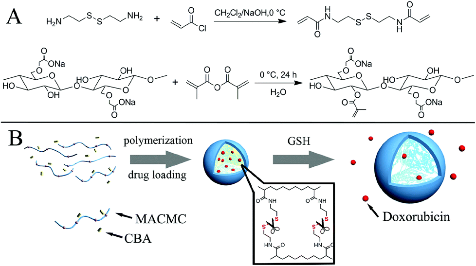 | ||
| Scheme 1 (A) Synthesis of CBA and MACMC. (B) Schematic diagram of the synthesis of MACMC–CBA nanogels and the release of DOX triggered by GSH. | ||
 | ||
| Fig. 1 1H NMR spectrum of (A) MACMC and (B) CBA. The structure in (A) represents only one of the possible outcomes for the chemical modification processes we performed. | ||
Next, reductively sensitive nanogels were prepared by copolymerization of MACMC with the disulfide-bond containing cross-linker, CBA, in a complete aqueous solution, as shown in Scheme 1B, and the nanogels formed were analyzed by a series of characterization techniques. DLS and zeta-potential measurements indicate that the MACMC–CBA nanogels in a PBS solution (10 mM, pH = 7.4) have a diameter of 193 ± 2 nm and a zeta-potential of −25.7 ± 1.6 mV. TEM and SEM images show that these nanogels have a well-defined spherical morphology, as shown in Fig. 2A and 2B. The diameter determined by TEM (103 nm, based on an analysis of 100 nanogels) is smaller than that measured by DLS, which is attributed to the dehydration effect of the nanogels in the dry state. Fig. 2D shows the FT-IR spectra of CBA, MACMC and MACMC–CBA nanogels. For nanogels, the main absorption bands at 3287 cm−1 for the O–H of cellulose and 1543 cm−1 for the N–H of CBA are displayed, thus indicating that the nanogels are composed of MACMC and CBA. In addition, elemental analysis of the nanogels shows that the compositions of the nanogels are 65% for MACMC and 35% for CBA. Since the colloidal stability of the carrier has a crucial influence on the drug release, biodistribution and targeting ability of the nanocarriers, the stability of nanogels in a salt solution was evaluated. As shown in Fig. 2E, the diameter of nanogels in salt-free aqueous medium (CNaCl = 0) is 310 nm, suggesting that the nanogels are in a totally swelling state. The degree of swelling of the nanogels, which is defined as the ratio of the diameter in solution determined by DLS to the one in the dry state from TEM, is calculated to be 301%. With increasing the concentration of NaCl to 6 mg mL−1, the nanogels shrink, resulting in a decrease in nanogel size from 310 nm to 192 nm, and the degree of swelling decreases to 186%. While continuously increasing the salt concentration to 18 mg mL−1 of NaCl, the diameter of the nanogels is kept nearly invariable. The diameter of nanogels under physiological conditions (CNaCl = 9 mg mL−1) is consistent with that determined in PBS (0.01 M, pH = 7.4). The water content of the swelling nanogels under physiological conditions is estimated to be about 84% (water content = 1 − Vd/Vs, Vd and Vs are the volume of the dried and saturated nanogels, respectively). This high water content of the nanogels combined with their rubbery consistency results in low interfacial tension, which contributes to fewer tendencies to absorb proteins from body fluids.34,35 These results indicate that the cross-linked nanogels are stable even in the aqueous medium with high salt concentration. To evaluate the sensibility of the nanogels containing disulfide bonds to reducing agents, the nanogels were incubated in PBS with 10 mM GSH for 24 h and the nanogel size was monitored by DLS (Fig. 2F). It is interesting that the nanogels after treatment present a bimodal size distribution: one peak at about 140 nm and another peak around 450 nm. We believe that both the peaks formed are most likely due to the reductive cleavage of the cross-linking disulfide bonds in the nanogels, leading to the disintegration of one part of nanogels to produce small sized particles and the swell of another part of the nanogels to form large sized ones. In comparison, there was no obvious change in the diameter when the MACMC–MBA nanogels were incubated in PBS containing 10 mM GSH for 24 h due to the absence of bio-reductive sensitivity (see the ESI, Fig. S1†). As mentioned above, there is a large difference in redox potential between the extracellular environment where GSH is found in the micromolar concentration and the intracellular cytosol where GSH is at around 10 mM. Thus, the reducing response test above suggests that the disulfide bond-contained nanogels may be de-cross-linked in a bio-reductive environment in the cytoplasm, which is in favour of intracellular drug release for drug-loaded nanogels.
 | ||
| Fig. 2 (A) TEM and (B) SEM images of the MACMC–CBA nanogels. (C) TEM image of the DOX-loaded MACMC–CBA nanogels. The scale bar in each figure is 200 nm. (D) FT-IR spectrum of MACMC (red), CBA (black) and the nanogels (blue). (E) Diameters, scattering intensity and degree of swelling of MACMC–CBA nanogels with different concentrations of NaCl. (F) Hydrodynamic diameter distribution of the CMCMA–CBA nanogels before (line) and after treatment with GSH for 24 h (dashed line) measured by DLS. | ||
DOX loading and drug release in vitro
For micelles self-assembled from block copolymers, the drugs are usually encapsulated in the hydrophobic sections. However, in most cases drug loading arising from hydrophobic interactions alone results in relatively low loading capacities.36 Unlike the micelles, MACMC–CBA nanogels containing meshed structures within the spherical matrix are expected to have promising application as high-loading drug carriers. To this end, doxorubicin was selected as a model of molecular drugs to assess the drug loading and release performance. DOX-loaded MACMC–CBA nanogels were prepared by an incubation method exploiting the electrostatic interactions between the protonated amino groups of DOX and carboxyl groups in MACMC. Unloaded DOX was removed by centrifugation at a speed of 12000 r min−1 for 40 min. The average diameter of DOX-loaded nanogels was determined to be 186.0 ± 1.6 nm by DLS, and the zeta-potential is −22.1 ± 1.2 mV. TEM observation confirms that the structure of nanogels does not change after drug encapsulation (Fig. 2C). Fig. 3A shows the drug loading efficiency and loading content of the nanogels prepared at variable values of pH. It is found that with an increase of medium pH from 4 to 10, the drug loading content of the nanogels monotonously increases from 16% to 40%, but the loading efficiency of DOX shows an initial increase from pH = 4 to pH = 8 and then a decrease at pH = 10. Thus, at pH = 8, the nanogels show optimal drug loading performance: the loading efficiency of DOX in the nanogels is 83% and the loading content reaches 36% (36 mg of DOX per 100 mg of DOX-loaded nanogels). The increase in drug loading content of the nanogel is probably attributed to the increase in the ionized degree and swelling of the nanogels with an increase of pH, which helps nanogels to capture DOX molecules. Compared to other drug delivery systems,37 the MACMC–CBA nanogels described here have much higher drug loading content. This is another feature of our nanogels in addition to the redox sensitivity. Hence, DOX-loaded nanogels with a loading content of 36% were chosen in the subsequent studies.
 | ||
| Fig. 3 (A) Loading efficiency and loading content of the DOX-loaded MACMC–CBA nanogels prepared at different pH values. (B) The stability of DOX-loaded MACMC–CBA nanogels in PBS (pH = 7.4, 10 mM). (C) In vitro release profiles of DOX from nanogels in FSB or PBS (pH = 4.5, 6.5, 7.4, 10 mM) at 37 °C with or without GSH (10 mM). | ||
The stability of DOX-loaded nanogels is also an important parameter of drug delivery vehicles. The changes in the hydrodynamic diameter and light scattering intensity in PBS at pH 7.4 were monitored by DLS and are shown in Fig. 3B. It is found that the prepared DOX-loaded nanogels are stable within the 14 days of the monitoring period and show no appreciable change in the hydrodynamic diameter.
Fig. 3C shows the in vitro release of DOX from nanogels in FBS and various kinds of buffer solutions. The release profiles were traced using a UV-Vis spectrometer. It can be seen that only 10% of the encapsulated DOX is released in FBS within 96 h, indicating the stability of the disulfide bond in serum. So the DOX loaded nanogels are highly stable in the blood and the premature DOX release is minimal. The in vitro release of DOX from nanogels was also examined by dialysis against PBS (10 mM, pH = 7.4, 6.5, 4.5) or PBS (10 mM, pH = 7.4) with 10 mM GSH at 37 °C. In the absence of GSH, only about 20% of the DOX loaded is released within 24 h at pH 7.4, and no tendency of further release is observed thereafter until 96 h, as shown in Fig. 3C. With decreasing medium pH, the amount of accumulative release of DOX from the nanogels increases dramatically. At pH 6.5 and 4.5, the release amounts of DOX from the nanogels are ca. 45% and 70% within 96 h, respectively, indicating that the release of DOX from the nanogels speeds up with the decrease in the pH value of the system. This is primarily ascribed to the weakening of electrostatic interaction between the cellulose and DOX; accordingly, the DOX is diffused from the nanogel matrix more easily at low pH. On the other hand, in the presence of 10 mM GSH in PBS (pH = 7.4) that mimicked a cytosolic environment, 70% of the loaded DOX is released in the initial 24 h and this value becomes 80% in 96 h (Fig. 3C), demonstrating that GSH triggered de-cross-linking of nanogels leads to an acceleration of the DOX release from disulfide-bond-contained nanogels.
Cytotoxicity of DOX-loaded nanogels in vitro
To evaluate the pharmacological activity of DOX released and the potential toxicity of the empty nanogels, the in vitro cytotoxicity of the DOX-loaded MACMC–CBA nanogels was evaluated using MTT assays. Our study involved two kinds of cell lines: human neuroblastoma SH-SY5Y and the murine hepatic carcinoma cell line H22. Both cancer cells were treated with either free drug or DOX-loaded nanogels. Fig. 4A and 4B shows the cell inhibition of the DOX-loaded nanogels against SH-SY5Y and H22 cell lines after 24 h of incubation. A dose dependent cytotoxicity is observed for both free DOX and the DOX-loaded nanogels. When the cells were treated with DOX-loaded MACMC–CBA nanogels at the concentration of 16 μg mL−1, 70% of SH-SY5Y cell proliferation and 65% of H22 cell proliferation are inhibited. The slight difference in the cytotoxicity of the two cell lines may be attributed to the differences in their genetic background and biological behaviour since H22 is the suspension liver cancer cell line from mouse, while SH-SY5Y are adherent human cells. | ||
| Fig. 4 In vitro cytotoxicity of free DOX and DOX-loaded nanogels against (A) SH-SY5Y and (B) H22 cell lines after 24 h of incubation. *Represents P < 0.05. (C) Viability of SH-SY5Y and H22 cell lines after incubation with MACMC–CBA nanogels for 24 h. (D) CLSM images of SH-SY5Y cells incubated with (D) FITC labeled MACMC–CBA nanogels (green). Endosomes/lysosomes of the cells are marked by Lyso Tracker (red) and the nuclei were stained with DAPI (blue). (E) and (F) CLSM images of SH-SY5Y cells incubated with DOX (red)-loaded, FITC (green) labeled (E) CMCMA–CBA and (F) CMCMA–MBA nanogels. The nuclei were stained with DAPI (blue) for 4 h at 37 °C. The scale bar in each figure is 20 μm. | ||
However, the inhibition of cellular proliferation is 57% when the SH-SY5Y cells were treated with DOX-loaded MACMC–MBA nanogels, which is lower than that incubated with bio-reductive responsive MACMC–CBA nanogels. This difference is significant (P < 0.05 at the drug concentrations of 4, 8 and 16 μg mL−1). The result indicated that the relatively faster release of DOX from MACMC–CBA nanogels in the reductive environment of the cytoplasm can induce more inhibition of cell proliferation, leading to an improved antitumor efficiency in vitro.
For comparison, empty MACMC–CBA nanogels were also tested. The cell viability of the empty nanogels is about 97% (SH-SY5Y) and 93% (H22) within the concentration up to 400 μg mL−1, demonstrating that the MACMC–CBA nanogels are nontoxic and cytocompatible (Fig. 4C). This result indicates that DOX still keeps its pharmacological activity when encapsulated into nanogels. Inhibition of cell proliferation is induced by the DOX encapsulated in the nanogels.
Cellular uptake and intracellular trafficking of nanogels
Tracing the cellular uptake and intracellular distribution of the MACMC–CBA nanogels is critical for assessing the biological activities of chemotherapeutic formulations.38 After FITC-labeled MACMC–CBA nanogels were incubated with SH-SY5Y cells for 4 h at 37 °C and the nuclei were selectively stained blue with DAPI, the cellular uptake of the nanogels was observed by CLSM. To investigate the mechanism of nanogel cellular internalization, Lyso Tracker (red) was used as an endosomal/lysosomal marker in the meantime. As shown in Fig. 4D, the red and green punctate fluorescence signals from the marker and nanogels are visible in the cytosol, respectively, indicating that the marker can well point out the endosomal/lysosomal positions, and nanogels can be efficiently internalized by the cells. The co-localization of Lyso Tracker red with FITC-nanogels demonstrates that the majority of nanogels are localized in endosomes or lysosomes, and minority nanogels are in the cytosol after 4 h of incubation. This result indicates that the endocytosis is the most likely pathway of cellular uptake of MACMC–CBA nanogels.It is also important to determine the fate of DOX in addition to the delivery vehicle. The intracellular DOX delivery of the nanogels was examined in SH-SY5Y cells using CLSM after co-incubation for 4 h. The red fluorescence arising from DOX is observed in two regions of the cell: the cytoplasm and the nuclei (Fig. 4E). It can also be seen that the red fluorescence observed in the cytoplasm is co-localized with the FITC labeled nanogels, identified as the unreleased DOX in the nanogels. On the other hand, the released DOX is located at the nuclei and overlapped with the blue nuclei to show a purple color, demonstrating that the nanogels can effectively deliver DOX into the cell nuclei.
For the non-bio-reductive sensitive MACMC–MBA nanogels, the fluorescence intensity of DOX in the cytoplasm is higher than the nuclei (Fig. 4F), which means a larger portion of DOX is located in the nanogels. Thus, considering the similar cellular uptake efficacy of the two nanogels (see the ESI, Fig. S2†), the release of DOX from MACMC–MBA nanogels is slower than that from bio-reductive responsive MACMC–CBA nanogels, which is well consistent with the results of the MTT assay. As a result, the DOX-loaded MACMC–MBA nanogels are less cytotoxic in vitro than MACMC–CBA nanogels (Fig. 4A).
As mentioned before, the nanogels are internalized by SH-SY5Y cells through an endocytosis pathway to localize in the endosomes or lysosomes. Thus, the low pH in the endosomes (pH = 5.5–6) and lysosomes (pH = 4–5) favors DOX release from the nanogels.39 Meanwhile, the cleavage of the disulfide linkages of nanogels occurring in the endosome40 also drives an efficient release of DOX from the nanogels. These results suggest that the red fluorescence from DOX in the cytoplasm and the nuclei is a result of cellular uptake of the nanogels, intracellular DOX release and subsequent trafficking of DOX to the nucleus.
Tracking the nanogels in vivo by NIR fluorescence imaging
To investigate the fate and tumor accumulation of the MACMC–CBA nanogels in vivo, a real time NIR fluorescence imaging technique was utilized. The nanogels labeled with a NIR fluorescence dye, NIR-797, were injected into subcutaneous hepatic H22 tumor-bearing mice via the tail vein and NIR fluorescence images at different times were acquired (Fig. 5). After 1 h post-injection (p.i.), accumulation of the MACMC–CBA nanogels at the tumor site appears and the intensity of the NIR signal increases as the time elapses. At 24 h post-injection, nanogel accumulation in the tumor reaches a maximum. Then, a slight decrease of fluorescence intensity in the tumor region is observed from 24 to 120 h. In contrast, the fluorescence intensity of nanogels in the liver and intestine is strong in the initial 4 h, and then decreases rapidly, indicating that a portion of the nanogels is rapidly recognized by the phagocyte and the reticuloendothelial system (RES) within the initial time, while the residual nanogels possess the stealth property. This result demonstrates that the MACMC–CBA nanogels have a prolonged circulation time and a significant EPR effect-driven passive targeting ability toward tumor accumulation. | ||
| Fig. 5 In vivo NIR fluorescence imaging of H22 tumor-bearing mice following intravenous injection of NIR-797 labeled MACMC–CBA nanogels. The dashed circle indicates the location of tumor tissue. The different fluorescence intensities are represented by different colors as shown in the color histogram. | ||
Biodistribution of DOX
To investigate the performance of the in vivo drug delivery system of MACMC–CBA nanogels, the biodistribution of DOX delivered by the nanogels was examined at different time intervals following a single injection of the DOX-loaded nanogels via the tail vein in subcutaneous hepatic H22 tumor bearing mice (DOX dose: 5 mg kg−1). The DOX content in tumors, organs and blood was determined by a spectrofluorometric method. Analysis of the distribution of DOX in major organs shows that drug-loaded nanogels are distributed throughout the organs of mice rapidly via blood circulation. Compared with free DOX, the nanogel formulation exhibits a higher plasma concentration at each time point. As displayed in Fig. 6, the DOX concentration in plasma at 1 h and 12 h p.i. is about 12.5% and 4.2% of the injected dose per milliliter of blood (% ID mL−1), respectively, which is 3 times higher than that of free DOX (4.0% and 1.4% ID mL−1 at 1 and 12 h p.i., respectively). Commonly, the blood circulation half-life for free DOX in a mouse model is in tens of minutes.41 Notably, the blood sampling reveals a significantly longer circulation time of DOX-loaded nanogels than free DOX. The increased circulation time results in more opportunities for tumor accumulation via the EPR effect, which is also evidenced by the results of NIR fluorescence imaging. | ||
| Fig. 6 Biodistribution of (A) DOX from the MACMC–CBA nanogels and (B) free DOX in different organs of H22 tumor-bearing mice at various time points after i.v. injection. The values are presented as the percentage of ID per gram of collected organs based on three mice per group. | ||
The tumor uptake of DOX for nanogel formulation is about 3% of the injected dose per gram of tissue (% ID g−1) at 4 h after injection and remains ca. 2% in the next 24 h (Fig. 6A), demonstrating that the drug-loaded nanogels remain in the tumor area for a long time. For free DOX, it is only 1.0% ID g−1 at 4 h p.i. (Fig. 6B) and decreases to 0.5% after 72 h p.i. The concentration of DOX in the tumor for the nanogel formulation is 3–4 fold higher than that of free DOX.
At the same time, the tumor uptake of DOX for MACMC–MBA nanogels was also investigated (see the ESI, Fig. S3†). The maximum level of DOX in the tumor among all the time points is about 3% ID g−1 at 4 h p.i. The distribution of DOX in the tumor for MACMC–MBA nanogels is similar to the distribution of DOX-loaded MACMC–CBA nanogels, since there are no obvious differences in diameter, zeta-potential and surface constitution between the two kinds of nanogel formulations.
Besides, a reduced DOX accumulation of nanogels in heart, spleen and lung is observed as compared with free DOX, suggesting that the nanogel formulation helps in reducing the cardiotoxicity of DOX and adverse effects on the other organs. However, the higher uptake of DOX is found in the liver and kidney, and the maximum levels of DOX of nanogels in liver and kidney among all the test time points are about 17% ID g−1 of wet tissue and 19% ID g−1 at 1 h p.i., respectively. The high DOX accumulation of nanogels in liver and spleen suggests that RES uptake occurs via blood circulation because of the binding of proteins in the plasma to nanogels, i.e. opsonization, which is also well consistent with the results of NIR fluorescence imaging. These results suggest that the DOX-loaded nanogels remain in the tumor for a longer action time with a relatively higher DOX concentration due to the EPR effect. Thus the MACMC–CBA nanogel drug delivery system is promising for a high treatment efficacy for cancer therapy.
In vivo antitumor efficacy of DOX-loaded nanogels
The in vivo antitumor efficacy of DOX-loaded MACMC–CBA nanogels was evaluated using subcutaneous hepatic H22 tumor bearing mice as animal model. DOX-loaded MACMC–CBA nanogels were given by intravenous administration at a dose of 5 mg kg−1 DOX equivalent. Saline, blank nanogels, as well as free DOX (5 mg kg−1), were also given as controls. All the samples were injected as a solution in 0.3 mL of saline. On the basis of the tumor volume measurements performed every other day (Fig. 7A), it can be seen that the tumors grow rapidly for the groups treated with saline and blank nanogels. The tumor volume increases to 4016 ± 708 mm3 (saline) and 4159 ± 851 mm3 (blank nanogels) on the 15th day. Meanwhile, the free DOX group exhibits some degree of antitumor efficacy, leading to a mean tumor volume of 3230 ± 583 mm3 on the 15th day. In contrast, a higher inhibition of tumor growth is observed within the group treated with DOX-loaded MACMC–CBA nanogels. On the termination of the experiment, the tumor volume is 2681 ± 592 mm3. The differences in tumor size between the nanogel group and the saline group (P < 0.05 since day 7) and between the nanogel group and the free DOX group (P < 0.05 since the 11th day) are highly significant. Although there is significant antitumor activity for DOX-loaded MACMC–CBA nanogels, the tumor growth inhibition (TGI) is only 33%. To improve the therapeutic efficacy of DOX-loaded nanogels, the multiple dose trial was employed in further experiments. DOX-loaded nanogels were given by intravenous administration at a dose of 5 mg kg−1 DOX equivalent every third day for 3 times (on days 1, 4, 7). Multiple injections of free DOX and DOX-loaded non-bio-reductive sensitive MACMC–MBA nanogels (3 times, 5 mg kg−1 in each injection) were also conducted as controls. For the treatment of multi-injected free DOX, the average tumor volume of the mice is 2369 ± 571 mm3 and the TGI is 50%. In comparison, a more significant inhibition of tumor growth is observed within the groups treated with both the DOX-loaded nanogel formulations. The tumor volumes of the mice are only 1724 ± 258 mm3 (DOX-loaded MACMC–MBA nanogels) and 1070 ± 308 mm3 (DOX-loaded MACMC–CBA nanogels) on the 15th day respectively. The difference in tumor size between the DOX-loaded MACMC–CBA nanogel group and saline group is highly significant (P < 0.05 since day 7). It can also be observed that bio-reductive responsive DOX-loaded MACMC–CBA nanogels show better antitumor activity than the DOX-loaded MACMC–MBA control (P < 0.05 since day 9). Although both the nanogel formulations exhibit similar biodistributions, the accelerated release of DOX in response to the bio-reductive environment of the tumor results in a higher TGI (77%) at the termination of the experiment for DOX-loaded MACMC–CBA nanogels, while it is 64% for the DOX-loaded MACMC–MBA group, indicating a key role of bio-reductive nanogels. The better antitumor efficacy of the multiple dose trial with DOX-loaded MACMC–CBA nanogels should be assigned to the higher DOX accumulation and longer exposure time of DOX on the target. More importantly, once the nanogels enter the tumors and are internalized by cancer cells, the loaded DOX is released fast with the trigger of GSH and intercalate DNA to disrupt the cell growth and division process.42 As a result, efficient delivery to the tumor site and the rapid release of the DOX from nanogels responding to the reductive environment enable an effective suppression of tumor growth in vivo.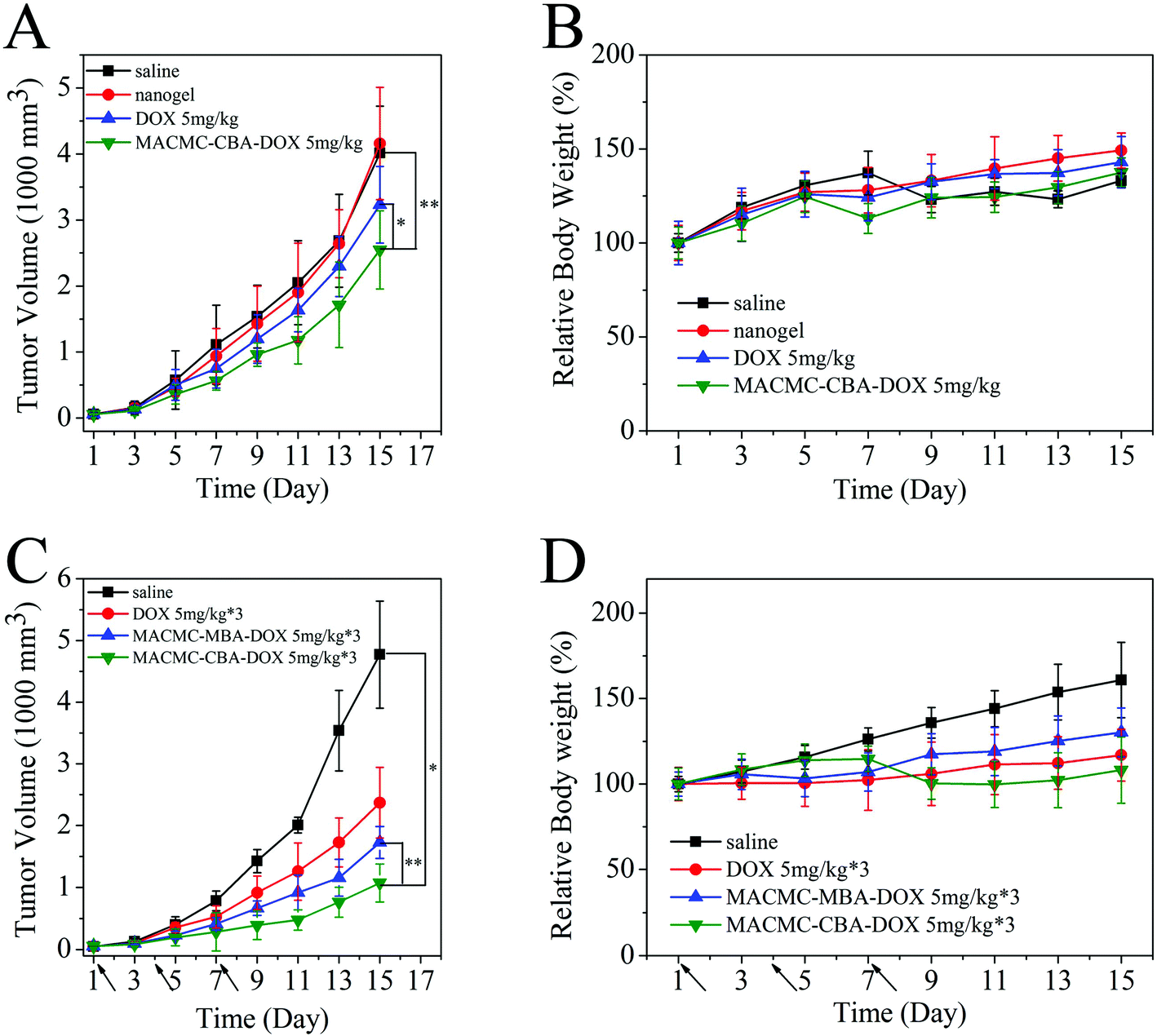 | ||
| Fig. 7 In vivo tumor growth curves of H22 tumor-bearing mice that received different treatments. (A) Single injection; (C) multiple dose treatment. The same DOX dose (5 mg kg−1) was injected for free DOX and DOX-loaded nanogels. Multiple dose treatment was administered on days 1, 4, and 7, marked by arrows. * and ** represent P < 0.05. (B) and (D) Evolution of body weight of each group during the experiments, expressed as the average relative body weights of mice compared to that on day 1. | ||
The body weights of each tested groups were also counted and the varied curves are displayed in Fig. 7B and 7D. The multiple dose group shows a slight decrease of body weight on the 9th day post-injection but recovered soon after this period. No serious decrease in the body weight is observed during the monitoring period, indicating the well-tolerated dose level of the drug and negligible toxicity of nanogels.
Histochemical observation
DOX is a chemotherapy drug which is known to interact with DNA and induce apoptosis in the treated tumor cells. Thus, to verify the therapeutic effects of DOX-loaded MACMC–CBA nanogels, the extent of apoptosis induced in tumor tissue was determined using TUNEL staining on day 8 after injection (for the multiple dose group, the next day of the last injection).43 The apoptotic cells in brown color are microscopically visualized in Fig. 8A and the degree of apoptosis is quantified by counting the percent of positive cells in 10 random fields. The data indicate that both free DOX and equivalent doses of DOX formulated in nanogels cause an apoptosis in tumor tissue. Tumor cells treated with multiple dose injection of DOX-loaded nanogels exhibit the highest degree of cell apoptosis (ca. 62.4%) compared to the groups of free DOX (34.1%) or a single dose (40.5%), indicating that multiple dose injection induces the highest degree of apoptosis of tumor cells (Fig. 8C). PCNA staining was also conducted to determine cell proliferation.44 The percentage of PCNA positive (proliferating) cells is significantly lower in the group which received a multiple dose trial compared with tumors of the other groups (Fig. 8B and 8C). In contrast, the percentages of proliferating cells in saline-treated and blank nanogel-treated tumors were ∼70%, indicating that the majority of the cells are proliferating. The decreased cell proliferation and increased apoptosis in the tumor cells further substantiated the superior antitumor efficacy of DOX-loaded nanogels compared with the other formulations. It could also be confirmed that multiple dose treatment using nanogels result in a continuous and efficient delivery of DOX to the tumor region and pharmacological efficacy. | ||
| Fig. 8 (A) and (B) Immunohistochemical staining for H22 tumor from mice that received different treatments indicated for (A) TUNEL and (B) PCNA. (C) Quantification of TUNEL staining- and PCNA staining-positive cells in H22 tumors from mice in various groups. The error bars were based on 10 random microscope fields for each group. | ||
Conclusions
In this work, a methacrylation strategy to functionalize CMC and prepare redox-responsive nanogels is demonstrated. The nanogels are stable against high concentration of NaCl and can be de-integrated in a bio-reducing environment containing 10 mM glutathione. When used as drug carriers, these nanogels can highly efficiently carry doxorubicin (DOX) with 36% loading content, and be internalized by the cancer cells through endocytosis for intracellular delivery. By using non-invasive live body fluorescence imaging technology, it was found that the nanogels could passively target to the tumor area by the EPR effect. The results of in vivo biodistribution demonstrate that DOX loaded nanogels have a significantly prolonged circulation time and enhanced drug accumulation in the tumor site. Compared with free DOX, the DOX-loaded nanogels exhibit superior antitumor efficacy on hepatic H22 tumor-bearing mice by inducing apoptosis of tumor cells. We believe that these cytocompatible and biodegradable nanogels have tremendous potential in drug delivery for cancer therapy.Acknowledgements
This study was supported by the National Natural Science Foundation of China (no. 51033002 and 51273090).Notes and references
- R. A. Petros and J. M. DeSimone, Nat. Rev. Drug Discovery, 2010, 9, 615 CrossRef CAS PubMed.
- R. S. Dothager and D. Piwnica-Worms, Cancer Res., 2011, 71, 5611 CrossRef CAS PubMed.
- A. T. Reza and S. B. Nicoll, Acta Biomater., 2010, 6, 179 CrossRef CAS PubMed.
- H. Takahashi, Y. Tahara, S. Sawada and K. Akiyoshi, Biomater. Sci., 2013, 1, 842–849 RSC.
- N. Morimoto, S. Hirano, H. Takahashi, S. Loethen, D. H. Thompson and K. Akiyoshi, Biomacromolecules, 2013, 14, 56 CrossRef CAS PubMed.
- H. Sun, B. Guo, X. Li, R. Cheng, F. Meng, H. Liu and Z. Zhong, Biomacromolecules, 2010, 11, 848 CrossRef CAS PubMed.
- I. Otsuka, K. Fuchise, S. Halila, S. Fort, K. Aissou, I. Pignot-Paintrand, Y. Chen, A. Narumi, T. Kakuchi and R. Borsali, Langmuir, 2010, 26, 2325 CrossRef CAS PubMed.
- R. Novoa-Carballal, D. V. Pergushov and H. E. Müller, Soft Matter, 2013, 9, 4297 RSC.
- G. Saravanakumar, K. H. Min, D. S. Min, A. Y. Kim, C.-M. Lee, Y. W. Cho, S. C. Lee, K. Kim, S. Y. Jeong, K. Park, J. H. Park and I. C. Kwon, J. Controlled Release, 2009, 140, 210 CrossRef CAS PubMed.
- Y. Guo, X. Wang, Z. Shen, X. Shu and R. Sun, Carbohydr. Polym., 2013, 92, 77 CrossRef CAS PubMed.
- F. Reyes-Ortega, F. J. Parra-Ruiz, S. E. Averick, G. Rodríguez, M. R. Aguilar, K. Matyjaszewski and J. S. Román, Polym. Chem., 2013, 4, 2800 RSC.
- S. Ganesh, A. K. Iyer, D. V. Morrissey and M. M. Amiji, Biomaterials, 2013, 34, 3489 CrossRef CAS PubMed.
- Y. Chen, D. Ding, Z. Mao, Y. He, Y. Hu, W. Wu and X. Jiang, Biomacromolecules, 2008, 9, 2609 CrossRef CAS PubMed.
- X.-H. Peng, Y. Wang, D. Huang, Y. Wang, H. J. Shin, Z. Chen, M. B. Spewak, H. Mao, X. Wang, Y. Wang, Z. Chen, S. Nie and D. M. Shin, ACS Nano, 2011, 5, 9480 CrossRef CAS PubMed.
- Y. Hu, X. Jiang, Y. Ding, H. Ge, Y. Yuan and C. Yang, Biomaterials, 2002, 23, 3193 CrossRef CAS.
- M. J. Ernsting, W.-L. Tang, N. MacCallum and S.-D. Li, Bioconjugate Chem., 2011, 22, 2474 CrossRef CAS PubMed.
- Y. H. Bae and H. Yin, J. Controlled Release, 2008, 131, 2 CrossRef CAS PubMed.
- Y.-L. Li, L. Zhu, Z. Liu, R. Cheng, F. Meng, J.-H. Cui, S.-J. Ji and Z. Zhong, Angew. Chem., Int. Ed., 2009, 48, 9914 CrossRef CAS PubMed.
- R. K. O'Reilly, C. J. Hawker and K. L. Wooley, Chem. Soc. Rev., 2006, 35, 1068 RSC.
- A. Sannino, M. Madaghiele, F. Conversano, G. Mele, A. Maffezzoli, P. A. Netti, L. Ambrosio and L. Nicolais, Biomacromolecules, 2004, 5, 92 CrossRef CAS PubMed.
- T. Ito, Y. Yeo, C. B. Highley, E. Bellas, C. A. Benitez and D. S. Kohane, Biomaterials, 2007, 28, 975 CrossRef CAS PubMed.
- A. Sannino, C. Demitri and M. Madaghiele, Materials, 2009, 2, 353 CrossRef CAS.
- Y. Yu and Y. Chau, Biomacromolecules, 2012, 13, 937 CrossRef CAS PubMed.
- K. H. Bae, H. Mok and T. G. Park, Biomaterials, 2008, 29, 3376 CrossRef CAS PubMed.
- Z. Yue, F. Wen, S. Gao, M. Y. Ang, P. K. Pallathadka, L. Liu and H. Yu, Biomaterials, 2010, 31, 8141 CrossRef CAS PubMed.
- F. Quero, M. Nogi, K.-Y. Lee, G. V. Poel, A. Bismarck, A. Mantalaris, H. Yano and S. J. Eichhorn, ACS Appl. Mater. Interfaces, 2011, 3, 490 CAS.
- N. Ballatori, S. M. Krance, S. Notenboom, S. Shi, K. Tieu and C. L. Hammond, Biol. Chem., 2009, 390, 191 CrossRef CAS PubMed.
- F. Q. Schafer and G. R. Buettner, Free Radical Biol. Med., 2001, 30, 1191 CrossRef CAS.
- P. Kuppusamy, H. Li, G. Ilangovan, A. J. Cardounel, J. L. Zweier, K. Yamada, M. C. Krishna and J. B. Mitchell, Cancer Res., 2002, 62, 307 CAS.
- Z. Ge and S. Liu, Chem. Soc. Rev., 2013, 42, 7289–7325 RSC.
- Y. Sun, X. Yan, T. Yuan, J. Liang, Y. Fan, Z. Gu and X. Zhang, Biomaterials, 2010, 31, 7124 CrossRef CAS PubMed.
- J. A. Burdick, C. Chung, X. Jia, M. A. Randolph and R. Langer, Biomacromolecules, 2005, 6, 386 CrossRef CAS PubMed.
- C. Zhang, W. Wang, T. Liu, Y. Wu, H. Guo, P. Wang, Q. Tian, Y. Wang and Z. Yuan, Biomaterials, 2012, 33, 2187 CrossRef CAS PubMed.
- A. Nishiguchi, H. Yoshida, M. Matsusaki and M. Akashi, Chem. Lett., 2010, 1184 CrossRef CAS.
- N. Sanson and J. Rieger, Polym. Chem., 2010, 1, 965 RSC.
- A. V. Kabanov and S. V. Vinogradov, Angew. Chem., Int. Ed., 2009, 48, 5418 CrossRef CAS PubMed.
- R. T. Chacko, J. Ventura, J. Zhuang and S. Thayumanavan, Adv. Drug Delivery Rev., 2012, 64, 836 CrossRef CAS PubMed.
- V. P. Torchilin, Adv. Drug Delivery Rev., 2005, 57, 95 CrossRef CAS PubMed.
- H. Takemoto, A. Ishii, K. Miyata, M. Nakanishi, M. Oba, T. Ishii, Y. Yamasaki, N. Nishiyama and K. Kataoka, Biomaterials, 2010, 31, 8097 CrossRef CAS PubMed.
- W. Gao, R. Langer and O. C. Farokhzad, Angew. Chem., Int. Ed., 2010, 49, 6567 CrossRef CAS PubMed.
- P. Chytil, T. Etrych, Č. Koňák, M. Šírová, T. Mrkvan, J. Bouček, B. Říhová and K. Ulbrich, J. Controlled Release, 2008, 127, 121 CrossRef CAS PubMed.
- F. A. Fornari, J. K. Randolph, J. C. Yalowich, M. K. Ritke and D. A. Gewirtz, Mol. Pharmacol., 1994, 5, 649 Search PubMed.
- Z. Liu, K. Chen, C. Davis, S. Sherlock, Q. Cao, X. Chen and H. Dai, Cancer Res., 2008, 68, 6652 CrossRef CAS PubMed.
- H. Okino, R. Maeyama, T. Manabe, T. Matsuda and M. Tanaka, Clin. Cancer Res., 2003, 9, 5786 CAS.
Footnote |
| † Electronic supplementary information (ESI) available: Changes in diameter after being treated with GSH and biodistribution of DOX in the tumor region of mice of non-bio-reductive sensitive MACMC–MBA nanogels. See DOI: 10.1039/c3bm60176e |
| This journal is © The Royal Society of Chemistry 2014 |