Other origins for the fluorescence modulation of single dye molecules in open-circuit and short-circuit devices†
Jefri S.
Teguh
a,
Michael
Kurniawan
b,
Xiangyang
Wu
a,
Tze Chien
Sum
b and
Edwin K. L.
Yeow
*a
aDivision of Chemistry and Biological Chemistry, School of Physical and Mathematical Sciences, Nanyang Technological University, 21 Nanyang Link, Singapore 637371, Singapore. E-mail: edwinyeow@ntu.edu.sg; Tel: +65 6316 8759
bDivision of Physics and Applied Physics, School of Physical and Mathematical Sciences, Nanyang Technological University, 21 Nanyang Link, Singapore 637371, Singapore
First published on 8th November 2012
Abstract
Fluorescence intensity modulation of single Atto647N dye molecules in a short-circuit device and a defective device, caused by damaging an open-circuit device, is due to a variation in the excitation light focus as a result of the formation of an alternating electric current.
Photoexcited single dye molecules,1,2 quantum dots3 and conjugated polymers4,5 display fluorescence intensity modulation when subjected to an external electric field (EF). For dye molecules, such as squaraine-derived rotaxanes, the emission intensity of single chromophores varies with the amplitude of the modulating EF.1,2 It has been proposed that this arises from an EF-induced change in the electron transfer efficiency between molecules and traps found in the surrounding nano-environment (e.g., polymer film).1,2 Furthermore, single dye molecules in a spectroelectrochemical cell coupled to a microscope undergo reversible redox reaction to form weakly emitting species, such that the observed fluorescence modulates with the cyclic voltammetric potential scan.6 Apart from the influence of EF, electric current (EC) has also been shown to cause fluorescence fluctuation for single dye molecules (e.g., 1,1′-dioctadecyl-3,3,3′,3′-tetramethylindodicarbocyanine) on indium tin oxide (ITO) in a short-circuit device.7 A plausible reason for this behavior is the adjustment of the Fermi level of ITO by the applied EC which leads to a change in the interfacial electron transfer dynamics between the two moieties.7
The majority of single-molecule fluorescence detection experiments are conducted on a scanning confocal fluorescence microscope where the excitation light is focused by an objective lens to a tight spot on a molecule located at the focal plane, and the fluorescence is subsequently collected using the same objective lens. Therefore, an out-of-focus illuminating light will result in a decrease in the recorded emission intensity. In this communication, the effects of an applied EF and EC on the focus of an excitation light in an open-circuit device and a short-circuit device, respectively, are investigated. Before understanding the roles played by intrinsic photophysical processes (e.g., charge transfer) in single-molecule fluorescence modulation, it is necessary to first elucidate any possible contributions from EF- or EC-induced out-of-focus excitation light to the emission intensity change.
The dye used here is Atto647N; a photo-stable chromophore commonly employed in single-molecule detection studies (Fig. S1, ESI†).8,9 Two types of devices were constructed as shown in Fig. 1. The short-circuit device was prepared by painting a thin U-shaped silver (Ag) electrode on top of a glass cover slip, and single dye molecules were subsequently deposited at the centre of the cover slip (Fig. 1a). The open-circuit device, shown in Fig. 1b, was prepared by spin coating in sequence a polystyrene (PS) film on an ITO-coated glass cover slip, a thin polyvinyl alcohol (PVA) film containing single Atto647N molecules, and a poly(methyl methacrylate) (PMMA) film. An aluminium (Al) electrode was then deposited on top of the PMMA film by thermal evaporation. The device structure is similar to the ones described in the literature5 where the PS layer is used as an insulator. The two parallel arms of the electrode in the short-circuit device and the ITO and Al electrodes in the open-circuit device were connected to a power generator that provided a sinusoidal voltage at 0.1 Hz (Fig. 2a, 4a and 6a). Experimental details are provided in the ESI.†
 | ||
| Fig. 1 (a) A short-circuit device fabricated by painting a U-shaped silver paste (grey) on top of a glass cover slip and single Atto647N molecules are deposited at the center of the device, and (b) an open-circuit device where the thicknesses of the polystyrene PS, polyvinyl alcohol PVA, poly(methyl methacrylate) PMMA and Al layers are 450, 30, 55 and 100 nm, respectively. Single Atto647N molecules are embedded in the PVA matrix (˙ = Atto647N molecule). The width and thickness of the Ag electrode are 2 mm and ∼30 μm, respectively. | ||
 | ||
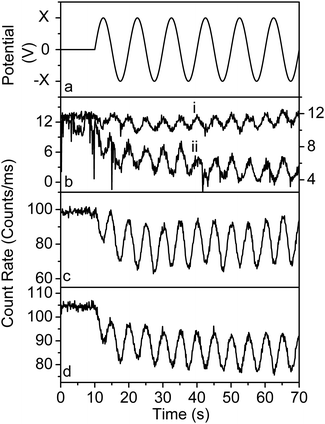
| Fig. 2 (a) Sinusoidal external potential (frequency of 0.1 Hz) applied to a short-circuit device. (b) Fluorescence intensity time trace of a single molecule in a short-circuit device subjected to a potential ranging from (i) +2 V to −2 V (left-axis) and (ii) +3 V to −3 V (right-axis). (c) Fluorescence intensity time trace of a bulk concentration of Atto647N and (d) BEL intensity of a short-circuit device subjected to a potential ranging from +3 V to −3 V. The external potential was applied 10 s after the start of the measurements and an oil objective lens was used. | ||
Typical fluorescence intensity time traces of single Atto647N molecules in a short-circuit device (Fig. 1a) are provided in Fig. 2b for an external sinusoidal potential (at frequency of 0.1 Hz) ranging from (i) +2 V to −2 V (maximum EC of 0.06 A generated), and (ii) +3 V to −3 V (maximum EC of 0.09 A). We note that the fluorescence intensity of the single molecules decreases when a voltage is initially applied at 10 s (Fig. 2b). The emission intensity time trace subsequently displays a sinusoidal oscillation at a frequency of 0.2 Hz. Minimum fluorescence (i.e., trough of the wave) is observed when either a +2 V/+3 V or −2 V/−3 V voltage is applied; corresponding to an EC flow of 0.06/0.09 A in the electrode. The fluorescence intensity increases when the EC decreases and reaches a maximum (i.e., crest of the wave) at zero potential (i.e., zero EC). Similar oscillatory behaviour in the fluorescence intensity time trace of an ensemble of Atto647N molecules in a short-circuit device is also observed (Fig. 2c). Since the dye molecules are not in contact with the Ag electrode, their photophysical properties such as fluorescence quantum yield are not influenced by the alternating EC. It is worth mentioning that the fluorescence modulation behaviour observed here has also been reported for dye molecules subjected to an external electric current.7
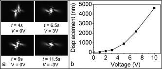
The effects of EC on the focus of the excitation light were investigated by recording the backscattered excitation light (BEL) intensity and the corresponding beam spot image from a short-circuit device. For potential ranging from +3 V to −3 V, the intensity of the BEL undergoes a sinusoidal modulation at a frequency of 0.2 Hz (Fig. 2d). Minimum (trough) and maximum (crest) intensities are observed at +3 V/−3 V and 0 V, respectively; similar to the single-molecule fluorescence modulation seen in Fig. 2b. In addition, the beam spot image of the BEL changes its shape pattern according to the amount of applied potential (see Fig. 3a and Movie S1, ESI†). In particular, the beam spot pattern is most diffused at voltages +3 V (t = 6.5 s in Fig. 3a) and −3 V (t = 11.5 s), and regains sharpness whenever the voltage approaches zero. This clearly demonstrates that the excitation light becomes increasingly out-of-focus with respect to the focal plane (i.e., in the z-direction) when the intensity of the EC increases, and regains focus when the EC is reduced to zero. Therefore, the fluorescence modulation observed in Fig. 2b is ascribed to changes in the focus of the excitation beam. The modulation in the BEL was observed when using both oil and air objective lenses.
 | ||
| Fig. 3 (a) Beam spot images from the BEL from a short-circuit device subjected to a sinusoidal potential ranging from V = +3 V to −3 V (Movie S1, ESI†). The time when the potential is first applied after the start of the experiment is t = 4 s. (b) Plot of initial displacement of device from the focused position, measured using AFM, vs. maximum applied voltage. | ||
We next discuss the possible reasons for the out-of-focus excitation light when an EC flows in a short-circuit device. From ellipsometry measurements, the refractive index of the glass substrate (1.537) does not change before and after the application of an external potential. This means that an EC in the Ag electrode does not influence the refractive index of the glass substrate. Another plausible explanation for the out-of-focus excitation beam is the displacement of the device away from the objective lens when a current is flowing in the electrode. We propose that when an EC is generated, a mechanical jerk in the electrode lifts the whole device upwards and away from the objective lens. The device subsequently relaxes back to a position closer to the focused position when the EC reduces to zero. At high potential bias, there is a significant drop in the BEL intensity when the voltage is initially applied (e.g., +3 V in Fig. 2d and +10 V in Fig. 4b), and the intensity does not return to its original value recorded before the application of the potential; suggesting the presence of a relatively big initial displacement from the focused position by the large Ag electrode. A control experiment, conducted using a short-circuit device fabricated from a significantly heavier glass substrate (i.e., base of a petri dish), did not show any drastic drop in the BEL intensity (Fig. 4b) when a potential is applied; indicating that the mechanical jerk was not strong enough to cause a large displacement of the heavy glass substrate. In addition, a device fabricated using a quartz cover slip with a significantly lower thermal expansion coefficient (α = 0.59 × 10⁻⁶ °C−1 at 20 °C) showed a smaller degree of initial drop in the BEL intensity and hence displacement when compared to a normal glass cover slip (α = 9 × 10⁻⁶ °C−1 at 20 °C) (Fig. 4b). A heat-induced mechanical jerk in the electrode due to current flow and thermal expansion of the upper surface of the glass substrate in contact with the electrode may collectively aid in displacing the whole device.
 | ||
| Fig. 4 (a) Sinusoidal external potential (frequency of 0.1 Hz) applied to a short-circuit device, as depicted in Fig. 1a. (b) BEL intensity time traces of a short-circuit device fabricated from (i) glass cover slip, (ii) quartz cover slip and (iii) glass Petri dish and subjected to a potential ranging from +10 V to −10 V. The BEL intensity time traces were measured using an oil objective lens. | ||
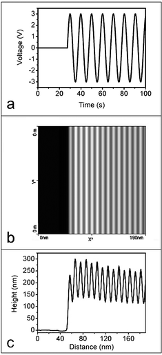
From AFM measurements, we note that the initial displacement of the device increases as the maximum EC generated increases (Fig. 3b). For an example, we observed that for a short-circuit device, an initial upward displacement of 235 nm from the original focused position (before application of an external potential) occurs when a sinusoidal potential ranging from +3 V to −3 V is applied (Fig. 5). The device does not return to its original position at zero current, and the displacement between the positions of the device at maximum EC (at +3 V/−3 V) and zero current (at 0 V) during the application of a modulating potential is ∼150 nm (Fig. 5c); which is in agreement with the results of Fig. 2.
 | ||
| Fig. 5 (a) Sinusoidal external potential ranging from +3 V to −3 V (at 0.1 Hz) is applied to a short-circuit device at ∼27 s after the start of the AFM scan. (b) AFM image of a single scan (100 s) across 190 nm of the glass substrate and (c) corresponding height profile which correlates with the applied potential. During the application of a sinusoidal potential bias, the grey strips in (b) correspond to a lower height and occur at zero current (at 0 V) whereas the white strips correspond to a higher height and occur at maximum EC (at +3 V/−3 V). | ||
A typical fluorescence intensity time trace of a single Atto647N molecule in an open-circuit device (Fig. 1b) that is subjected to a sinusoidal potential (at 0.1 Hz) ranging from +10 V to −10 V is presented in Fig. 6b. The molecule experiences a maximum EF of 1.9 × 10⁵ V cm−1 when the voltage is adjusted to +10 V/−10 V. The emission intensity of the molecule does not modulate with the EF and is similar to those previously reported for single Atto647N molecules immobilized on a glass substrate8,9 and PVA matrix.10 Furthermore, the BEL intensity does not show any significant variation with the applied potential (Fig. 6e) and is not EF-dependent. Varying the EF does not cause a sinusoidal-type modulation of either the excitation light focus or fluorescence intensity of Atto647N.
 | ||
| Fig. 6 (a) Sinusoidal external potential (frequency of 0.1 Hz) applied to an open-circuit device (b and e) and a defective device (c, d, f and g). (b) Fluorescence and (e) BEL intensity time traces of a single molecule in an intact open-circuit device subjected to a potential ranging from +10 V to −10 V. Fluorescence and BEL intensity time traces of a single molecule in a defective device subjected to a potential ranging from +5 V to −5 V (c and f, respectively) and +10 V to −10 V (d and g, respectively). The external potential was applied 10 s after the start of the measurements and an oil objective lens was used. | ||
The multi-layer open-circuit device in Fig. 1b is easily converted to a short-circuit device (i.e., defective device) when the polymer layers between the ITO and Al electrodes are lightly scratched to allow contact between the two electrodes. Single-molecule fluorescence modulation is again observed as illustrated in Fig. 6c for an Atto647N molecule in a defective device that is subjected to a sinusoidal potential ranging from +5 V to −5 V. In this case, an alternating EC from 0 to 0.14 A flows across the device at areas where the two opposite electrodes are in contact. Both the fluorescence intensity time trace (Fig. 6c) and BEL intensity (Fig. 6f) display a sinusoidal modulation with minimum (trough) and maximum (crest) intensities occurring at the highest and zero EC, respectively. Out-of-focus excitation light is thus responsible for the EC-dependent modulation of the single molecule’s fluorescence in a defective device. Furthermore, the fluorescence and BEL intensities are restored at zero potential (or zero EC) to values that are close to those measured before the application of the potential bias (i.e., when the device is in a focused position). This means that the relatively small areas of contact between the ITO and Al electrodes are not sufficiently large to cause a significant initial displacement of the defective device.
The fluorescence and BEL intensity time traces of a defective device with an applied voltage ranging between +10 V and −10 V exhibit complex behaviour (Fig. 6d and g). For the first 30 s of the measurement, the fluorescence intensity of the single Atto647N molecule in Fig. 6d shows consistent behaviour (i.e., crest of intensity trace at zero potential and trough at +10 V/−10 V potential). However, a dip in the emission intensity at zero EC is observed at 35 s. The intensity at zero EC gradually decreases over the next few cycles (i.e., at 40 s, 45 s, 50 s, etc.) whereas the intensity at +10 V and −10 V increases, such that after 50 s, the trough and crest of the modulating fluorescence intensity time trace correspond to zero and +10 V/−10 V, respectively. This means that an enhancement in the fluorescence intensity is now observed when the maximum EC (0.3 A) is flowing in the electrode relative to the fluorescence at zero current. Similar behaviour was observed in the BEL intensity (Fig. 6g) where the intensity undergoes a transition from being minimum to maximum at zero potential after 30 s. In this case, the excitation beam becomes out-of-focus at zero current and restores its focus at maximum EC. Several mechanisms such as opposite mechanical movements at zero and maximum currents and types of electrode may be responsible for this behaviour. We are currently conducting further experiments to elucidate the exact nature of the phenomenon observed here.
Conclusions
In this communication, we have unambiguously demonstrated that the drop in the detected fluorescence intensity of single dye molecules in either a short-circuit device (Fig. 1a) or a defective device, caused by damaging an open-circuit device (Fig. 1b), is due to an out-of-focus excitation beam arising from a displacement of the device away from its focused position by the generated EC in the electrode. For an open-circuit device such as the one depicted in Fig. 1b, it is often very easy to damage the thin polymer/active layers so that the two opposite electrodes come into contact with each other. For an example, we have found that an open-circuit device can readily lose its integrity during fabrication and when connecting wires from the generator to the electrodes; hence causing current leaks and out-of-focus excitation to occur. When studying the EF-induced fluorescence modulation of single fluorescent systems, it is imperative to ensure that the open-circuit device remains intact during fabrication, and before and after application of a potential bias. This is necessary to avoid any possible misinterpretation of results in the event that the device is found to be defective. Furthermore, experiments aimed at examining the effects of EC on the fluorescence of single molecules should also be carefully designed to ensure the absence of an out-of-focus beam.Acknowledgements
We gratefully acknowledge the financial support from the Singapore Ministry of Education AcRF Tier 1 grants (RG45/07 and RG49/08), SPMS Collaborative Research Award (M58110090) and NTU start-up grant (M58110068). T. C. S. acknowledges the funding support from the Singapore National Research Foundation (NRF) through the Competitive Research Program (NRF-CRP5-2009-04).References
- F. Zhang, G. Zhang, R. Chen, X. Wang, L. Siao and S. Jia, Phys. Scr., 2012, 82, 055303 CrossRef.
- R. Chen, G. Zhang, Y. Gao, L. Siao and S. Jia, Appl. Phys. Lett., 2012, 100, 203118 CrossRef.
- S.-J. Park, S. Link, W. L. Miller, A. Gesquiere and P. F. Barbara, Chem. Phys., 2007, 341, 169–174 CrossRef CAS.
- P. R. Hania and I. G. Scheblykin, Chem. Phys. Lett., 2005, 414, 127–131 CrossRef CAS.
- P. R. Hania, D. Thomsson and I. G. Scheblykin, J. Phys. Chem. B, 2006, 110, 25895–25900 CrossRef CAS.
- C. Lei, D. Hu and E. J. Ackerman, Chem. Commun., 2008, 5490–5492 RSC.
- G. Zhang, L. Xiao, R. Chen, Y. Gao, X. Wang and S. Jia, Phys. Chem. Chem. Phys., 2011, 13, 13815–13820 RSC.
- X. Wu, G. Xing, S. L. Tan, R. D. Webster, T. C. Sun and E. K. L. Yeow, Phys. Chem. Chem. Phys., 2012, 14, 9511–9519 RSC.
- X. Wu, T. D. M. Bell and E. K. L. Yeow, Angew. Chem., Int. Ed., 2009, 48, 7379–7382 CrossRef CAS.
- J. N. Clifford, T. D. M. Bell, P. Tinnefeld, M. Heilemann, S. M. Melnikov, J. Hotta, M. Sliwa, P. Dedecker, M. Sauer, J. Hofkens and E. K. L. Yeow, J. Phys. Chem. B, 2007, 111, 6987–6991 CrossRef CAS.
Footnote |
| † Electronic supplementary information (ESI) available. See DOI: 10.1039/c2cp43284f |
| This journal is © the Owner Societies 2013 |
