DOI:
10.1039/C2RA20399E
(Paper)
RSC Adv., 2012,
2, 7875-7885
Received
5th March 2012
, Accepted 17th June 2012
First published on 20th June 2012
Abstract
The present study describes the controlled synthesis of lanthanum hexaboride nanostructures with efficient field emission properties. The synthesis is mediated by a nanostructured lanthanum hydroxide precursor, which is controlled by varying the capping agent and pH using a hydrothermal route. The effect of charge on the capping agent (surfactant) strongly affects the shape and size of the precursor (neutral surfactants lead to the formation of nanorods while a cationic surfactant results in the formation of particles). This precursor mediated route leads to lanthanum hexaboride nanostructures at much lower temperatures (∼500 °C lower than the conventional solid state route) and allows for variation of morphology of nanostructured films. Vertically aligned nanorods (30 nm × 200–400 nm), nanoparticles (25 nm) and sub-micron particles (0.2–0.25 microns) could be precisely obtained. Field emission studies of these vertically aligned nanorods show a very high field enhancement factor (4191), which is required for an efficient field emitter.
1. Introduction
Lanthanum hexaboride is widely used for high electron density cathodes because of its high melting point of 2500 °C,1 high electrical conductivity,2 low work function (∼2.6 eV)3 and low vapour pressure at high temperatures.4 Recent reviews on the field emission properties of various metal oxides, nitrides and borides5–7 suggest that the metal hexaborides show superior performance with a high field enhancement factor, as well as good current stability. Among the known hexaboride based field emitters, lanthanum hexaboride shows the most promising performance compared to other metal hexaborides and hence it has been used in most of the applications as an electron source. Lanthanum hexaboride has been synthesized via the solid state route,8–13 hydrothermal route,14,15 gas phase reaction (CVD and PVD) and magnetic sputtering.16–23 The synthesis of lanthanum hexaboride via the solid state route tends to give micron-sized particles due to the formation of LaB6 at high temperature (∼1800 °C),1,2,6 while the gas phase reactions like CVD are quite expensive and are not used for commercial purposes. In addition, the fabrication of LaB6 films has earlier been reported via chemical vapor deposition and magnetic sputtering,16–23 which require expensive equipment and chemicals. These techniques do not enable fine control of the shape and size of micron-sized particles of lanthanum hexaboride. Chemical processes like spin coating are cost effective and simple, and can be used routinely for deposition of nanostructured films of oxide, selenide and sulphides.24–26 However, there is no report for fabrication of metal boride films via low temperature chemical routes. Therefore, the motivation of the present study is to obtain vertically aligned nanorods of lanthanum hexaboride, which would be efficient field emitters, since anisotropic nanostructures with sharp tips enhance the field emission properties. We have optimized the synthetic process mediated by nanostructured lanthanum hydroxide precursors (particles and nanorods with various aspect ratios), which efficiently react with boron at much lower temperatures (∼500 °C or less) than earlier reports to yield nanostructured lanthanum hexaboride. Furthermore, films of lanthanum hexaboride were fabricated by the spin coating route using optimal conditions. We have investigated the field emission of the LaB6 nanostructures where the vertically aligned nanorods offer excellent properties with a high field enhancement factor of 4191 compared to the maximum reported field enhancement factor of 3585.18,19
2. Results and discussion
The present study is based on investigation of the critical parameters involved in controlling the shape and size of lanthanum hydroxide using the hydrothermal method and further heating this precursor with boron to yield lanthanum hexaboride. In the first stage, we optimize conditions for control of the shape and size of lanthanum hydroxide precursors via the hydrothermal route using various surfactants (cationic (CTAB), anionic (AOT) and non-ionic (Tergitol)) and also the pH of the solution. In the second stage, the as-obtained lanthanum hydroxide precursors (with various sizes and morphologies) were reacted with boron to synthesize pure LaB6 at a low temperature of 1300 °C (normally lanthanum hexaboride is synthesized at 1800 °C). In the third stage lanthanum hexaboride films on silicon substrate are fabricated by the spin coating route. We then investigated the field emission properties of these nanostructured lanthanum hexaboride films.
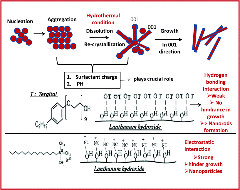
The PXRD studies of the lanthanum hydroxide precursor, obtained via the hydrothermal route using Tergitol as a non-ionic surfactant and sodium hydroxide at varying concentrations (0.1 M, 0.2 M and 0.3 M), show that the products are poorly crystalline and all the reflections could be indexed on the basis of a hexagonal cell of lanthanum hydroxide with space group P63/m (Fig. 1a–c, JCPDS No. 060585). A similar hydrothermal reaction in the presence of a cationic surfactant (CTAB) and sodium hydroxide (0.1 M) also resulted in the formation of lanthanum hydroxide (Fig. 1d). Transmission electron microscopy studies of these precursors show a drastic change in morphology. TEM studies of lanthanum hydroxide precursors obtained using CTAB–NaOH (0.1 M, pH 8), Tergitol–NaOH (0.1 M, pH 8), Tergitol–NaOH (0.2 M, pH 10) and Tergitol–NaOH (0.3 M, pH 13) show the formation of particles with an average diameter of 0.4 μm (Fig. 2a), nanorods with d = 8–10 nm, l = 120 nm and AR = 12 (Fig. 2b), nanorods with d = 8–10 nm, l = 240 nm and AR = 24 (Fig. 2c) and nanorods with d = 8–10 nm, l = 300–500 nm, AR = 30–50 (Fig. 2d), respectively. It may be noted that the above mentioned pH is measured before initiating the hydrothermal reaction. After completion of the reaction (2 days at 120 °C), the observed pH was found to be lower (pH = 7), which is due to the consumption of hydroxyl ions to form lanthanum hydroxide. The change in morphology of lanthanum hydroxide (from nanorods to nanoparticles) can be attributed to two factors, the surfactant charge and the initial pH of the reaction. Tergitol is a non-ionic surfactant, containing a surface hydroxyl group, which is adsorbed to the surface of lanthanum hydroxide via hydrogen bonding and facilitates anisotropic growth in the favored crystallographic directions. Earlier reports on lanthanum hydroxide nanorods via the hydrothermal route in the absence of a surfactant also show27 the formation of anisotropic nanostructures of La(OH)3, which is the thermodynamically favored crystal morphology (it has a hexagonal crystal system). On the contrary, in the presence of the cationic surfactant (CTAB), spheres of 0.4 μm in diameter are formed. Investigation of the time-dependent hydrothermal reaction shows27 that the growth of the lanthanum hydroxide nanorods at 150 °C involves four different steps: nucleation, aggregation, dissolution–recrystallization and the growth process. Lanthanum hydroxide nanoparticles form during the nucleation–dissolution process, while during the growth–recrystallization process, the anisotropic nanostructures crystallize. During the recrystallization and growth process, the stabilization of the isotropic morphology may be explained due to the strong binding of CTAB, being a cationic surfactant, to the negatively charged La(OH)3 surface by electrostatic interaction, which is much stronger than the hydrogen bonding interaction. This uniformly capped moiety does not favour the orientational growth and hence leads to the formation of spherical particles instead of nanorods (Scheme 1). Earlier reports on lanthanum hydroxide27 describe much thicker nanorods (20 nm × 200 nm) with a small aspect ratio AR = 10–12. The thickness of the nanorods shown in our method is 8–10 nm and it also provides excellent control over the aspect ratio (AR = 12, 24 and 30–50). Varying the initial pH by increasing the concentration of hydroxyl ions in the reaction led to an increase in the aspect ratio of the nanorods from 12 to 30 (Fig. 2b–d). Thus, under hydrothermal conditions where the surfactant acts as a capping agent, cationic surfactants facilitate the growth of lanthanum hydroxide particles, while non-ionic surfactants help in the growth of anisotropic nanostructures of lanthanum hydroxide, and the aspect ratio can be controlled by a change in initial pH (Fig. 2e).
 |
| | Fig. 1 PXRD studies of the lanthanum hydroxide precursor. | |
 |
| | Fig. 2 Transmission electron microscopic study of the lanthanum hydroxide precursor obtained at 120 °C using (a) CTAB, (b) Tergitol/initial pH = 8, (c) Tergitol/initial pH = 10, (d) Tergitol/initial pH = 13 and (e) variation of the aspect ratio of the lanthanum hydroxide precursor with pH in the presence of the Tergitol surfactant. | |
The reaction of the lanthanum hydroxide precursor (obtained using Tergitol as a capping agent) with boron at 1300 °C leads to the formation of pure lanthanum hexaboride (cubic, Pm![[3 with combining macron]](https://www.rsc.org/images/entities/char_0033_0304.gif) m, a = 4.159 Å, (#JCPDS: 340427)) (Fig. 3a–c) (in some batches 5–10% LaBO3 impurity was observed and was removed by acid wash). Earlier reports on the synthesis of lanthanum hexaboride via the solid state route suggest that under ambient pressure, the formation of LaB6 is only possible at 1800 °C, in which La2O3 and boron have been used as starting materials.9 Other solid state routes via the carbothermal and the aluminium flux method (not the cleanest method) suggest that the stabilization of LaB6 is possible at 1700 °C and 1500 °C, respectively.10–12 In the borothermal process, as in the present study, high purity LaB6 can be obtained by the reduction of the nanostructured lanthanum hydroxide precursor at the much lower temperature of 1300 °C; the morphology and size of the lanthanum hexaboride nanostructures depends on the initial precursor yielding nanorods (30 nm × 200–400 nm) and other nanostructures. Transmission electron micrographs of LaB6 show the presence of lanthanum hexaboride nanorods (30 nm × 200–400 nm) (Fig. 4a), nanoparticles (25 nm) (Fig. 4b) and much larger particles of 0.2–0.25 μm (Fig. 4c). The nanostructured lanthanum hexaborides are single crystalline, as shown by the lattice fringes corresponding to the (110) planes (Fig. 5a–c) and sharp spots in the electron diffraction pattern (Fig. 6a–c). Fig. 6a shows the electron diffraction pattern with a row of well-defined spots running along the a*- axis. Also, there are weak diffraction spots with streaks running nearly normal to the nanorod length axis. These kinds of diffraction pattern in the electron diffraction studies have been commonly observed for nanorods of oxides.28 However, the electron diffraction patterns of LaB6 nanoparticles 25 nm and 250 nm show the presence of sharp spots forming rings due to their nanocrystalline nature. So, from the electron diffraction study, we can clearly decipher the presence of highly uniform nanorods (Fig. 4a), and their growth direction (Fig. 6a) and nanoparticles (Fig. 4b–c, 6b–c) of lanthanum hexaborides. Earlier, the formation of LaB6 nanowires of 100 nm in diameter and micron sized length has been reported by Zhang et al.17,19 and there is no report so far that suggests the formation of thin nanorods (30 nm) of LaB6 as obtained here.
m, a = 4.159 Å, (#JCPDS: 340427)) (Fig. 3a–c) (in some batches 5–10% LaBO3 impurity was observed and was removed by acid wash). Earlier reports on the synthesis of lanthanum hexaboride via the solid state route suggest that under ambient pressure, the formation of LaB6 is only possible at 1800 °C, in which La2O3 and boron have been used as starting materials.9 Other solid state routes via the carbothermal and the aluminium flux method (not the cleanest method) suggest that the stabilization of LaB6 is possible at 1700 °C and 1500 °C, respectively.10–12 In the borothermal process, as in the present study, high purity LaB6 can be obtained by the reduction of the nanostructured lanthanum hydroxide precursor at the much lower temperature of 1300 °C; the morphology and size of the lanthanum hexaboride nanostructures depends on the initial precursor yielding nanorods (30 nm × 200–400 nm) and other nanostructures. Transmission electron micrographs of LaB6 show the presence of lanthanum hexaboride nanorods (30 nm × 200–400 nm) (Fig. 4a), nanoparticles (25 nm) (Fig. 4b) and much larger particles of 0.2–0.25 μm (Fig. 4c). The nanostructured lanthanum hexaborides are single crystalline, as shown by the lattice fringes corresponding to the (110) planes (Fig. 5a–c) and sharp spots in the electron diffraction pattern (Fig. 6a–c). Fig. 6a shows the electron diffraction pattern with a row of well-defined spots running along the a*- axis. Also, there are weak diffraction spots with streaks running nearly normal to the nanorod length axis. These kinds of diffraction pattern in the electron diffraction studies have been commonly observed for nanorods of oxides.28 However, the electron diffraction patterns of LaB6 nanoparticles 25 nm and 250 nm show the presence of sharp spots forming rings due to their nanocrystalline nature. So, from the electron diffraction study, we can clearly decipher the presence of highly uniform nanorods (Fig. 4a), and their growth direction (Fig. 6a) and nanoparticles (Fig. 4b–c, 6b–c) of lanthanum hexaborides. Earlier, the formation of LaB6 nanowires of 100 nm in diameter and micron sized length has been reported by Zhang et al.17,19 and there is no report so far that suggests the formation of thin nanorods (30 nm) of LaB6 as obtained here.
 |
| | Fig. 3 PXRD studies of lanthanum hexaboride obtained using a lanthanum hydroxide precursor using (a) Tergitol/initial pH = 8, (b) Tergitol/initial pH = 10 and (c) Tergitol/initial pH = 13. | |
 |
| | Fig. 4 Transmission electron microscopic study of lanthanum hexaboride obtained at 1300 °C using a lanthanum hydroxide precursor synthesized using (a) Tergitol/initial pH = 8, (b) Tergitol/initial pH = 10 and (c) Tergitol/initial pH = 13. | |
 |
| | Fig. 5 HRTEM of lanthanum hexaboride obtained at 1300 °C using a lanthanum hydroxide precursor synthesized using (a) Tergitol/initial pH = 8, (b) Tergitol/initial pH = 10 and (c) Tergitol/initial pH = 13. | |
 |
| | Fig. 6 Electron diffraction studies of lanthanum hexaboride obtained at 1300 °C starting from a lanthanum hydroxide precursor synthesized using (a) Tergitol/initial pH = 8, (b) Tergitol/initial pH = 10 and (c) Tergitol/initial pH = 13. | |
Further, LaB6 films were fabricated by spin coating using a dispersion of LaB6 powder (obtained by the hydrothermal route). Glancing angle X-ray diffraction of LaB6 films was successfully indexed on the basis of a cubic cell with a = 4.141 Å (#JCPDS: 340427) (Fig. 7a–c). Atomic force microscopy study of the film fabricated using polycrystalline LaB6 nanorods (30 nm × 200–400 nm), LaB6 nanoparticles (∼25 nm) and LaB6 particles (200–250 nm) shows that the nanorods are vertically aligned (Fig. 8a) (the inset of Fig. 8a shows that the surface consists of well aligned nanorods) while the nanoparticles are homogenously distributed over the Si substrate. The vertical assembly of nanorods is of significance for applications and very limited methods are known to obtain such alignment without using a template. The solvent evaporation technique has been shown to control the orientational order of nanorods (∼96% in the case of CdS).24 We could optimize the spin coating methodology using a suspension of LaB6 nanorods to fabricate films made up of vertically oriented LaB6 nanorods. The various spin coating parameters such as spin rate and spinning time, as well as the suspension density, play a crucial role in obtaining such an orientation of the LaB6 nanorods.
The field emission characteristics of the LaB6 films obtained using the hydrothermal route are shown in Fig. 8. The current density (J) is given by the Fowler–Nordheim equation29
| |  |
(1)
|
where
E is the applied field strength (in V μm
−1), Φ is the work function of the emitters,
β is the field enhancement factor and can be expressed as
β =
h/
r,
30 (
h is the height and
r is the radius of curvature of an emitting center). For good field emitters, apart from the low work function, the shape and size of the materials are also important and it is known that the anisotropic
nanostructures are better field emitters
30–32 compared to other morphologies. The current density
vs. applied field behaviour of
nanorods and
nanoparticles has been shown in
Fig. 9 and
Table 1. LaB
6 films and bare Si show an increase in emission current density with increase in applied field as expected. The
nanorods (30 nm × 200–400 nm) show much higher (nearly 8 fold) current density of 51.66 μA cm
−2 compared to the 25 nm
nanoparticles (6.63 μA cm
−2). The films with larger particles (200–250 nm) show an even lower current density of 2.10 μA cm
−2. The comparison of the current density was carried out at a field strength of 12 V μm
−1. The LaB
6 film consisting of
nanorods shows low turn-on fields of 3.06 V μm
−1 (measured at 1 μA cm
−2), while the LaB
6 film with smaller
nanoparticles (25 nm) shows a turn-on field of 3.15 V μm
−1 compared to the larger particles of 200–250 nm (turn on field 8.28 V μm
−1). The
nanorod-based film offers high brightness and current density compared to the nanoparticulated film. The Fowler–Nordheim (F–N) plots (
Fig. 10 a–c) show linear behavior, which indicates that emission from the LaB
6 film follows the tunneling mechanism. The
nanorods were homogeneously distributed in the films as confirmed by transmission electron and atomic force micrographs earlier (
Fig. 4a–c and
Fig. 8a–c). As shown in
Fig. 10 a–c, the F–N plot is linear, from which we obtain the field enhancement factor (
β), which is equal to
bΦ
3/2/slope, where
b is a constant with a value of 6.83 × 10
3 V eV
−3/2 μm
−1 and Φ is the work function of the emitter (2.6 eV). The field enhancement factors obtained from the ln(
J/
E2)
vs. 1/
E plot in the high field region (1/
E = 0.08–0.20 μm V
−1) for the
nanorods (30 nm × 200–400 nm),
nanoparticles (25 nm) and submicron particles (200–250 nm) are 1289, 1240 and 1216 respectively (
Fig. 10 a–c). However, in the low field region (1/
E = 0.25–0.70 μm V
−1), the corresponding field enhancement factors are 4191 (
nanorods, 30 nm × 200–400 nm), 4009 (
nanoparticles, 25 nm) and 3652 (particles, 200–250 nm). Note that these values are much higher than the maximum reported field enhancement factor of 3585 (in low field range of 1/
E = 0.2–0.55 μm V
−1).
15,16 The mechanism of the two-slope field enhancement behaviour of LaB
6 is not yet clear. However, for
zinc oxide and metal borides,
33,34 the two-slope behavior has been reported earlier, which is due to space charge,
35 surface adsorbate
36 and damage of emission site
37 at high voltage. The high field enhancement factor obtained in our study is mainly attributed to three factors (a) small size of
nanorod, (b) uniform size with sharp tip and (c) most importantly the vertical orientation of
nanorods.
 |
| | Fig. 9 Field emission study of films fabricated using polycrystalline LaB6 (a) nanorods (30 nm × 200–400 nm), (b) nanoparticles (25 nm), (c) particles (200–250 nm) and (d) bare Si. | |
 |
| | Fig. 10 The F–N plot for the lanthanum hexaboride film fabricated using polycrystalline LaB6 (a) nanorods (30 nm × 200–400 nm), (b) nanoparticles (25 nm) and (c) particles (200–250 nm). | |
| Reaction conditions La3+ (0.1 M) + OH− (x molar) + 10% surfactant hydrothermal process: 120 °C/2 days |
Shape and size of the lanthanum precursor |
Size and morphology of lanthanum hexaboride |
Surface investigation of lanthanum hexaboride film using atomic force microscopic study |
Field emission study |
| 1. Surfactant: cationic, CTAB |
Particles |
— |
— |
— |
|
x = 0.1 M |
[Fig. 2a] |
| 2. Surfactant: anionic, AOT |
Stable dispersion was formed |
— |
— |
— |
|
x = 0.1 M |
| 3. Surfactant: non- ionic, Tergitol |
Dia. 8–10 nm and length 120 nm |
Nanorods (dia. 30 nm, length 200–400 nm, aspect ratio 6.6) |
Vertically aligned nanorods |
Turn on field (at 1 μA cm−2) = 3.06 V μm−1, maximum current density at 12 V μm−1 = 51.66 μA cm−2 |
|
x = 0.1 M. |
[Fig. 2b] |
[Fig. 4a] |
[Fig. 8a] |
Field enhancement factor, β1 = 1289, β2 = 4191 [Fig. 9,10a] |
| 4. Surfactant: non- ionic, Tergitol |
Dia. 8–10 nm and length 240 nm |
Spherical particles 25 nm |
Flat spherical particles 25 nm |
Turn on field (at 1 μA cm−2) = 3.15 V μm−1 |
| Maximum current density at 12 V μm−1 = 6.63 μA cm−2 |
|
x = 0.2 M |
[Fig. 2c] |
[Fig. 4b] |
[Fig. 8b] |
Field enhancement factor β1 = 1240, β2 = 4009 [Fig. 9,10b] |
| 5. Surfactant: non- ionic, Tergitol |
Dia. 8–10 nm and length 300–500 nm |
Nanoparticles ∼200–250 nm |
Flat spherical particles 0.2–0.25 μm |
Turn on field (at 1 μA cm−2) = 8.28 V μm−1 |
| Maximum current density at 12 V μm−1 = 2.10 μA cm−2 |
| Field enhancement factor, β1 = 1216, β2 = 3652 |
|
x = 0.3 M |
[Fig. 2d] |
[Fig. 4c] |
[Fig. 8c] |
[Fig. 9,10c] |
3. Experimental
Lanthanum nitrate hexahydrate (Spectrochem, 99.98%, India), Tergitol (Sigma Aldrich, 99.99%), NaOH (99%, Fischer Scientific, India), CTAB (cetyltrimethyl ammonium bromide) (99.99% Spectrochem, India) and boron (99.98%, Loba Chem., India) were used as starting materials. Refluxing in acid medium was carried out to further purify the boron powder.
To an aqueous solution of lanthanum nitrate (0.1 M, 250 ml) containing 10% surfactant, 250 ml of NaOH (0.1 M) containing 10% surfactant (CTAB or Tergitol) was added and subsequently stirred for 30 min. The resulting solutions were further loaded into a one litre Teflon lined autoclave and heated at 120 °C for two days. Three different surfactants (CTAB or Tergitol or AOT) with different charges were used to investigate the effect of surfactant charge on the morphology of the lanthanum hydroxide precursor. The resulting white products were separated by centrifugation, followed by washing with acetone, and dried in air. It may be noted that the use of AOT as a surfactant leads to the formation of stable dispersion and hence the product could not be isolated. The effect of pH in the reaction has been studied by varying the concentration of sodium hydroxide (0.2 M and 0.3 M) in the presence of non ionic surfactant Tergitol.
The as-obtained lanthanum hydroxide precursors, obtained using 0.1 M, 0.2 M and 0.3 M NaOH, were mixed with boron in 1![[thin space (1/6-em)]](https://www.rsc.org/images/entities/char_2009.gif) :9, 1:19 and 1:23 weight ratios, respectively, and annealed under an argon atmosphere at 800 °C for 6 h and subsequently at 1300 °C for 6 h. The as-obtained product was further purified by washing with concentrated acid and then with water and acetone and dried in a vacuum desiccator.
50 mg of as-obtained lanthanum hexaborides (obtained using Tergitol surfactant at various pH) were dispersed in 2 ml of a mixture of ethanol and ethylene glycol (1:1 volume ratio). 200 μl of the resulting suspension was subjected to spin coating on the Si substrate (1 inch × 1 inch) at 3000 rpm for 1 min. The procedure was repeated twice. After spin coating, the samples were subjected to drying at 200 °C via a slow drying process (with slow heating rate and cooling rate of 20 °C h−1) under an argon atmosphere.
:9, 1:19 and 1:23 weight ratios, respectively, and annealed under an argon atmosphere at 800 °C for 6 h and subsequently at 1300 °C for 6 h. The as-obtained product was further purified by washing with concentrated acid and then with water and acetone and dried in a vacuum desiccator.
50 mg of as-obtained lanthanum hexaborides (obtained using Tergitol surfactant at various pH) were dispersed in 2 ml of a mixture of ethanol and ethylene glycol (1:1 volume ratio). 200 μl of the resulting suspension was subjected to spin coating on the Si substrate (1 inch × 1 inch) at 3000 rpm for 1 min. The procedure was repeated twice. After spin coating, the samples were subjected to drying at 200 °C via a slow drying process (with slow heating rate and cooling rate of 20 °C h−1) under an argon atmosphere.
3.4 Characterization
X-ray diffraction studies were carried out on a Bruker D8 advance X-ray diffractometer using Ni-filtered Cu-Kα radiation. A step size of 0.02 deg. with a step time of 1 s was used for the 2-theta range 10°–70°. The raw data was subjected to background correction, and Kα2-reflections were removed by a stripping procedure to obtain accurate lattice constants. The grain size was evaluated from slow scans with a step size of 0.002° in 2-theta and a step time of 2 s. TEM studies were carried out with a Technai G220 electron microscope operated at 200 kV. A drop of ethanol with the dispersed powder was taken onto a porous carbon film supported on a copper grid, and then dried under vacuum. Characterization of the CeB6 film on the silicon substrate was carried out by glancing-angle X-ray diffraction (GAXRD) studies using a Phillips X'Pert, PRO-PW 3040 diffractometer operating in Bragg–Brentano geometry with Cu-Kα radiation. The diffractograms were obtained for 2θ ranges between 10° and 70° at a fixed glancing angle of 0.5 °C. The ellipsometry studies were performed to measure the refractive index in the range 200–1000 nm using a M-2000F ellipsometer (J. A. Woollam Co., Inc.). The experimentally determined ψ (psi) and Δ (delta) values were found for angles of incidence at 55°, 65° and 75°, and the data were fitted using optical models based on WVASE32 software. The atomic force microscopy investigation was carried out using Digital Instruments/Nanoscope-IIIa AFM. Field emission (FE) measurements were performed in an indigenously developed UHV FE set-up using cathode anode planar geometry, with a base pressure of ∼2.5 × 10⁻⁶ Pa at room temperature. An APLAB high voltage DC power supply (0.5–5 kV; 20 mA) and a Keithley multimeter were used to measure current–voltage (I–V) characteristics. The applied electric field was estimated by dividing the applied voltage by the inter-electrode gap (typically, 250 μm). The turn-on field (Ft) is taken as the field strength where the emission current density (J) reaches 1 μA cm−2. High voltage was stepped up and down three times to check the repeatability of the field emission (FE) results.
4. Conclusions
A novel route to control the anisotropy of lanthanum hexaboride nanostructures mediated by a hydroxide precursor has been developed. The precursor mediated route leads to boride nanostructures at much lower temperature and has the potential for application to a wide variety of metal borides. The hydrothermally grown hydroxide precursor plays a significant role in obtaining the desired morphology, which, combined with the spin coating, was effectively used to obtain highly uniform vertically aligned nanorods that show significantly superior field emission properties. The next step would be to scale up the synthesis so as to obtain large scale production of the precursor (lanthanum hydroxide) with an appropriate aspect ratio, and also to further lower the temperature of the borothermal reaction along with large scale synthesis of the lanthanum hexaboride powder.
Acknowledgements
AKG thanks the CSIR and DST, Govt. of India for the financial support. Menaka thanks UGC, Govt. of India for a fellowship.
References
- J. M. Lafferty, J. Appl. Phys., 1951, 22, 299 CrossRef CAS.
- B. Post, D. Moskowitz and F. W. Glaser, J. Am. Chem. Soc., 1956, 78, 1800 CrossRef CAS.
- M. Gesley and L. W. Swanson, Appl. Surf. Sci., 1984, 146, 583 CAS.
- P. R. Davis, M. A. Gesley, G. A. Schwind and L. W. Swanson, Appl. Surf. Sci., 1989, 37, 381 CrossRef CAS.
- L. Li, Y. Zhang, X. Fang, T. Zhai, M. Liao, X. Sun, Y. Koide, Y. Bando and D. Golberg, J. Mater. Chem., 2011, 21, 6525 RSC.
- L. Li, Y. Zhang, X. Fang, T. Zhai, M. Liao, X. Sun, Y. Koide, Y. Bando and D. Golberg, Adv. Mater., 2010, 22, 3161 CrossRef CAS.
- L. Li, T. Zhai, H. Zeng, X. Fang, Y. Bando and D. Golberg, J. Mater. Chem., 2011, 21, 40 RSC.
- J. R. Rea and E. Kostiner, J. Cryst. Growth, 1971, 11, 110 CrossRef CAS.
- T. Niemyski, I. Procka, J. Jun and J. Paderno, J. Less Common Met., 1968, 15, 97 CrossRef CAS.
- S. Otani, H. Nakagawa, Y. Nishi and N. Kieda, J. Solid State Chem., 2000, 154, 238 CrossRef CAS.
- P. Peshev, J. Solid State Chem., 1997, 133, 237 CrossRef CAS.
- S. Otani, S. Honma and Y. Ishizawa, J. Alloys Compd., 1993, 193, 286 CrossRef CAS.
- T. Tanaka, E. Bannai and S. Kawai, J. Cryst. Growth, 1975, 30, 193 CrossRef CAS.
- M. Zhang, L. Yuan, X. Wang, H. Fan, X. Wang, X. Wu, H. Wang and Y. Qian, J. Solid State Chem., 2008, 181, 294 CrossRef CAS.
- L. Wang, L. Xu, Z. Ju and Y. Qian, CrystEngComm, 2010, 12, 3923 RSC.
- V. Craciun and D. Craciun, Appl. Surf. Sci., 2005, 247, 384 CrossRef CAS.
- H. Zhang, Q. Zhang, J. Tang and L. C. Qin, J. Am. Chem. Soc., 2005, 127, 2862 CrossRef CAS.
- D. J. Late, K. S. Date, M. A. More, P. Misra, B. N. Singh, L. M. Kukreja and C. V. Dharmadhikari, Nanotechnology, 2008, 19, 265605 CrossRef.
- H. Zhang, J. Tang, Q. Zhang, G. Zhao, G. Yang, J. Zhang, O. Zhou and L. C. Qin, Adv. Mater., 2006, 18, 87 CrossRef CAS.
- K. Harada, H. Nagata and R. Shimizu, J. Electron. Microsc., 1991, 40, 1 Search PubMed.
- G. Schell, H. Winter, H. Rietschel and F. Gompf, Phys. Rev. B, 1982, 25, 1589 CrossRef CAS.
- M. Ishii, M. Aono, S. Muranaka and S. Kawai, Solid State Commun., 1976, 20, 437 CrossRef CAS.
- K. Segawa, A. Tomita, K. Iwashita, M. Kasaya, T. Suzuki and S. Kunii, J. Magn. Magn. Mater., 1992, 104–107, 1233 CrossRef CAS.
- J. L. Baker, A. Widmer-Cooper, M. F. Toney, P. L. Geissler and A. P. Alivisatos, Nano Lett., 2010, 10, 195 CrossRef CAS.
- Y. Kang, N. G. Park and D. Kim, Appl. Phys. Lett., 2005, 86, 113101 CrossRef.
- M. Zanella, R. Gomes, M. Povia, C. Giannini, Y. Zhang, A. Riskin, M. V. Bael, Z. Hens and L. Manna, Adv. Mater., 2011, 23, 2205 CrossRef CAS.
- T. Xia, J. Wang, N. Lin, L. Huo, H. Zhao and G. Mountrichas, J. Alloys Compd., 2010, 507, 245 CrossRef CAS.
- H. Zhang, M. Feng, F. Liu, L. Liu, H. Chen, H. Gao and J. Li, Chem. Phys. Lett., 2004, 389, 337 CrossRef CAS.
- R. H. Fowler and L. Nordheim, Proc. R. Soc. London, Ser. A, 1928, 119, 173 CrossRef CAS.
- A. G. Rinzler, J. H. Hafner, P. Nikolaev, L. Lou, S. G. Kim, D. Tomanek, P. Nordlander, D. T. Colbert and R. E. Smalley, Science, 1995, 269, 1550 CAS.
- W. A. D. Heer, A. Chatelain and D. Ugarte, Science, 1995, 270, 1179 Search PubMed.
- B. Varghese, T. C. Hoong, Z. Yanwu, M. V. Reddy, B. V. R. Chowdari, A. T. S. Wee, T. B. C. Vincent, C. T. Lim and C. H. Sow, Adv. Funct. Mater., 2007, 17, 1932 CrossRef CAS.
- Menaka, R. Patra, S. Ghosh and A. K. Ganguli, J. Solid State Chem, 2012 DOI:10.1016/j.jssc.2012.04.051.
- Menaka, R. Patra, S. Ghosh and A. K. Ganguli, J. Mater. Chem., 2012, 22, 6356 RSC.
- P. Y. Chen, T. C. Cheng, J. H. Tsai and Y. L. Shao, Nanotechnol., 2009, 20, 40520 Search PubMed.
- D. R. Penn, Phys. Rev. B: Solid State, 1974, 9, 844 CrossRef.
- P. W. May, S. Höhn, W. N. Wang and N. A. Fox, Appl. Phys. Lett., 1998, 72, 2182 CrossRef CAS.
|
| This journal is © The Royal Society of Chemistry 2012 |
Click here to see how this site uses Cookies. View our privacy policy here.