Mechano- and magneto-optical sensitivity of YIG buffer systems
J.
Griesbauer
*a,
T.
Körner
a,
T.
Wehlus
a,
A.
Heinrich
a,
B.
Stritzker
a,
J.
Simon
b and
W.
Mader
b
aUniversity of Augsburg EP4, 86135, Augsburg, Germany. E-mail: Josef.Griesbauer@physik.uni-augsburg.de
bUniversity of Bonn, Institut fuer anorganische Chemie, 53117, Bonn, Germany
First published on 9th September 2010
Abstract
Thin garnet films can be used as magneto-optical sensors or for the realization of optical isolators or modulators in integrated optics. For this purpose, it is desirable to use cheap substrates like SiO2, instead of, e.g., crystalline Y3Fe5O12 (YIG). In the following, we describe the ablation of thin YIG films on SiO2 substrates (with cuts of (11![[2 with combining macron]](https://www.rsc.org/images/entities/char_0032_0304.gif) 0), (10
0), (10![[1 with combining macron]](https://www.rsc.org/images/entities/char_0031_0304.gif) 0), (001) and amorphous) using pulsed laser deposition, after which an additional annealing step was used to crystallize the garnet. A previously undescribed anomaly occurred in the produced samples. When measuring the rotation of the direction of polarization (PR) of light transmitting through them, we observed large oscillations in the wavelength spectra of the PR. This did not occur with all types of substrates and depended on the applied magnetic field, but due to the huge rotation angles observed (up to 2° for a 160 nm thin YIG layer), this cannot be ascribed to the Faraday effect (FE). Here, we show that this behavior can be explained by using a simple layer system and taking the birefringence of the substrates into account. TEM images of the real samples were used to set up the layer system, which consists of a thin YIG layer, an intersection area between YIG and the substrate and the crystalline SiO2. Further investigations concerning the dependence on the magnetic field or the qualitative mechanical tension can be assigned to the proposed model in a satisfactory way, by using a simple mathematical matrix model for layered systems.
0), (001) and amorphous) using pulsed laser deposition, after which an additional annealing step was used to crystallize the garnet. A previously undescribed anomaly occurred in the produced samples. When measuring the rotation of the direction of polarization (PR) of light transmitting through them, we observed large oscillations in the wavelength spectra of the PR. This did not occur with all types of substrates and depended on the applied magnetic field, but due to the huge rotation angles observed (up to 2° for a 160 nm thin YIG layer), this cannot be ascribed to the Faraday effect (FE). Here, we show that this behavior can be explained by using a simple layer system and taking the birefringence of the substrates into account. TEM images of the real samples were used to set up the layer system, which consists of a thin YIG layer, an intersection area between YIG and the substrate and the crystalline SiO2. Further investigations concerning the dependence on the magnetic field or the qualitative mechanical tension can be assigned to the proposed model in a satisfactory way, by using a simple mathematical matrix model for layered systems.
I. Introduction
The deposition of iron garnets on readily available cheap substrates like Si or SiO2 is a central goal for the usage of these garnets in magneto-optical sensor technology, although their direct ablation is not possible on most of these materials.1 For example, the quite prominent magneto-optical material Bi3Fe5O12 (BIG) only grows on already existing garnet structures far from thermodynamic equilibrium and thus cannot be crystallized by an additional annealing step, as is possible for Y3Fe5O12 (YIG). Therefore, by using an additional buffer layer of annealed YIG, we attempted to deposit materials like BIG onto non-garnet substrates, such as SiO2.Here, only the prepared thin YIG buffer layers on different SiO2 substrates are of interest in that they revealed sinusoidal oscillations in their wavelength-dependent PR spectra. This “oscillatory behavior” (OB) only occurred for certain substrate cuts of crystalline SiO2, whereas the OB had a characteristic dependence of its amplitudes on the applied magnetic field and the mechanical stress applied to the sample. Considering stress birefringence and (Fabry–Perot) interferences, a model was set up to give a satisfactory explanation for the entire identified behavior of the OB.
II. Experimental details
All samples were prepared in the same way: YIG layers were deposited with PLD performed at a 246 nm wavelength and a 5 J cm−2 energy density, whereas the corresponding substrates were heated to 550 °C in an atmosphere of O2 at a pressure of 0.03 mbar. To obtain a polycrystalline YIG buffer layer, the resulting samples were annealed at 1100 °C. Eventually, different rare iron garnets could be deposited on the samples.2,3Because four different types of substrates were used, we want to focus on four typical samples: the characteristic properties can be seen in Table 1.
| Sample | Substrate | SiO2 cut | YIG layer |
|---|---|---|---|
| 1 | Crystalline SiO2 | (100) |
∼1 nm |
| 2 | Crystalline SiO2 | (110) |
160 nm |
| 3 | Crystalline SiO2 | (001) | ∼1 nm |
| 4 | Amorphous quartz | — | 12 nm |
III. Experimental results
Influence of the crystal cut
Fig. 1 shows the PR spectra of the introduced samples in a magnetic field of about 0.15 T. The spectra were measured by using linearly polarized light transmitted through the samples, and eventually, the wavelength-dependent PR spectra (and ellipticity) were determined with a simple spectrometer (explained in detail in ref. 9). | ||
| Fig. 1 Typical measurement of the PR of annealed YIG layers on different substrates in a magnetic field of 0.15 T. There is a clear sinusoidal behavior in cases of sample 1 and 2. For samples 3 and 4, almost no rotation of polarization can be seen. | ||
For both samples 1 and 2, OB is shown, whereas sample 3 and sample 4 produced hardly any PR, in good agreement with the expected low Faraday rotation of the thin YIG layers. As can be seen, the sinusoidal behavior only occurs for (100)- or (110)-cut SiO2 substrates and not for (001)-cut SiO2 or amorphous quartz.
Firstly, the most striking fact at this point is that the high PR values of samples 1 and 2 cannot be explained by the typical Faraday effect of thin YIG layers, which would predict only a few hundredths of a degree (0.01°) of rotation for the given layer thicknesses. Secondly, not only does the OB propose Fabry–Perot interferences,4 but the transmission spectra as well; Fig. 2 shows a typical spectrum for sample 2, revealing the same periodicity of the oscillations in wavelength both for PR and transmission.
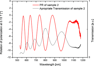 | ||
| Fig. 2 Transmission and PR spectra of sample 2. The detailed coincidence of the sinusoidal form suggests that the origin of the oscillations in the PR spectra lies in the Fabry–Perot interferences of a multilayer system. | ||
Influence of stress
Astonishingly, the OB also depends on the mechanical stress applied to the samples, qualitatively shown in Fig. 3 for sample 2: for having no quantitative access to the stress produced by our sample holder, in Fig. 3, nearly no stress means that the samples were loosely fixed in the holder; “normal” stress describes the common slight force that fixed the samples during measurements, whereas a large stress means that the samples were quite rapidly jammed into the holder (it should be mentioned here that the seeming break-down or inversion of the actual curves at 45° happens due to amplitudes larger than 45° and the method of calculation for the PR spectra9).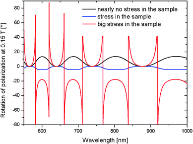 | ||
| Fig. 3 Measurement of the PR at 0.15 T with different stresses on sample 2. Remarkable differences in shape and even in amplitude can be seen, whereas as a trend, larger stresses correspond to larger OB amplitudes. | ||
The large variation of amplitudes with the applied stress in Fig. 3 indicates that the main origin of the OB has to be ascribed to a mechano-optical effect, which most prominently is the stress birefringence of the crystalline SiO2 (a classical optical effect10). The YIG buffer layer itself is polycrystalline and therefore will not show this type of birefringence.
Influence of the magnetic field
As Fig. 4 shows, samples 1 and 2 have a clear non-linear dependency on the magnetic field, in contrast to an overall linear behavior, which would be expected if the Faraday effect of the YIG buffer layer was the main reason for the OB. For sample 1, a quite distinct quadratic dependency of the maximal amplitude of the OB is shown, whereas for sample 2, a quadratic fit is not completely sufficient and cubic functions may have to be taken into account.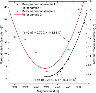 | ||
| Fig. 4 Dependence of the maximal amplitudes of the oscillations in the PR on the magnetic field for samples 1 and 2. For sample 1, there is a clear quadratic dependence, whereas sample 2 seems to have even cubic dependencies. A detailed explanation is given in Section VIII. | ||
For a comprehensive explanation, a simple method for the calculation of the PR of layered systems will be introduced in the following section, and stress birefringence will be explained in more detail. Finally, a model system for the layer structure of the samples will be set up and used to explain all dependencies, whether on substrate type or on the magnetic field.
IV. Basic outline of the 4 × 4 matrix formalism by Yeh
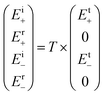
The 4 × 4 matrix formalism by Yeh6 is an easily feasible and, in most cases numerical, calculus for multilayer systems with birefringent properties describing both transmitted and reflected light. It is quite comparable and understandable in the sense of a simple, well-known classical analogon of 1941: the Jones formalism.11 In the case of the 4 × 4 matrix formalism, every border between two layers of an optical system is assigned a 4 × 4 matrix. Typically, these matrices consist of eight entries containing the optical properties of the border layers (refractive indices for left and right circularly polarized light, layer thicknesses and the wavelengths of the transmitted light). For more details, the reader may be referred to ref. 7, which gives a detailed explanation and derivation of these matrices noted as T(n−1, n) in the following text.The matrix T(n−1, n) is marked as (n − 1, n) and describes the border between the (n − 1) and the (n) layers of the system. For a whole system of border layers, the matrix  is used and represents all essential attributes needed for the calculation of transmitting or reflecting light. Eventually, to obtain the properties of light hitting a system T, one has to solve the following linear equation:
In the present case, we used this method to set up the matrix T for an objected layer system and solved the above-described linear equation system for every single wavelength (it may be mentioned here that having layer systems consisting of more than two layers will cause the matrix T to be quite complex, which makes it unreasonable to calculate an explicit formula f(λ) for the PR).
V. Stress birefringence of a crystal
As described above, the stress birefringence of the SiO2 is the main reason for the OB not only due to the dependence on mechanical stress, but also because the crystallinity and crystal direction have a strong influence. The “normal” birefringence of crystalline SiO2 is well known and is explained by the different lengths of the crystal axis, whereas the influence of mechanical forces was first explained by Brewster in 1816,5 where, additionally, the stress in the crystal and its birefringence are correlated (in a simple explanation, mechanical stress on the crystal strains the crystal axis further, so that the birefringent character is amplified).
Fig. 5 illustrates the situation in the present SiO2 substrates and their hexagonal lattice. Only the crystal axis perpendicular to the z-axis differs in length, namely, for the short edges in the x2–x3-plane compared to the long z-direction edge. Thus, a priori, only light incident perpendicular to the z-axis, the principal axis of the crystal, can experience birefringence that may be amplified by the external mechanical stress. Speaking in terms of the crystal cuts used, only incoming light in the (110)- and (100)-cut substrates will pass the SiO2 perpendicular to the principal crystal axis and therefore produce PR, whereas both of the other substrate types used will not be able to show that birefringent characteristic, whether due to a wrong cut ((001)) or the lack of a crystalline substrate (amorphous quartz).
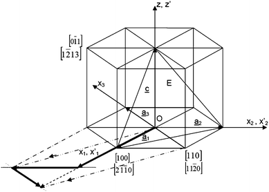 | ||
| Fig. 5 Illustration of the crystal directions of a hexagonal lattice, as found in crystalline SiO2. The z-axis represents the principal axis of the crystal, meaning that birefringence will only occur when light hits the crystal perpendicular to that axis. | ||
VI. Explanation model
The proposed Fabry–Perot effect requires a simple multilayer system (two or more layers) that has at least one strong birefringent part to account for the dependency on the mechanical stress and the large amplitudes of the OB. As discussed above, the latter part shall be represented by the SiO2 substrates, whereas the assumed multilayer system is represented in Fig. 6 and consists of: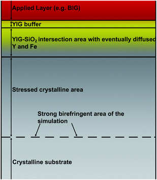 | ||
| Fig. 6 Layer system model for the explanation of the OB. The additional (red) layer plays no essential part in the investigated samples. The main areas are the polycrystalline YIG, the YIG–SiO2 intersection area and the crystalline SiO2 with a birefringent reflection border. | ||
• A small polycrystalline YIG buffer area.
• An amorphous YIG–SiO2 intersection area.
• A stressed SiO2 area, which is condensed in one birefringent border for the 4 × 4 matrix formalism. Because birefringence is a reciprocal effect (in contrast to the Faraday effect), the PR of light transmitting doubly in two opposite directions through a birefringent area will not be changed. Only when reflected at a birefringent border can a rotation of PR occur? For this reason, the many reflections that may take place all over the stressed SiO2 area in the present case can be combined into one birefringent reflecting border of a certain depth as a good approximation;
• The “bulk” crystalline SiO2 of the substrate.
The whole system will be reproduced by a TEM image later on, but it will be used to calculate the OB in the following.
VII. Calculation of the oscillations
For the described 4 × 4 matrix formalism, the thicknesses of the different layers and the corresponding refractive indices for the left and right circularly polarized light are needed. To obtain the latter indices for the garnet structures, Faraday rotation spectra of pure YIG films on Gd3Ga5O12 were first used to calculate the Verdet constant:
B: magnetic field, d: layer thickness, V(λ): Verdet constant, ΘF(λ): Faraday rotation angle, and λ: wavelength.
Now, the refractive indices n+and n− for left and right circularly polarized light can be stated as:
For stress-birefringent SiO2, the term Δn: = V(λ) × (B × (λ/2π)) (Δn: “birefringence” of SiO2) can be replaced by a constant value (about 0.1 for the following calculations; one of two fitting parameters) when only used in a single border area with zero thickness, as shown in Fig. 6. Moreover, for n(λ) and the complex parts of the refractive indices, k(λ) adequate models were used, which are described in detail in ref. 8.
For sample 1, a layer system with the following thicknesses was implemented:
1. YIG-buffer: 1 nm (approximate average thickness according to TEM and Rutherford Backscattering Spectroscopy (RBS));
2. YIG–SiO2 intersection area: 24 nm (according to TEM);
3. Depth of the strong birefringent area: 1680 nm (fitted to measurement; together with the stress birefringence of SiO2, the two fitting parameters).
Fig. 7 shows the results of the numerical calculation. Many details of the measurement such as the decreasing amplitude at longer wavelengths are reproduced, whereas the assumed depth and birefringence of the border area in the SiO2 (see Fig. 6) have reasonable values.
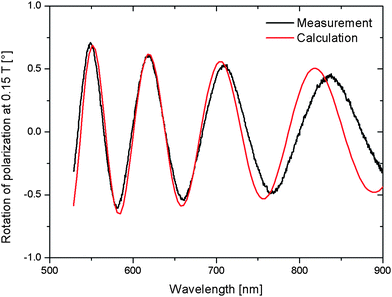 | ||
| Fig. 7 Comparison of measured and calculated spectra. A very good agreement is shown, underlining the correctness of the assumed model for the calculations, whose main parts are shown in Fig. 6. | ||
VIII. Quadratic dependence on the magnetic field
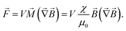
For explaining the quadratic dependence on the magnetic field, the YIG–SiO2 intersection area shall be investigated in more detail. As proposed before and as will be suggested by the ensuing TEM analysis, parts of the YIG buffer diffuse into the SiO2, which loses its crystallinity, setting up a small intersection area (about 24 nm thick). Therefore, a gradient of the magnetic susceptibility will be situated in this area, resulting in a force perpendicular to the sample surface when brought into a magnetic field.Using the force ![[F with combining right harpoon above (vector)]](https://www.rsc.org/images/entities/i_char_0046_20d1.gif) = −∇(
= −∇(![[small mu, Greek, vector]](https://www.rsc.org/images/entities/i_char_e0e9.gif)
![[B with combining right harpoon above (vector)]](https://www.rsc.org/images/entities/i_char_0042_20d1.gif) ) on a magnetic dipole with
) on a magnetic dipole with ![[M with combining right harpoon above (vector)]](https://www.rsc.org/images/entities/i_char_004d_20d1.gif) = /V (V volume) and = χ
= /V (V volume) and = χ![[H with combining right harpoon above (vector)]](https://www.rsc.org/images/entities/i_char_0048_20d1.gif) , one obtains:
, one obtains:
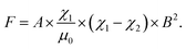
For a border between two susceptibilities χ1 and χ2 of the surface A, the force perpendicular to the surface is given by:
Following this for a continuous gradient of susceptibility χ, the resulting force can be stated as:
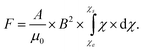
The integral  gives a constant value depending on the configuration of the border area (here, the YIG–SiO2 intersection area), and eventually, the force on the sample surface is proportional to B2. Using the stress optic law,12 one can relate this force on the SiO2 crystal to its birefringence Δn:
C: stress optic coefficient and σ1/2: first and second principal stress.
In the present case, the second principal stress may be assumed to be zero, and the first principal stress may assumed to be directly proportional to the force acting on the surface of the sample. Therefore,
| Δn ∝ B2. |
Using the 4 × 4 matrix formalism, it is easy to show that Δn is directly proportional to the amplitude of the OB, so that at least the quadratic dependence is well explained by the proposed model system.
To summarize the above calculations, in a static magnetic field, the gradient of magnetic susceptibility (YIG–SiO2 intersection area) produces a force perpendicular to the sample surface, which additionally stresses the crystalline SiO2 and therefore amplifies its birefringent character and consequently the amplitude of the OB.
Concerning the nearly cubic dependency on the magnetic field of sample 2, no sufficient explanation can be given here, but the thicker YIG layer on sample 2 and its ferromagnetic attributes may introduce some cubic terms into the force on the substrate.
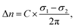
IX. TEM investigation of the layer structure
Using the TEM method, a picture of the layer system of sample 1 is shown in Fig. 8. As described above, the YIG–SiO2 intersection area can be clearly identified, whereas a determination of the overall thickness of the YIG buffer is best done by RBS rather than by using the small section shown in this TEM picture. Nevertheless, the most critical assumption of an intersection area of YIG and SiO2 is definitely proven.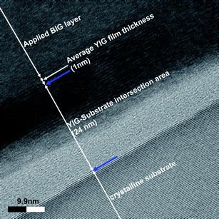 | ||
| Fig. 8 TEM picture of the layer system of sample 1. The structure shown in Fig. 6 can be clearly identified, giving the final proof for the assumed model system by the direct analysis of a given sample. | ||
In addition, for identifying the crystallinity of the SiO2 substrate, a diffraction pattern using TEM was produced (Fig. 9), which shows hexagonal reflexes resembling the hexagonal lattice of the SiO2.
 | ||
| Fig. 9 Diffraction pattern of the border area shown in Fig. 8. The clear reflexes of the crystalline hexagonal lattice of SiO2 can be seen. The additional sprinkled reflexes may be attributed to the polycrystalline YIG or the intersection area of YIG–SiO2. | ||
X. Conclusion and outlook
The OB of the presented samples is explained by a simple layer model, mainly consisting of a polycrystalline YIG buffer, a YIG–SiO2 intersection area and the crystalline SiO2 substrates. The large amplitudes of the PR are ascribed to the stress birefringence of the SiO2, whereas all observed dependencies on the magnetic field, crystal cut and mechanical stress can be explained in a comprehensive way.Not only due to their mechanical dependencies but also because of their strong magnetic dependence, these samples represent a sufficient realization of combined mechanical and magnetic sensors. Additionally, the further clarification of the magnetic dependencies (cubic) could lead to further insight into the magnetic structures of YIG and may present a possibility for measuring magnetic fields very sensitively. In this respect, the strong mechano-sensitive behavior may give new insights into the long known effect of stress birefringence, when analyzed more quantitatively. All together, this description and the explanation of the samples here only present the very basics of an exciting group of samples combining the different areas of mechano- and magneto-optical effects.
References
- H. Dötsch, N. Bahlmann, O. Zhuromskyy, M. Hammer, L. Wilkens, R. Gerhardt and P. Hertel, J. Opt. Soc. Am. B, 2005, 22, 240 Search PubMed.
- R. Lux, A. Heinrich, S. Leitenmeier, T. Körner, M. Herbort and B. Strizker, J. Appl. Phys., 2006, 100, 113511 CrossRef.
- S. Leitenmeier, A. Heinrich, J. K. N. Lindner and B. Strizker, J. Appl. Phys., 2006, 99, 08M704 CrossRef.
- S. Kahl, V. Popov and A. M. Grishin, J. Appl. Phys., 2003, 94, 5688 CrossRef CAS.
- D. Brewster, Philos. Trans. R. Soc. London, 1816, 106, 156 CrossRef.
- P. Yeh, Optical Waves in Layered Media, Wiley-Intersience Publication, 1988 Search PubMed.
- S. Visnovsky, K. Postava and T. Yamaguchi, Opt. Express, 2001, 9(3), 158 CrossRef CAS.
- S. Kahl, PhD thesis, Bismuth Iron Garnet Films for Magneto-optical Photonic Crystals, University of Augsburg, 2004 Search PubMed.
- S. Leitenmeier, PhD thesis, Zum Wachstum von Magnetooptischen Bismutdotierten Seltenerdeisengranatfilmen, 2007 Search PubMed.
- R. W. Pohl, Optik und Atomphysik, Springer, 1958 Search PubMed.
- R. Clark Jones, J. Opt. Soc. Am., 1941, 31, 488–493.
- S. Haussühl, Physical Properties of Crystals, Wiley-VCH, 2007 Search PubMed.
| This journal is © The Royal Society of Chemistry 2011 |