Nanoscale of biomimetic moth eye structures exhibiting inverse polarization phenomena at the Brewster angle
Shang-Yu
Chuang
a,
Hsuen-Li
Chen
*a,
Jiann
Shieh
*b,
Chun-Hung
Lin
c,
Chao-Chia
Cheng
d,
Hao-Wei
Liu
a and
Chen-Chieh
Yu
a
aDepartment of Materials Science and Engineering, National Taiwan University, Taipei, Taiwan, ROC. E-mail: hsuenlichen@ntu.edu.tw; Fax: +886-2-23634562; Tel: +886-2-23634562
bNational Nano Device Laboratory, Hsinchu, Taiwan, ROC. E-mail: jshieh@ndl.org.tw
cInstitute of Electro-Optical Science and Engineering, National Cheng Kung University, Taiwan, ROC
dDepartment of Physics, National Central University, Taiwan, ROC
First published on 8th March 2010
Abstract
The unique functionalities of nanoscale structures in the natural world are an inspiration to the development of new nano-manufacturing techniques. For example, the cornea of the moth's eye features a sub-wavelength natural antireflective architecture. To date, almost all optical research into moth eye structures has been focused on their antireflective properties. No studies of inverse polarization phenomena at the Brewster angle have been reported, especially in biomimetic structures. For the first time, we discovered a unique inverse polarization phenomenon on moth eye structures that arises from TM-polarized light having a higher reflectance than TE-polarized light on moth eye structures at angles of incidence near the Brewster angle, unlike the behavior of polarized light on flat interfaces. Herein, we report a one-step colloidal lithography process that allows the fabrication of several kinds of moth eye structures. We characterized these moth eye structures experimentally and through rigorous coupled-wave analysis to understand the mechanism underlying this inverse polarization phenomenon in both visible and near infrared ray (NIR) regimes. Controlling the structural height and degree of non-close-packing of the moth eye structures had a dramatic effect on the extent of inverse polarization. This study is potentially important for various polarization-dependent devices and measurements.
Introduction
Several unique optical phenomena exist in biological systems exhibiting nanoscale architectures, such as the photonic band gap structures on the wings of butterflies and beetles,1 and the antireflection properties on the cornea of moths’ eyes.2–6 Inspired by the latter, sub-wavelength structures (SWS) serving as antireflection coatings have attracted much attention this decade.5–16 The outer surface of the moth's eye features hexagonal arrays of non-close-packed (NCP) nipples, which have a height and separation of less than 300 nm; this structural array reflects to a lesser degree.5,7,9 Such natural antireflection structures are a superior solution toward suppressing Fresnel reflection. The presence of a refractive index gradient allows moth eye structures to achieve much lower reflectance at the surface between two optical media. Because of the excellent antireflective ability of moth eyes, the relationship between SWS architectures and optical antireflection has attracted a tremendous amount of attention. By combining a refractive index gradient with light trapping, an SWS can maintain its antireflective ability over a broad range of incident angles and wavelengths of incoming light. In addition to SWS being vastly superior as antireflective structures relative to commonly used dielectric multilayer coatings, SWS also eliminate several of the problems arising from multilayer coatings, e.g., the selection of appropriate materials, layer-to-layer adhesion, and thermal mismatch or diffusion of one material into another.6,7,9 As a result, many kinds of SWS manufacturing techniques have been developed to achieve not only fast and cheap fabrication but also low reflectance.6,8Colloidal lithography is a low cost and relatively high throughput technique for fabricating SWS.5,7,9,17,18 Its main advantage in nanofabrication is that the large-area self-assembly of colloids into well-ordered structures can be performed without the need for expensive instruments. Several nanostructures, including nanoscale rings, dots, and rods, have been fabricated recently using colloidal lithography. Jiang et al. developed a templating approach—involving an NCP colloid template dispersed in the monomer—for the fabrication of NCP cylindrical pillars.17 They encountered difficulties, however, in controlling the spacing of the NCP-SWS. Recently, we developed one- and two-step colloidal lithography processes for the fabrication of moth eye, close-packed pyramidal arrays and NCP cylindrical pillar arrays structures.19 For our optimized material, the reflectance over the wavelength range from 350 to 850 nm was less than 2%. Most, if not all, major studies on SWS have been focused on their antireflection properties against non-polarized light. Investigations into the characteristics of polarized light interacting with moth eye structures are relatively rare.4,20–22
Brewster discovered a special phenomenon in the Fresnel reflection of linearly polarized light at large incident angles: When the incoming light has its electric field aligned parallel to the incident plane (i.e., TM-polarized light), its reflected wave will vanish at a certain incident angle, known as the Brewster angle.23,24 This phenomenon is not observed when the electric field of the light is aligned perpendicular to the incident plane (i.e., TE-polarized light). In this study, we used a colloidal lithography process to fabricate several kinds of moth eye structures with various base diameters and NCP spacings to investigate the effects of angle-dependent polarization. We observed a unique inverse polarization phenomenon at the Brewster angle for sub-100 nm biomimetic moth eye structures in both visible and near infrared ray (NIR) regimes.
Experimental
(100)-Oriented Si wafers were made hydrophilic. Using a dip coater, the substrates were coated with a monolayer of close-packed PS spheres. Solutions of PS spheres (average diameter: 350 nm) were diluted to 5% for the assembly of the monolayer arrays. Using a reactive ion etcher (Anelva, ECR-6001), the patterns of the close-packed PS spheres were transferred to the Si substrate. The reactive gases for reactive ion etching were Cl2, SF6, and O2 (flow rates: 90, 5, and 5 sccm, respectively). The cross-section profiles were observed using a JEOL JSM-6500F scanning electron microscope; the optical properties were measured using a Hitachi U-4100 various-angle optical spectrometer. RCWA was used to simulate the angle-dependent reflection properties of various periodic nanostructures. For near-field analysis, the finite difference time domains (FDTD) method was used to investigate the optical behavior of the polarized light transmission through the moth-eye structures.Results and discussion
Fig. 1 displays the one-step etching process that we used to control the base diameter and NCP spacing. We used a dip coater to apply a monolayer of close-packed polystyrene (PS) spheres onto a silicon substrate as the template (Fig. 1a). The pattern of the PS spheres was transferred to the Si substrate through reactive ion etching. The reactive gas O2 was used to minify the close-packed PS spheres and the reactive gases Cl2 and SF6 were used to remove the underlying Si substrates. The one-step etching process involved minifying the close-packed PS spheres and etching the underlying Si substrate simultaneously. As the size of the PS spheres decreased gradually, the etched area of the underlying Si increased gradually as well. Thus, sub-wavelength moth eye structures were readily obtained using this one-step etching process. In the early stages of the etching process, the PS spheres remained above the incompletely etched structure, providing a close-packed moth eye structure with a flat top (Fig. 1b). After a particular etching time, the PS sphere templates were worn away entirely to yield a completely etched, close-packed moth eye structure featuring sharp tips (Fig. 1c). Beyond this point, further etching resulted in NCP moth eye structures. Fig. 1d displays NCP moth eye structures exhibiting different spacings that we obtained after over-etching for various durations. Fig. 1e and 1f present SEM images of close-packed and NCP moth eye structures, respectively, each having a period of 350 nm. Fig. 1g indicates that etched Si having a height of ca. 310 nm and a small, flat roof appeared when we performed the etching process for 45 s. Upon increasing the etching duration, the top of the etched Si became sharper and the width narrower. Progressing from Fig. 1g to 1j, we observe that the base diameter decreased upon increasing the duration of etching. Fig. 1k displays the base diameter and structural heights with respect to the etching duration. The height of the moth eye structure remained at ca. 420 ± 50 nm, but the base diameter of the etched Si decreased from 350 to 245 nm at an etching duration of 100 s, and then decreased further to 90 nm after 150 s.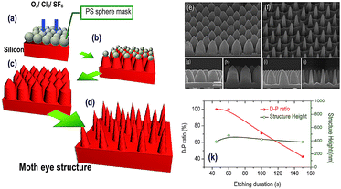 | ||
| Fig. 1 (a–d) Schematic representations of one-step etching processes: (a) a flat silicon substrate with close-packed PS spheres; (b) under-etching; (c) complete-etching; (d) over-etching. (e, f) SEM images of (e) a close-packed pyramid structure having a period of 350 nm and (f) an NCP moth eye structure having a period of 350 nm. (g–j) Cross-sectional SEM images of the textured profiles obtained after etching for (g) 30, (h) 60, (i) 100, and (j) 150 s using the one-step etching process. (k) Structural heights and base diameter plotted with respect to the etching duration. | ||
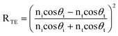
Fig. 2 displays the angle-dependent polarization of a flat Si substrate and an NCP moth eye structure with a base diameter of 245 nm. We measured the reflectance as the function of incident angle to study the polarization of TM- and TE-polarized light at the wavelength of 1250 nm. The incident angles ranged from 5 to 75° for both the TE- and TM-polarized light (electric fields perpendicular and parallel to the incident plane, respectively). Fig. 2a displays the measured reflectance of a flat Si substrate. The reflectance of the TE-polarized light increased from 30 to 50% when the incident angle increased from 5 to 75°; this behavior is consistent with that calculated from the Fresnel equation for TE-polarized light:
 | (1) |
 | (2) |
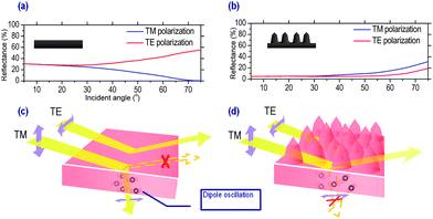 | ||
| Fig. 2 (a, b) Reflectance of TE- and TM-polarized light plotted with respect to the angle of incidence for (a) flat Si and (b) NCP moth eye structures. (c, d) Schematic representations of the reflective behavior of polarized light and the oscillating electric dipole in (c) flat Si and (d) NCP moth eye structures. | ||
NCP moth eye structures formed after over-etching. We fabricated a moth eye structure formed after over-etching, with a period of 350 nm and a base diameter of 245 nm, after etching for 100 s (Fig. 2b). The reflectances of TE- and TM-polarized light were both less than 5% at an incident angle of 5°. When we increased the incident angle, the reflectances of the TE- and TM-polarized light both increased slightly. In contrast to the reflectance of the flat Si substrate, the TE- and TM-polarized light had lower and higher reflectances, respectively, at large incident angles. An obvious inverse polarization phenomenon appeared for the NCP moth eye structure having a period of 350 nm and a base diameter of 245 nm.
Here, we propose a mechanism for this unique inverse polarization phenomenon. We observed two key factors relating to the inverse polarization phenomenon of the artificial moth eye structure: a reduction in the reflectance of TE-polarized light and an increase in the reflectance of TM-polarized light at large incident angles. For TE-polarized light, the SWS moth eye structure served as an antireflective structure that featured a lower reflectance relative to that of the flat Si substrate. Moth eye structures provide a gradual transition in the effective index of refraction, from air to textured Si, and thereby suppress reflection. For the same moth eye structure, however, the reflectance of TM-polarized light exhibits a different trend. To explain this phenomenon, let us consider the electron oscillator model.24 For a flat substrate, the electric field of the incident light drives the bound electrons at the interface of two media oscillating as electric dipoles. The energy is then re-radiated through the oscillation of these electric dipoles. The re-radiated energy appears in the form of reflected and refracted light. The electric dipoles, however, cannot re-radiate energy in the direction in which they are oscillating. As illustrated in Fig. 2c, when the incident light is linearly polarized and its electric field is parallel to the plane of incidence (i.e., TM-polarized), the electric dipoles will oscillate in the direction of the reflected wave at the Brewster angle and, as a result, no reflected light will be observed. In contrast, Fig. 2d illustrates the oscillating dipoles in the moth eye structure at the Brewster angle. The electric dipoles in the textured Si substrate are randomized by the moth eye structure; therefore, they oscillate in various directions. The randomized dipoles allow energy to be re-radiated in various directions as reflected light. Therefore, the Brewster angle phenomenon disappears for TM-polarized light in the moth eye structure. This rationale explains why, relative to a flat Si substrate, an increase in the reflectance of TM-polarized light occurs around the Brewster angle.
We also used the finite difference time domain (FDTD) method to investigate the mechanism leading to the randomized electric dipoles—by analyzing the propagation of light within the near-field regime—in the SWS moth eye structure. Fig. 3 displays the results obtained from an oblique incident TM-polarized plane wave having a wavelength of 1250 nm propagating from 1 μm above a moth eye structure having a period of 350 nm, a height of 400 nm, and a base diameter of 245 nm. Fig. 3a demonstrates the continuous wavefront of a plane wave propagating toward the surface of the moth eye structure. In Fig. 3b, the directly transmitted plane wavefront was distorted significantly after the plane wave had propagated through the SWS moth eye structure. This distorted transmission plane wavefront randomizes the oscillating direction of the electric dipoles in the textured Si, resulting in the elimination of the Brewster angle effect. Therefore, the reflectance of TM-polarized light would not be expected to decrease dramatically at the Brewster angle.
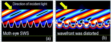 | ||
| Fig. 3 FDTD diagrams of the wavefront of a plane wave (a) before and (b) after entering a moth eye structure. | ||
We used rigorous coupled-wave analysis (RCWA) simulations to recognize the tendency of increasing TE-polarized reflectance and decreasing TM-polarized reflectance that were responsible for the inverse polarization phenomena. Using this approach, we simulated the reflection of light from flat Si and NCP moth eye structures (having a base diameter of 245 nm) with heights ranging from 100 to 400 nm. Fig. 4a displays the simulated angle-dependent reflectance of TE-polarized light at a wavelength of 1250 nm calculated by RCWA. The reflectance of TE-polarized light from the flat Si substrate was ca. 31.6% at normal incidence. In contrast, the SWS moth eye structures of various heights all exhibited reflectances lower than that of the flat Si substrate because of the refractive index gradient. Upon increasing the height of the moth eye structures from 100 to 400 nm, the reflectance at normal incidence decreased from 22.3 to 0.68%, due to the improved refractive index gradient. At large incident angles, the reflectance of the flat Si increased enormously (ca. 82.1% at 75°). In contrast, because of the refractive index gradient, broadband antireflection properties existed for the moth eye structure regardless of the incident angle. Fig. 4b displays the simulated angle-dependent reflectance of TM-polarized light on the moth eye structures at a wavelength of 1250 nm. In this case, the moth eye structures also demonstrated reflectances relatively lower than that of the flat Si surface at normal incidence. We observed an unusual phenomenon, however, at large incident angles. The Brewster angle phenomenon was evident for the flat Si substrate, whereas it began to disappear for the moth eye structure having a height of 200 nm, and it vanished completely when the structure height was 300 nm. The reflectance of TM-polarized light increased when increasing the height of the moth eye structure, because the Brewster angle effect of the flat interface was entirely eliminated.
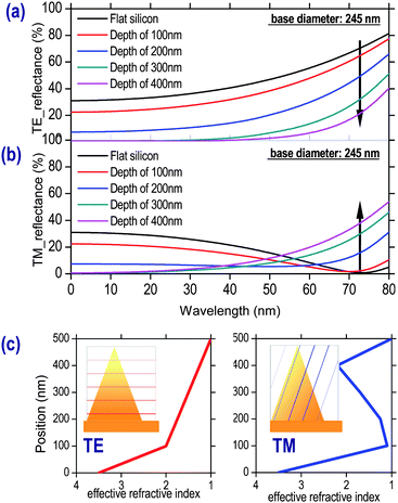 | ||
| Fig. 4 RCWA simulations of a flat Si substrate and moth eye structures having a period of 350 nm, a base diameter of 200 nm, and heights ranging from 100 to 400 nm. (a) TE- and (b) TM-polarized light. (c) Effective refractive index profiles for TE- and TM-polarized light between air and the Si structure interface. | ||
Fig. 4c displays profiles of the effective refractive index of polarized light at large incident angles. The effective refractive index was estimated at different positions to which the incident light propagated from air to the silicon substrate, and the position of the bulk silicon surface was denoted as zero. Under TE-polarized light, the NCP structure exhibited a gradual change in its effective index of refraction from 1 to that of Si (n = 3.5). In contrast, the effective refractive index under TM-polarized light first increased from that of air (n = 1), but then fluctuated up and down as the light propagated through the moth eye structure. Unlike the TE-polarized light, the TM-polarized light would not encounter a superior refractive index gradient in the moth eye structures. For the larger effective refractive index variation, the reflectance of TM-polarized light is, therefore, larger than that of TE-polarized light. In this study, we fabricated moth eye structures featuring different base diameters and NCP spacings using colloidal lithography for various etching durations to investigate the inverse polarization phenomena.
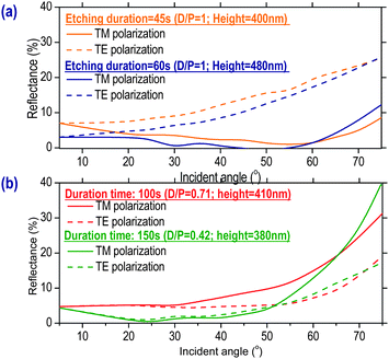
The one-step colloidal lithography process allowed us to readily fabricate structures of fixed height but various base diameters and NCP spacings. Because our RCWA simulations revealed that inverse polarization occurred for the NCP moth eye structure having a height about 400 nm, we fabricated NCP moth eye structures having heights of around 400 nm and various base diameters to study their inverse polarization phenomena. Fig. 5a displays the angle-dependent reflectance of close-packed moth eye structures having a fixed period of 350 nm and heights of 400 and 480 nm. For the samples etched for 45 (orange line) and 60 s (blue line), the base diameter was equal to the period of moth eye, was meaning that they were close-packed structures. The reflectance of TE-polarized light (dash line) was greater than that of TM-polarized light (solid line), indicating that no inverse polarization phenomenon occurred in this type of close-packed structure. Increasing the etching duration to 100 and 150 s provided NCP moth eye structures having base diameters of 245 and 90 nm, respectively (period = 350 nm).
 | ||
| Fig. 5 Measured angle-dependent reflectances of moth eye structures prepared for etching durations of (a) 45 and 60 s and (b) 100 and 150 s. | ||
Fig. 5b reveals the presence of an inverse polarization phenomenon at large angles of incidence; it was more evident for the NCP moth eye structure than for the close-packed structure.
Next, to quantify the degree of non-closed packing, we define the “D–P ratio”, where P and D represent the period and base diameter of the moth eye structures. We studied the inverse polarization phenomena of three kinds of nanostructures: (i) an NCP moth eye structure (D–P ratio = 0.5–0.8; height = 200–600 nm),4,6,11–13,20,21 (ii) a close-packed pyramid structure (D–P ratio = 1; height =200–600 nm),5,7,9,22 and (iii) a close-packed nano-cone structure (D–P ratio = 1; height = 1–2 μm).25–27 Each structure featured a fixed period of 350 nm. We used the RCWA method to simulate the reflectance behavior of these structures as a function of the angle of incidence. For the NCP moth eye structure (Fig. 6a) under 1250 nm light at an angle of incidence of ca. 40°, the reflectance was ca. 10% for TM-polarized light and 1% for TE-polarized light; i.e., inverse polarization emerged. With its small D–P ratio, the inverse polarization effect of the NCP moth eye structure was clearly observable even for a shallow NCP structure having a height of 400 nm. We define this cutoff height as the “inversion height”; it represents the height of the moth eye structure at which inverse polarization was appreciable. For the close-packed pyramid structure of the same height (400 nm), the reflectance of TE-polarized light was higher than that of TM-polarized light when the incident angle was greater than 20° (Fig. 6b); i.e., inverse polarization was not evident. For the close-packed nano-cone structure having a height of 1.5 μm (Fig. 6c), the reflectances of TM- and TE-polarized light were 12.5 and 7.7%, respectively, at an angle of incidence of 75°; therefore, inverse polarization is also observable in close-packed nano-cone structures having high aspect ratios. Note that the close-packed pyramid structures in Fig. 6b had the same D–P ratio as that of the nano-cone structure (i.e., D–P = 1), yet they exhibited distinctly different behavior. RCWA simulations suggested that the cutoff inversion height for the close-packed pyramid structure would be ca. 1.5 μm, much higher than the height of the pyramid structure in Fig. 6b; therefore, we would not expect to observe inverse polarization for this particular structure. Fig. 6d displays the cutoff inversion heights and incident angles plotted with respect to the D–P ratio. For the structure having a D–P ratio of 0.71, the cutoff inversion height was ca. 400 nm. When the D–P ratio increased from 0.91 to 1, the inversion height increased from 900 nm to 1.5 μm. The cutoff inversion angle also increased upon increasing the D–P ratio. For a D–P ratio of 0.71, inverse polarization began at an incident angle of 10°. Upon increasing the D–P ratio from 0.71 to 1, the cutoff inversion angle also increased from 10 to 62°. Thus, inverse polarization phenomena appeared at large incident angles when the moth eye structures were more close-packed. The inverse polarization phenomenon exists for both NCP moth eye structures and close-packed nano-cone structures having high aspect ratios; it is dependent on the cutoff inversion height and inversion angle. Because the close-packed nano-cone structure features a large cutoff inversion angle and relatively low reflectances for both TM- and TE-polarized light, the inverse polarization effect in such high-aspect-ratio nano-cones is more difficult to observe than that in NCP moth eye structures. Furthermore, we also studied a non-close-packed cylindrical nanostructure28–30 featuring nanowires having an average diameter and length of ca. 100 nm and ca. 1 μm, respectively. Neither our experimental optical measurements nor our RCWA simulations revealed the presence of inverse polarization phenomena in such cylinder-like nanowire structures.
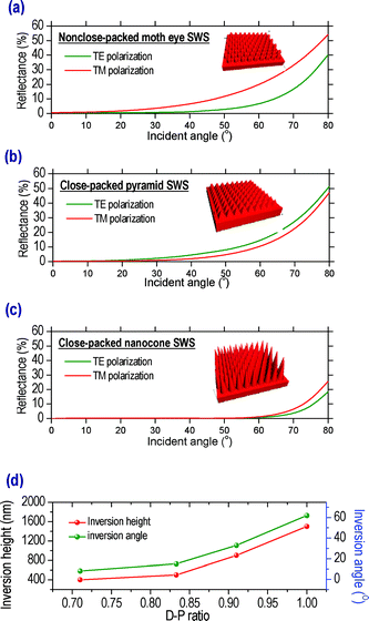 | ||
| Fig. 6 (a–c) Measured angle-dependent reflectances of (a) NCP moth eye, (b) close-packed pyramid, and (c) high-aspect-ratio close-packed nano-cone nanostructures. (d) Inversion heights and angles plotted with respect to the D–P ratio. | ||
The experimental results discussed above were focused on the wavelength of 1250 nm. We further discuss the inverse polarization phenomenon in the visible regime at the wavelength of 633 nm. Fig. 7 displays the measured angle-dependent reflectances under 633 nm light for moth eye structures having base diameters of 245 and 90 nm. As indicated in Fig. 7a, the inverse polarization phenomenon was not obvious on the moth eye structure having a base diameter of 245 nm, that was in contrast to the apparent inverse polarization phenomenon observed at 1250 nm. As described above, the moth eye structure having a base diameter of 90 nm would demonstrate the inverse polarization phenomenon at the wavelength of 1250 nm, and we still observed the inverse polarization phenomenon at the wavelength of 633 nm, as indicated in Fig. 7b. This gave us an inspiration that the inverse polarization phenomenon would occur in the visible regime only if the structure had a smaller base diameter. To further verify our assumptions, RCWA was used to simulate the inverse polarization phenomenon at the wavelength of 633 nm. We used the reflectance ratio of TM-polarized light to TE-polarized light (RTM/RTE) to observe the inverse polarization phenomenon. For a flat silicon substrate, the reflectance of TM-polarized light would be lower than TE-polarized light due to the Brewster angle effect, leading to a reflectance ratio of RTM/RTE smaller than one. However, for a structure that performed the inverse polarization phenomenon, the reflectance ratio would be greater than unity. Fig. 8 displays the reflectance ratio of moth eye structures having a base diameter of 90 nm and a larger moth eye structure with a fixed D–P ratio and aspect ratio as the former. For the moth eye structure with a base diameter of 90 nm and a height of 100 nm, the reflectance ratio of RTM/RTE was smaller than one, which represented that the inverse polarization phenomenon did not occur. As the structure height increased, the inverse polarization phenomenon occurred (RTM/RTE > 1). Clearly, the inverse polarization phenomenon was successfully demonstrated in the visible regime on the moth eye structure having a base diameter of 90 nm and a height of 150 nm. When the nanoscale of the moth eye structure was enlarged three times (base diameter = 270 nm, height = 450 nm), the reflectance ratio of RTM/RTE decreased to less than one, indicating that the inverse polarization phenomenon disappeared. Therefore, we found that the inverse polarization phenomenon would occur in the visible regime only if the moth eye structure had a base diameter in the sub-100 nm scale.
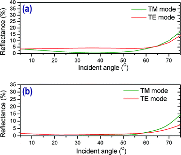 | ||
| Fig. 7 Measured angle-dependent reflectances of moth eye structures having a base diameter of (a) 245 nm (b) 90 nm. | ||
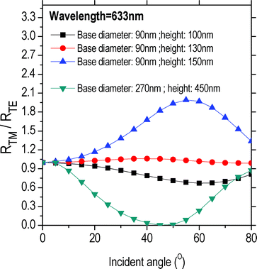 | ||
| Fig. 8 Angle-dependent reflectance ratios of TM-polarized light to TE-polarized light (RTM/RTE) for moth eye structures having various base diameters and heights. | ||
Conclusions
We observed a unique inverse polarization phenomenon on biomimetic moth eye structures in both near infrared ray and visible regimes. Biomimetic moth eye structures exhibit the inverse polarization phenomenon relating to the Brewster angle because (i) the Brewster angle effect is eliminated by the moth eye structures via randomization of the direction of oscillation of the electric dipoles and (ii) TE- and TM-polarized light encounter different refractive index gradient profiles at large angles of incidence. Because of a larger variation in effective refractive index, the reflectance of TM-polarized light is larger than that of TE-polarized light at large incident angles. One-step colloidal lithography is a convenient technique for fabricating such sub-wavelength close-packed and NCP moth eye structures. Under optimized etching conditions, we could control both the heights, base diameters and NCP spacings of these moth eye structures. The inverse polarization phenomena were more evident in moth eye structures having smaller base diameters and taller structural features. From a survey of several kinds of nanostructures, we observed inverse polarization for NCP moth eye structures and nano-cone structures having high-aspect ratios, but not for close-packed pyramidal structures or nanowire structures. The inverse polarization phenomenon occurred in our biomimetic NCP moth eye structures because of the small cutoff inversion angle and inversion height. We also found that the inverse polarization phenomenon would occur in the visible regime only if the moth eye structure had a base diameter in the sub-100 nm scale. This study is potentially important for various polarization-dependent devices and measurements.Acknowledgements
We thank the National Science Council, Taiwan, R.O.C., for supporting this study under contracts NSC-97-2221-E-002-046-MY3 and NSC-97-2623-7-002-008-ET.Notes and references
- L. P. Biró, Zs. Bálint, K. Kertész, Z. Vértesy, G. I. Márk, Z. E. Horváth, J. Balázs, D. Méhn, I. Kiricsi, V. Lousse and J.-P. Vigneron, Phys. Rev. E: Stat., Nonlinear, Soft Matter Phys., 2003, 67, 021907 CrossRef CAS.
- D. G. Stavenga, S. Foletti, G. Palasantzas and K. Arikawa, Proc. R. Soc. London, Ser. B, 2006, 273, 661–667 CrossRef CAS.
- S. J. Wilson and M. C. Hutley, Optica acta, 1982, 29, 993–1009 Search PubMed.
- Y. F. Huang, S. Chattopadhyay, Y. J. Jen, C. Y. Peng, T. A. Liu, Y. K. Hsu, C. L. Pan, H. C. Lo, C. H. Hsu, Y. H. Chang, C. S. Lee, K. H. Chen and L. C. Chen, Nat. Nanotechnol., 2007, 2, 770–774 CrossRef CAS.
- C. H. Sun, P. Jiang and B. Jiang, Appl. Phys. Lett., 2008, 92, 061112 CrossRef.
- P. Lalanne and G. M. Morris, Nanotechnology, 1997, 8, 53–56 CrossRef CAS.
- C. H. Sun, B. Ho, J. B. Jiang and P. Jiang, Opt. Lett., 2008, 33, 2224–2226 Search PubMed.
- E. Rephaeli and S. Fan, Appl. Phys. Lett., 2008, 92, 211107 CrossRef.
- W. L. Min, A. P. Betancourt, P. Jiang and B. Jiang, Appl. Phys. Lett., 2008, 92, 141109 CrossRef.
- S. Wang, X. Z. Yu and H. T. Fan, Appl. Phys. Lett., 2007, 91, 061105 CrossRef.
- H. Sai, H. Fujii, K. Arafune, Y. Ohshita, Y. Kanamori, H. Yugami and M. Yamaguchi, Jpn. J. Appl. Phys., 2007, 46, 3333–3336 CrossRef CAS.
- J. Shieh, C. H. Lin and M. C. Yang, J. Phys. D: Appl. Phys., 2007, 40, 2242–2246 CrossRef CAS.
- H. Sai, H. Fujii, K. Arafune, Y. Ohshita and M. Yamaguchi, Appl. Phys. Lett., 2006, 88, 201116 CrossRef.
- K. Hadobás, S. Kirsch, A. Carl, M. Acet and E. F. Wassermann, Nanotechnology, 2000, 11, 161–164 CrossRef CAS.
- Y. Kanamori, K. Hane, H. Sai and H. Yugami, Appl. Phys. Lett., 2001, 78, 142–143 CrossRef CAS.
- W. H. Southwell, J. Opt. Soc. Am. A, 1991, 8, 549–553 CrossRef CAS.
- W. L. Min, B. Jiang and P. Jiang, Adv. Mater., 2008, 20, 3914–3918 CrossRef CAS.
- C. H. Sun, W. L. Min, N. C. Linn, P. Jiang and B. Jiang, Appl. Phys. Lett., 2007, 91, 231105 CrossRef.
- H. L. Chen, S. Y. Chuang, C. H. Lin and Y. H. Lin, Opt. Express, 2007, 15, 14793–14803 CrossRef.
- Y. Kanamori, M. Sasaki and K. Hane, Opt. Lett., 1999, 24, 1422–1424 Search PubMed.
- Y. Kanamori, H. Kikuta and K. Hane, Jpn. J. Appl. Phys., 2000, 39, L735–737 CrossRef CAS.
- G. R. Lin, Y. C. Chang, E. S. Liu, H. C. Kuo and H. S. Lin, Appl. Phys. Lett., 2007, 90, 181923 CrossRef.
- S. P. F. Humphreys-Owen, Proc. Phys. Soc., 1961, 77, 949–957 CrossRef CAS.
- W. T. Doyle, Am. J. Phys., 1985, 53, 463–468 CrossRef.
- J. Zhu, Z. Yu, G. F. Burkhard, C. M. Hsu, S. T. Connor, Y. Xu, Q. Wang, Mi. McGehee, S. Fan and Y. Cui, Nano Lett., 2009, 9, 279–283 CrossRef CAS.
- K. Peng, Y. Xu, Y. Wu, Y. Yan, S. T. Lee and J. Zhu, Small, 2005, 1, 1062–1067 CrossRef CAS.
- K. Peng, A. Lu, R. Zhang and S. T. Lee, Adv. Funct. Mater., 2008, 18, 3026–3035 CrossRef CAS.
- W. K. Choi, T. H. Liew, M. K. Dawood, H. I. Smith, C. V. Thompson and M. H. Hong, Nano Lett., 2008, 8, 3799–3802 CrossRef CAS.
- Z. Huang, H. Fang and J. Zhu, Adv. Mater., 2007, 19, 744–748 CrossRef CAS.
- K. Peng, M. Zhang, A. Lu, N. B. Wong, R. Zhang and S. T. Lee, Appl. Phys. Lett., 2007, 90, 163123 CrossRef.
| This journal is © The Royal Society of Chemistry 2010 |