Three dimensional visualisation of human facial exposure to solar ultraviolet
Nathan
Downs
* and
Alfio
Parisi
Centre for Rural and Remote Area Health, University of Southern Queensland, Toowoomba, 4350, Australia. E-mail: downsn@usq.edu.au; Fax: 61 7463 12721; Tel: 61 7463 12727
First published on 18th October 2006
Abstract
A three dimensional computer model of the human face has been developed to represent solar ultraviolet exposures recorded by dosimeter measurements on a manikin headform under low cloud conditions and various solar zenith angles. Additionally, polysulfone dosimeters have been successfully miniaturised to provide the detailed measurements required across the face. The headform used in this research was scanned at 709 individual locations to make a wireframe mesh consisting of 18 vertical contours and 49 horizontal contours covering half the manikin's frontal facial topography. Additionally, the back of the headform and neck have also been scanned at 576 locations. Each scanned location has been used as a viable dosimeter position on the headform and represents a grid intersection point on the developed computer wireframe. A series of exposures recorded by dosimeters have been translated into three dimensional exposure ratio maps, representing ambient solar ultraviolet exposure. High dosimeter density has allowed for the development of individual topographic contour models which take into account complex variation in the face and improve upon previously employed techniques which utilise fewer dosimeters to interpolate exposure across facial contours. Exposure ratios for solar zenith angle ranges of 0°–30°, 30°–50°, and 50°–80° have been developed.
Introduction
The frequency and distribution of environmental ultraviolet radiation (UV) on exposed sites of the human body has been linked to the occurrence of non-melanoma skin cancers.1 The increased risk of the development of cutaneous malignant melanoma, cataracts and premature ageing of the skin due to environmental exposures to UV is also well defined. The regular monitoring of solar UV radiation is however often related to horizontal surfaces. Additionally, the provision of the UV index is for a horizontal surface.2 Models that relate exposures of a horizontal surface to the topography of the human body can be used to better define the causative role of environmental UV to skin and ocular disorders. Previously, manikin headforms employing polysulfone dosimeters have been used to develop exposure models to various sites on the human face.3,4 These models provide biologically damaging exposures to the face based on the measurement of erythemal exposures to various sites located on the headform. While accurate at predicting environmental UV exposures to specific facial sites, these models rely on interpolation techniques to predict exposures between measurement sites.Developing accurate models to predict environmental UV exposure across complex shapes like the human face requires the chosen dosimeter sites to be as close together as possible to reduce interpolation errors between measurement sites while simultaneously accounting for variations in orientation and shading caused by facial topography. One approach to the measurement of UV exposure over a human body has been to measure with radiometers the UV irradiances to 27 differently inclined surfaces over a period of 2 min.5 These measurements, interpolated in two dimensions, allow visualization of the total UV exposure received over the human body. An alternate approach to allow the visualization of UV exposure over the human facial region, involves the measurement of solar UV exposures to an extensive number of facial sites. This approach has been developed and is described in this paper. By developing a model based on actual dosimeter measurements recorded in close proximity to one another, this research extends previous research3 to visualize UV exposures under various physical environments and conditions.
Materials and methods
Modelling the headform
The previously employed method3 represented two-dimensional UV exposures calculated from graphical interpolations of 16 measurement sites layered on top of a forward facing photograph. The method reported in this paper has extended the previous method to create a three dimensional model wireframe from 709 individual measurement locations plotted across an x–y–z grid of a manikin headform. The newly developed model utilises the same manikin headform employed previously.3 The physical life-size dimensions of the headform and the miniaturised dosimeters used in this research facilitate the advantage of being able to be studied in realistic environments including under tree shade and restrictive urban areas.Each measurement location on the newly developed model has been plotted along one of eighteen vertical contours each separated by 5 mm spanning from the centre of the headform to the ear to include the manikin's full frontal facial topography. Each vertical contour covering the vertex to the nose, the chin and upper neck is made up of 50 measurement sites. The number of measurement sites along vertical contours is reduced to 5 for sites measured along the ear. Measurement sites on each horizontal and vertical contour are spaced 5 mm apart relative to a horizontal and vertical plane. To represent the entire headform in three dimensional space each of the vertical contours have been mirrored so that the model consists of a symmetrical left and right side. The measurement sites recorded along each vertical contour were used to construct 49 horizontal contours which, when combined with the vertical contours, form the three dimensional grid. Intersections between vertical and horizontal contours represent individual measurement sites for dosimeters which have also been marked on the manikin headform. Fig. 1 compares the marked manikin headform with the 3D computer model.
 | ||
| Fig. 1 Comparison of the manikin headform with the computer model. The photograph indicates the position of viable dosimeter locations which correspond with model wireframe grid intersections. | ||
The headform model was developed using MATLAB version 7 (The MathWorks, Inc. 2004) and can be viewed from any angle and varying levels of magnification. The model has been divided into two halves. UV exposure maps developed from dosimeter measurements recorded at specific sites (grid intersections) are represented on the model's left side. Fig. 1 depicts the facial exposure wireframe developed for this research. The figure shows both the exposure wireframe and dosimeter locations used to develop a facial exposure map. It should be noted that variations in surface brightness on the computer model's reference surface skin (right side) do not indicate UV exposure levels. This surface has been included for reference. As the frontal facial model incorporates up to 709 viable dosimeter locations, the accuracy of any developed exposure map can be compared to the total number of dosimeter measurements used to create it.
Dosimeter miniaturisation
Using the developed wireframe model, a series of facial UV exposures expressed as percentages relative to a horizontal plane have been developed. Environmental UV exposure sets were recorded between solar zenith angle (SZA) ranges of 0°–30°, 30°–50° and 50°–80° on an open sporting ground at least 30 m from the nearest buildings. The manikin headform was placed on a base, completing approximately two revolutions every minute (Fig. 2). Rotating platforms have been used previously to investigate the influence of solar UV on various sites of the human body and to approximate the random movements of human subjects.6 | ||
| Fig. 2 Exposure location with manikin headform and rotating platform photographed in 60° increments showing the surrounding environment. The headform was placed in a vertical position at a height of approximately 30 cm rotating twice every minute. | ||
Small flexible polysulfone dosimeters were utilised so that they could be attached closely together on the manikin headform. The dosimeters employed for this research were manufactured from polysulfone sheets cast at the University of Southern Queensland and fit to small flexible rectangular holders measuring approximately 10 by 15 mm with a clear aperture of 6 mm onto which the polysulfone was adhered. A technique explaining the manufacture and subsequent absolute sensitivity of the polysulfone film utilised for this research has been given previously.7 The newly developed dosimeters have been made smaller compared to previously employed dosimeters in order to allow for the required density of measurement points over the face. Fig. 1 shows the positions of dosimeters placed along vertical contours 1, 7, 14 and 18 located on the centre of the face, through the eye, the side of the face and ear respectively. The change in optical absorbency due to UV exposure of each individual polysulfone dosimeter was measured at 330 nm in a UV spectrophotometer (model UV1601, Shimadzu Co. Kyoto) at four specific aperture locations and averaged over each individual dosimeter to be utilised as a single UV measurement point that can be located on the model grid.
UV Exposure ratios
Levels of relative exposure for each SZA are calculated as: | (1) |
Contour interpolation
Fig. 3 depicts the facial exposure ratios for the respective SZA ranges produced from the measured changes in absorbency for dosimeters placed along a series of vertical contours. Environmental UV exposure ratios measured on the manikin headform ranging from 0–100 are represented on the model wireframe by specific colours. Fig. 4 depicts the individual dosimeter locations used to produce each of the exposure ratio sets of Fig. 3. Exposure ratio data were collected at the same location for each listed SZA range at the University of Southern Queensland Toowoomba campus (27.5° S 151.9° E). The location is removed from an urban environment and has consistently low aerosol concentrations. Results were collected on a grass surface and the recorded UV albedo for all SZA ranges did not exceed 5%. The respective ozone levels for each SZA range were recorded by the Total Ozone Mapping Spectrometer10 SZA 0°–30°: 264 DU; SZA 30°–50°: 284 DU; SZA 50°–80°: 260 DU. Data was collected under clear sky conditions, with the exception of SZA 0°–30° in which the cumulus cloud cover reached a maximum of 4 oktas. | ||
| Fig. 3 (a) Exposure ratio at SZA range 0°–30°, 18 February, 10:20–12:05. (b) Exposure ratio at SZA range 30°–50°, 16 September, 12:00–14:30. (c) Exposure ratio at SZA range 50°–80°, 27 May, 13:00–16:00. | ||
 | ||
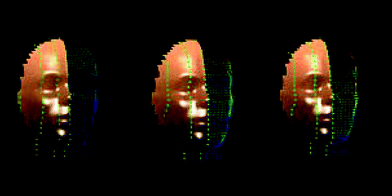
| Fig. 4 Dosimeter positions from left for SZA: 0°–30°; 30°–50°; 50°–80°. | ||
The developed 3D exposure grid has two significant advantages over previous facial exposure models. Firstly, coloured exposures are indicative of actual data values. There are no blending errors created by mixing coloured data with a background image of the headform. Secondly, as the grid accommodates closer dosimeter positions, the grid intersections represent actual measured exposures at specific dosimeter locations, reducing errors due to interpolating exposure ratios between dosimeter measurement points.
Exposure ratios and assigned colour values for each contour in the wireframe are represented as linear interpolated averages within each 5 mm segment separating dosimeter grid locations. Segments of a horizontal or vertical contour between any adjoining dosimeter intersection points are coloured on the wireframe mesh depending on the interpolated recorded exposure ratios of both dosimeters in an adjacent pair. Segments between adjacent grid intersections, for both vertical and horizontal contours are divided into 5 colour levels.
Results
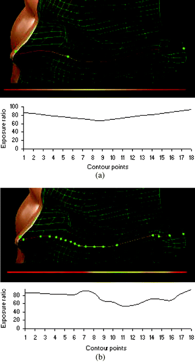
Fig. 5 illustrates the versatility and improvement in accuracy that can be achieved with future versions of the developed model when compared to previous models which have employed fewer dosimeters and linear interpolation techniques to represent large scale facial exposures. Fig. 5 provides example data which compare the predicted exposure along a horizontal contour connecting the ear, cheek and nose using only three measurement sites (Fig. 5(a)) compared to the same exposure contour calculated using 18 measurement sites (Fig. 5(b)). Note that the data provided for this contour comparison are intended for illustration purposes only and do not form part of the exposure data set used to produce the illustrations provided in Fig. 3. For clarity, Fig. 5 illustrates the change in colour along the contour underneath the 3D wireframe representation of the selected contour. Variations in the colour on this exposed contour clearly indicate that interpolation across varying facial topography is more accurate when a greater number of measurement locations are used. The comparison highlights the limitations of previous models which linearly interpolate exposures between widely spaced dosimeter sites. | ||
| Fig. 5 (a) Linearly interpolated exposure ratio map interpolated from three dosimeter measurements at positions CN1, CN9 and CN18 for the same horizontal contour plotted in Fig. 5(b). (b) Exposure ratio map represented across 18 dosimeter sites along a single horizontal contour connecting the nose, cheek and ear. | ||
By limiting the number of dosimeters used in creating a contour map, detail in the effective exposures along the contour can be lost if linear interpolation is used. Collection of further data along each horizontal and vertical contour used to create the entire facial wireframe is intended to be used with this model using the technique presented here to develop exposure profiles for each individual contour that match actual facial UV exposure distributions. The current computer model developed from this research allows accurate facial contour data to be collected under various conditions such that UV exposure profiles can be fitted to each contour which will allow measurement of exposures along contours that in the future utilise fewer dosimeters and simultaneously take into account variation in facial topography.
Fitting solutions to develop contour profiles
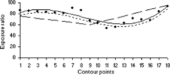
The modelled wireframe can be used to reduce the number of dosimeters required to plot an accurate facial exposure that takes into account facial topography. Fig. 6 represents a polynomial exposure profile fit to the horizontal contour extending from the ear through to the nose illustrated in Fig. 5(a). This exposure profile has been interpolated using a third order polynomial and was developed from 18 dosimeter exposures for a SZA in the range of 30° to 50° under clear sky conditions. Although a better fit to the data could be achieved with a higher order polynomial and could be used with the technique presented, a third order fit seems sufficient here for the purposes of illustration. Increasing the order of the polynomial fit to the data, while advantageous for this specific manikin headform may be unnecessary considering the detailed variation in individual facial topographies. Fig. 6 illustrates the benefit of using limited data collected at a later date within the same SZA range by fitting that data to the previously determined polynomial exposure profile, representing UV exposures based on facial topography rather than linear interpolation. | ||
| Fig. 6 Dot points—measured exposure ratios recorded along a single horizontal contour connecting the nose, cheek and ear. Solid line—third order polynomial fit exposure profile of the above data set. Small dashed line—example exposure profile developed from three dosimeter measurements utilising the previously measured exposure profile. Long dashed line—example linear interpolated exposure profile developed from the same three example dosimeter measurements. | ||
As an example, three exposures recorded at a later date at contour points CN1, CN9 and CN18 have been averaged and compared with the same three points that form part of the original data set which was used to develop the polynomial exposure profile for the contour illustrated in Fig. 5(a). The difference between the three averaged dosimeter points of the original data set and the average of the three example dosimeter measurements determines an offset value which is used to displace the original polynomial profile, representing the predicted exposure along the entire contour. Using this technique, three dosimeter measurements can estimate the exposure along a single contour based on previously collected data. In Fig. 6, the original data set polynomial profile (solid line) includes exposure ratios of 86 recorded at intersection point CN1, 67 recorded at intersection point CN9 and 94 recorded at intersection point CN18. Continuing the example, later measurements performed using only three dosimeters record exposure ratios of 76 at CN1, 60 at CN9 and 96 at CN18 resulting in a difference of −5 when averaged across all three dosimeter locations and compared with the same exposure locations of the original contour profile data. The resulting exposure contour estimate is plotted in Fig. 6 (small dashed line), displaced by −5 exposure ratio units from the solid polynomial profile.
Table 1 compares the effectiveness of linear interpolation with the averaged polynomial fitting technique described above to represent the estimated exposure across the horizontal contour of Fig. 5(a) when utilising only three measurement points. The estimated exposures of both the linear interpolation and averaged polynomial fit were compared at each point in the original data set. The comparison shows clearly that the error expressed relative to each of the original 18 measured dosimeter locations is significantly less for the polynomial fitting technique. As is also evident in Fig. 6, more advanced curve fitting techniques could be applied to the method outlined here to further reduce the relative error of future exposure profile estimates.
| Measured contour exposure ratio data provided from the original data set | Polynomial fit of CN1, CN9 and CN18 | Linear fit of CN1, CN9 and CN18 | ||
|---|---|---|---|---|
| Value | Relative error (%) | Value | Relative error (%) | |
| 86 | 81 | −6 | 76 | −12 |
| 85 | 80 | −6 | 74 | −13 |
| 84 | 79 | −6 | 72 | −14 |
| 83 | 78 | −6 | 70 | −16 |
| 82 | 77 | −6 | 68 | −17 |
| 81 | 76 | −6 | 66 | −19 |
| 91 | 86 | −5 | 64 | −30 |
| 86 | 81 | −6 | 62 | −28 |
| 67 | 62 | −7 | 60 | −10 |
| 64 | 59 | −8 | 64 | 0 |
| 54 | 49 | −9 | 68 | 26 |
| 56 | 51 | −9 | 72 | 29 |
| 63 | 58 | −8 | 76 | 21 |
| 72 | 67 | −7 | 80 | 11 |
| 70 | 65 | −7 | 84 | 20 |
| 68 | 63 | −7 | 88 | 29 |
| 84 | 79 | −6 | 92 | 10 |
| 94 | 89 | −5 | 96 | 2 |
Exposure ratio plots
Full facial exposure ratio plots include exposure data that have been averaged across different regions of the face to complete the graphical models presented in Fig. 3. For reference, Tables 2, 3 and 4 list the x–y–z grid intersection exposure ratio percentage levels used to plot the facial exposure ratio maps for each of the SZA ranges. Data in these tables, listed from the first row to the last correspond with exposure levels located on the top of the manikin headform through to the neck. The exposure percentage level has been arranged so that corresponding horizontal contour levels can also be read across each table. Currently, data on the 709 intersection point grid is averaged using linear interpolation between both vertical and horizontal contours. Measured dosimeter exposure values calculated from eqn (1) are listed in bold font, each of the other data points, representing grid intersections, listed in the tables are interpolated data. Fig. 3(b) (SZA 30°–50°, Table 3) and Fig. 3(c) (SZA 50°–80°, Table 4), did not utilise dosimeters placed on the ear. For simplicity, vertical contours CN15, CN16, CN17 and CN18 in Table 3 and Table 4 repeat exposures recorded along the side of the head measured by vertical contour CN14. This simplification would seem to be a reasonable estimate given all vertical contours after CN14 are orientated in approximately the same facial plane. Due to measurement density, dosimeters were also positioned along every second point of each measured vertical contour used to produce the plots of Fig. 3. The plots presented in Fig. 3 therefore illustrate the estimated exposure ratio for each of the atmospheric conditions listed previously within each SZA range. Future work will extend the model to include exposure data for all contours under clear sky and higher cloud cases.| CN1 nose | CN2 | CN3 | CN4 | CN5 | CN6 | CN7 eye | CN8 | CN9 | CN10 | CN11 | CN12 | CN13 | CN14 side | CN15 | CN16 | CN17 | CN18 ear |
|---|---|---|---|---|---|---|---|---|---|---|---|---|---|---|---|---|---|
| 100 | 100 | 100 | 100 | 100 | 100 | ||||||||||||
| 91 | 93 | 95 | 97 | 99 | 99 | 99 | 99 | ||||||||||
| 82 | 86 | 90 | 94 | 98 | 98 | 98 | 98 | 98 | 98 | ||||||||
| 73 | 77 | 81 | 84 | 88 | 84 | 81 | 77 | 73 | 70 | 66 | |||||||
| 64 | 68 | 71 | 75 | 78 | 75 | 73 | 70 | 68 | 65 | 63 | 63 | ||||||
| 55 | 58 | 61 | 64 | 67 | 65 | 64 | 63 | 62 | 60 | 59 | 59 | 59 | |||||
| 46 | 48 | 51 | 53 | 55 | 54 | 54 | 53 | 52 | 51 | 51 | 51 | 51 | 51 | ||||
| 52 | 53 | 53 | 54 | 55 | 52 | 50 | 48 | 46 | 44 | 42 | 42 | 42 | 42 | ||||
| 58 | 57 | 56 | 55 | 54 | 51 | 49 | 46 | 43 | 41 | 38 | 38 | 38 | 38 | 38 | |||
| 51 | 52 | 53 | 53 | 54 | 51 | 47 | 44 | 41 | 37 | 34 | 34 | 34 | 34 | 34 | |||
| 44 | 47 | 49 | 52 | 54 | 51 | 48 | 45 | 42 | 39 | 36 | 36 | 36 | 36 | 36 | |||
| 43 | 45 | 47 | 49 | 52 | 49 | 47 | 45 | 43 | 40 | 38 | 38 | 38 | 38 | 38 | 38 | ||
| 42 | 44 | 46 | 47 | 49 | 47 | 45 | 43 | 41 | 39 | 37 | 37 | 37 | 37 | 37 | 37 | ||
| 44 | 46 | 48 | 50 | 53 | 50 | 47 | 44 | 42 | 39 | 36 | 36 | 36 | 36 | 36 | 36 | ||
| 46 | 49 | 51 | 54 | 56 | 53 | 51 | 48 | 46 | 43 | 41 | 41 | 41 | 41 | 41 | 41 | ||
| 37 | 38 | 38 | 39 | 39 | 40 | 41 | 42 | 43 | 44 | 45 | 45 | 45 | 45 | 45 | 45 | ||
| 28 | 27 | 25 | 24 | 22 | 23 | 24 | 25 | 25 | 26 | 27 | 27 | 27 | 27 | 27 | 27 | ||
| 30 | 26 | 22 | 18 | 14 | 13 | 12 | 11 | 11 | 10 | 9 | 9 | 9 | 9 | 9 | 9 | ||
| 33 | 26 | 19 | 12 | 5 | 6 | 6 | 7 | 7 | 8 | 8 | 8 | 8 | 8 | 8 | 8 | ||
| 35 | 27 | 20 | 12 | 5 | 5 | 6 | 6 | 6 | 7 | 7 | 7 | 7 | 7 | 7 | 7 | ||
| 37 | 29 | 21 | 13 | 5 | 6 | 6 | 7 | 7 | 8 | 9 | 9 | 9 | 9 | 9 | 9 | ||
| 39 | 33 | 27 | 21 | 14 | 14 | 13 | 12 | 11 | 11 | 10 | 18 | 26 | 34 | 42 | 50 | 58 | |
| 42 | 37 | 33 | 28 | 24 | 23 | 23 | 23 | 23 | 22 | 22 | 26 | 30 | 33 | 37 | 41 | 45 | |
| 44 | 41 | 38 | 36 | 33 | 33 | 33 | 33 | 34 | 34 | 34 | 34 | 33 | 33 | 32 | 32 | 31 | 31 |
| 46 | 45 | 44 | 43 | 42 | 42 | 41 | 41 | 40 | 40 | 40 | 36 | 32 | 29 | 25 | 22 | 18 | 18 |
| 27 | 30 | 32 | 35 | 38 | 39 | 40 | 41 | 43 | 44 | 45 | 40 | 35 | 30 | 25 | 20 | 15 | 15 |
| 8 | 14 | 21 | 27 | 33 | 33 | 34 | 34 | 34 | 34 | 35 | 31 | 27 | 23 | 19 | 15 | 11 | 11 |
| 7 | 12 | 18 | 23 | 29 | 28 | 27 | 26 | 26 | 25 | 24 | 22 | 21 | 19 | 17 | 16 | 14 | 14 |
| 6 | 11 | 15 | 20 | 24 | 23 | 22 | 21 | 20 | 19 | 19 | 18 | 18 | 18 | 18 | 17 | 17 | |
| 18 | 21 | 24 | 27 | 30 | 27 | 24 | 22 | 19 | 16 | 13 | 14 | 14 | 15 | 16 | 16 | 17 | |
| 29 | 31 | 33 | 34 | 36 | 32 | 28 | 24 | 20 | 16 | 13 | 13 | 13 | 13 | 13 | 13 | ||
| 21 | 23 | 25 | 27 | 30 | 27 | 24 | 21 | 18 | 15 | 12 | 12 | 12 | 12 | 12 | 12 | ||
| 13 | 16 | 18 | 21 | 23 | 21 | 20 | 18 | 16 | 14 | 13 | 13 | 13 | 13 | 13 | 13 | ||
| 23 | 23 | 24 | 24 | 24 | 22 | 20 | 19 | 17 | 15 | 13 | 13 | 13 | 13 | 13 | |||
| 33 | 31 | 29 | 27 | 25 | 23 | 21 | 19 | 17 | 15 | 13 | 13 | 13 | |||||
| 20 | 19 | 19 | 18 | 18 | 17 | 16 | 15 | 14 | 13 | 12 | 12 | 12 | |||||
| 7 | 8 | 9 | 9 | 10 | 11 | 11 | 12 | 13 | 13 | 14 | 14 | 14 | |||||
| 13 | 14 | 15 | 16 | 18 | 17 | 17 | 17 | 17 | 16 | 16 | 16 | ||||||
| 18 | 20 | 22 | 23 | 25 | 23 | 21 | 20 | 18 | 16 | 14 | 14 | ||||||
| 24 | 23 | 22 | 22 | 21 | 20 | 18 | 17 | 15 | 14 | 12 | 12 | ||||||
| 29 | 26 | 23 | 20 | 17 | 16 | 15 | 15 | 14 | 13 | 12 | 12 | ||||||
| 19 | 17 | 16 | 14 | 13 | 13 | 13 | 13 | 12 | 12 | 12 | 12 | ||||||
| 8 | 8 | 9 | 9 | 9 | 10 | 11 | 12 | 13 | 14 | 16 | 16 | ||||||
| 5 | 6 | 7 | 8 | 9 | 10 | 12 | 14 | 16 | 17 | 19 | 19 | ||||||
| 2 | 4 | 5 | 7 | 9 | 10 | 12 | 14 | 16 | 17 | 19 | 19 | ||||||
| 3 | 4 | 5 | 7 | 8 | 10 | 12 | 14 | 15 | 17 | 19 | 19 | ||||||
| 3 | 4 | 6 | 7 | 8 | 10 | 12 | 14 | 16 | 18 | 20 | 20 | ||||||
| 5 | 6 | 7 | 8 | 10 | 11 | 13 | 15 | 17 | 18 | 20 | 20 | ||||||
| 6 | 7 | 9 | 10 | 11 | 13 | 15 | 17 | 19 | 21 | 23 | 23 | ||||||
| 6 | 7 | 9 | 10 | 11 | 13 | 16 | 18 | 20 | 23 | 25 | 25 |
| CN1 nose | CN2 | CN3 | CN4 | CN5 | CN6 | CN7 eye | CN8 | CN9 | CN10 | CN11 | CN12 | CN13 | CN14 side | CN15 | CN16 | CN17 | CN18 ear |
|---|---|---|---|---|---|---|---|---|---|---|---|---|---|---|---|---|---|
| 100 | 100 | 100 | 100 | 100 | 100 | ||||||||||||
| 100 | 100 | 100 | 100 | 100 | 100 | 100 | 100 | ||||||||||
| 93 | 94 | 95 | 97 | 98 | 99 | 100 | 100 | 100 | 100 | ||||||||
| 86 | 88 | 91 | 93 | 95 | 98 | 100 | 100 | 100 | 100 | 100 | |||||||
| 78 | 82 | 85 | 89 | 93 | 96 | 100 | 100 | 100 | 100 | 100 | 100 | ||||||
| 71 | 75 | 79 | 83 | 87 | 91 | 95 | 95 | 95 | 95 | 95 | 95 | 95 | |||||
| 68 | 72 | 75 | 79 | 83 | 86 | 90 | 90 | 89 | 89 | 89 | 89 | 88 | 88 | ||||
| 65 | 68 | 71 | 75 | 78 | 81 | 84 | 85 | 85 | 86 | 86 | 87 | 87 | 88 | ||||
| 64 | 67 | 69 | 72 | 74 | 77 | 79 | 79 | 79 | 79 | 79 | 79 | 79 | 79 | 79 | |||
| 62 | 63 | 64 | 66 | 67 | 68 | 69 | 69 | 69 | 69 | 70 | 70 | 70 | 70 | 70 | |||
| 64 | 63 | 62 | 61 | 60 | 59 | 58 | 59 | 59 | 60 | 60 | 61 | 61 | 62 | 62 | |||
| 66 | 66 | 67 | 67 | 67 | 68 | 68 | 66 | 64 | 62 | 59 | 57 | 55 | 53 | 53 | 53 | ||
| 67 | 69 | 70 | 72 | 74 | 75 | 77 | 73 | 70 | 66 | 63 | 59 | 56 | 52 | 52 | 52 | ||
| 69 | 71 | 72 | 74 | 76 | 77 | 79 | 75 | 71 | 67 | 63 | 59 | 55 | 51 | 51 | 51 | ||
| 67 | 70 | 72 | 75 | 77 | 80 | 82 | 77 | 72 | 67 | 61 | 56 | 51 | 46 | 46 | 46 | ||
| 66 | 63 | 60 | 57 | 53 | 50 | 47 | 46 | 45 | 44 | 43 | 42 | 41 | 40 | 40 | 40 | ||
| 51 | 45 | 38 | 32 | 25 | 19 | 12 | 16 | 20 | 24 | 29 | 33 | 37 | 41 | 41 | 41 | ||
| 35 | 31 | 28 | 24 | 20 | 17 | 13 | 17 | 21 | 25 | 30 | 34 | 38 | 42 | 42 | 42 | ||
| 37 | 33 | 29 | 25 | 21 | 17 | 13 | 18 | 22 | 27 | 31 | 36 | 40 | 45 | 45 | 45 | ||
| 38 | 35 | 31 | 28 | 24 | 21 | 17 | 21 | 26 | 30 | 35 | 39 | 44 | 48 | 48 | 48 | ||
| 60 | 54 | 47 | 41 | 34 | 28 | 21 | 24 | 28 | 31 | 35 | 38 | 42 | 45 | 45 | 45 | ||
| 82 | 73 | 64 | 55 | 45 | 36 | 27 | 29 | 31 | 33 | 36 | 38 | 40 | 42 | 42 | 42 | 42 | |
| 90 | 81 | 71 | 62 | 52 | 43 | 33 | 36 | 38 | 41 | 43 | 46 | 48 | 51 | 51 | 51 | 51 | |
| 98 | 91 | 85 | 78 | 71 | 65 | 58 | 58 | 59 | 59 | 59 | 59 | 60 | 60 | 60 | 60 | 60 | 60 |
| 77 | 78 | 79 | 80 | 81 | 82 | 83 | 79 | 75 | 71 | 66 | 62 | 58 | 54 | 54 | 54 | 54 | 54 |
| 56 | 58 | 60 | 62 | 63 | 65 | 67 | 64 | 61 | 58 | 56 | 53 | 50 | 47 | 47 | 47 | 47 | 47 |
| 32 | 35 | 38 | 42 | 45 | 48 | 51 | 49 | 48 | 46 | 45 | 43 | 42 | 40 | 40 | 40 | 40 | 40 |
| 9 | 15 | 21 | 27 | 33 | 39 | 45 | 43 | 41 | 39 | 38 | 36 | 34 | 32 | 32 | 32 | 32 | 32 |
| 20 | 23 | 26 | 30 | 33 | 36 | 39 | 38 | 37 | 36 | 35 | 34 | 33 | 32 | 32 | 32 | 32 | |
| 31 | 34 | 36 | 39 | 42 | 44 | 47 | 45 | 43 | 41 | 38 | 36 | 34 | 32 | 32 | 32 | 32 | |
| 49 | 50 | 51 | 52 | 52 | 53 | 54 | 51 | 48 | 45 | 41 | 38 | 35 | 32 | 32 | 32 | ||
| 67 | 64 | 60 | 57 | 54 | 50 | 47 | 45 | 42 | 40 | 38 | 36 | 33 | 31 | 31 | 31 | ||
| 39 | 39 | 39 | 40 | 40 | 40 | 40 | 37 | 34 | 31 | 27 | 24 | 21 | 18 | 18 | 18 | ||
| 12 | 16 | 20 | 24 | 28 | 32 | 36 | 32 | 27 | 23 | 19 | 15 | 10 | 6 | 6 | |||
| 31 | 31 | 31 | 31 | 31 | 31 | 31 | 31 | 31 | 31 | 31 | 31 | 31 | |||||
| 50 | 47 | 43 | 40 | 36 | 33 | 29 | 29 | 29 | 29 | 29 | 29 | 29 | |||||
| 30 | 29 | 29 | 28 | 27 | 27 | 26 | 26 | 26 | 26 | 26 | 26 | 26 | |||||
| 11 | 15 | 19 | 23 | 26 | 30 | 34 | 34 | 34 | 34 | 34 | 34 | ||||||
| 32 | 34 | 35 | 37 | 39 | 40 | 42 | 42 | 42 | 42 | 42 | 42 | ||||||
| 54 | 52 | 49 | 47 | 45 | 42 | 40 | 40 | 40 | 40 | 40 | 40 | ||||||
| 44 | 43 | 42 | 41 | 40 | 39 | 38 | 38 | 38 | 38 | 38 | 38 | ||||||
| 34 | 34 | 33 | 33 | 33 | 32 | 32 | 32 | 32 | 32 | 32 | 32 | ||||||
| 21 | 22 | 23 | 24 | 24 | 25 | 26 | 26 | 26 | 26 | 26 | 26 | ||||||
| 8 | 10 | 12 | 14 | 15 | 17 | 19 | 19 | 19 | 19 | 19 | 19 | ||||||
| 8 | 9 | 10 | 11 | 11 | 12 | 13 | 13 | 13 | 13 | 13 | 13 | ||||||
| 8 | 10 | 12 | 14 | 15 | 17 | 19 | 19 | 19 | 19 | 19 | 19 | ||||||
| 8 | 11 | 13 | 16 | 19 | 21 | 24 | 24 | 24 | 24 | 24 | 24 | ||||||
| 10 | 12 | 15 | 17 | 19 | 22 | 24 | 24 | 24 | 24 | 24 | 24 | ||||||
| 12 | 14 | 16 | 19 | 21 | 23 | 25 | 25 | 25 | 25 | 25 | 25 | ||||||
| 12 | 14 | 16 | 19 | 21 | 23 | 25 | 25 | 25 | 25 | 25 | 25 |
| CN1 nose | CN2 | CN3 | CN4 | CN5 | CN6 | CN7 eye | CN8 | CN9 | CN10 | CN11 | CN12 | CN13 | CN14 side | CN15 | CN16 | CN17 | CN18 ear |
|---|---|---|---|---|---|---|---|---|---|---|---|---|---|---|---|---|---|
| 100 | 100 | 100 | 100 | 100 | 100 | ||||||||||||
| 100 | 100 | 100 | 100 | 100 | 100 | 73 | 73 | ||||||||||
| 100 | 98 | 96 | 93 | 91 | 89 | 87 | 87 | 87 | 87 | ||||||||
| 100 | 100 | 100 | 100 | 100 | 100 | 100 | 100 | 100 | 100 | 100 | |||||||
| 100 | 99 | 97 | 96 | 95 | 93 | 92 | 92 | 92 | 92 | 92 | 92 | ||||||
| 89 | 88 | 87 | 87 | 86 | 85 | 84 | 84 | 84 | 84 | 84 | 84 | 84 | |||||
| 78 | 79 | 79 | 80 | 81 | 81 | 82 | 81 | 80 | 79 | 78 | 77 | 76 | 75 | ||||
| 86 | 85 | 84 | 83 | 82 | 81 | 80 | 78 | 77 | 75 | 74 | 72 | 71 | 69 | ||||
| 94 | 93 | 91 | 90 | 88 | 87 | 85 | 82 | 79 | 76 | 72 | 69 | 66 | 63 | 63 | |||
| 91 | 91 | 91 | 91 | 90 | 90 | 90 | 89 | 88 | 86 | 85 | 84 | 83 | 82 | 82 | |||
| 88 | 87 | 86 | 85 | 84 | 83 | 83 | 85 | 88 | 90 | 93 | 95 | 98 | 100 | 100 | |||
| 88 | 86 | 84 | 82 | 79 | 77 | 75 | 76 | 78 | 79 | 80 | 81 | 83 | 84 | 84 | 84 | ||
| 88 | 87 | 85 | 84 | 82 | 81 | 79 | 77 | 76 | 74 | 73 | 71 | 70 | 68 | 68 | 68 | ||
| 90 | 89 | 88 | 87 | 85 | 84 | 83 | 80 | 77 | 73 | 70 | 67 | 64 | 61 | 61 | 61 | ||
| 92 | 90 | 89 | 87 | 86 | 84 | 83 | 78 | 74 | 70 | 66 | 61 | 57 | 53 | 53 | 53 | ||
| 74 | 75 | 77 | 78 | 79 | 81 | 82 | 77 | 71 | 66 | 60 | 55 | 49 | 44 | 44 | 44 | ||
| 56 | 54 | 53 | 51 | 50 | 48 | 47 | 45 | 43 | 41 | 39 | 38 | 36 | 34 | 34 | 34 | ||
| 49 | 42 | 36 | 30 | 24 | 17 | 11 | 19 | 26 | 34 | 41 | 49 | 56 | 64 | 64 | 64 | ||
| 41 | 38 | 35 | 32 | 29 | 26 | 23 | 33 | 43 | 53 | 63 | 74 | 84 | 94 | 94 | 94 | ||
| 54 | 51 | 47 | 44 | 41 | 37 | 34 | 39 | 43 | 48 | 52 | 57 | 61 | 66 | 66 | 66 | ||
| 67 | 62 | 57 | 53 | 48 | 43 | 38 | 38 | 38 | 38 | 37 | 37 | 37 | 37 | 37 | 37 | ||
| 84 | 77 | 70 | 63 | 56 | 49 | 42 | 44 | 46 | 48 | 51 | 53 | 55 | 57 | 57 | 57 | 57 | |
| 100 | 92 | 84 | 75 | 67 | 59 | 51 | 54 | 58 | 62 | 66 | 69 | 73 | 77 | 77 | 77 | 77 | |
| 67 | 65 | 64 | 63 | 62 | 60 | 59 | 59 | 59 | 60 | 60 | 60 | 60 | 61 | 61 | 61 | 61 | 61 |
| 33 | 39 | 45 | 51 | 57 | 63 | 69 | 65 | 62 | 58 | 55 | 51 | 48 | 44 | 44 | 44 | 44 | 44 |
| 29 | 37 | 46 | 54 | 62 | 71 | 79 | 74 | 69 | 64 | 59 | 54 | 49 | 44 | 44 | 44 | 44 | 44 |
| 25 | 32 | 39 | 46 | 53 | 60 | 68 | 64 | 61 | 57 | 54 | 50 | 47 | 43 | 43 | 43 | 43 | 43 |
| 23 | 29 | 34 | 40 | 45 | 51 | 56 | 55 | 55 | 54 | 54 | 53 | 53 | 52 | 52 | 52 | 52 | 52 |
| 21 | 27 | 32 | 38 | 43 | 49 | 54 | 55 | 56 | 57 | 58 | 59 | 60 | 61 | 61 | 61 | 61 | |
| 43 | 45 | 46 | 48 | 49 | 51 | 52 | 51 | 50 | 49 | 48 | 47 | 46 | 46 | 46 | 46 | 46 | |
| 65 | 63 | 62 | 60 | 59 | 57 | 56 | 52 | 48 | 45 | 41 | 37 | 34 | 30 | 30 | 30 | ||
| 52 | 53 | 54 | 56 | 57 | 58 | 59 | 55 | 51 | 47 | 44 | 40 | 36 | 32 | 32 | 32 | ||
| 39 | 41 | 43 | 46 | 48 | 50 | 52 | 49 | 47 | 44 | 42 | 39 | 37 | 34 | 34 | 34 | ||
| 52 | 50 | 49 | 48 | 47 | 46 | 45 | 43 | 42 | 40 | 39 | 37 | 36 | 34 | 34 | |||
| 64 | 60 | 57 | 53 | 49 | 46 | 42 | 42 | 42 | 42 | 42 | 42 | 42 | |||||
| 43 | 42 | 42 | 41 | 40 | 40 | 39 | 39 | 39 | 39 | 39 | 39 | 39 | |||||
| 22 | 25 | 28 | 31 | 34 | 37 | 40 | 40 | 40 | 40 | 40 | 40 | 40 | |||||
| 29 | 31 | 33 | 35 | 37 | 39 | 41 | 41 | 41 | 41 | 41 | 41 | ||||||
| 36 | 39 | 41 | 44 | 47 | 49 | 52 | 52 | 52 | 52 | 52 | 52 | ||||||
| 49 | 51 | 54 | 56 | 58 | 61 | 63 | 63 | 63 | 63 | 63 | 63 | ||||||
| 62 | 59 | 56 | 53 | 50 | 47 | 44 | 44 | 44 | 44 | 44 | 44 | ||||||
| 46 | 42 | 39 | 35 | 32 | 28 | 25 | 25 | 25 | 25 | 25 | 25 | ||||||
| 29 | 27 | 25 | 23 | 21 | 19 | 17 | 17 | 17 | 17 | 17 | 17 | ||||||
| 18 | 16 | 15 | 13 | 11 | 10 | 8 | 8 | 8 | 8 | 8 | 8 | ||||||
| 7 | 7 | 6 | 6 | 5 | 5 | 4 | 4 | 4 | 4 | 4 | 4 | ||||||
| 1 | 1 | 1 | 1 | 0 | 0 | 0 | 0 | 0 | 0 | 0 | 0 | ||||||
| 7 | 7 | 7 | 8 | 8 | 8 | 8 | 8 | 8 | 8 | 8 | 8 | ||||||
| 13 | 14 | 14 | 15 | 15 | 16 | 16 | 16 | 16 | 16 | 16 | 16 | ||||||
| 21 | 22 | 23 | 24 | 25 | 26 | 27 | 27 | 27 | 27 | 27 | 27 | ||||||
| 28 | 30 | 31 | 33 | 34 | 36 | 37 | 27 | 37 | 37 | 37 | 37 |
Erythemal headform exposure
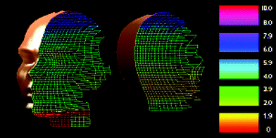
Apart from the development of exposure ratio estimates and contour exposure profiles, the developed model provides a suitable framework onto which biologically effective exposures can be directly plotted. To demonstrate the versatility of the model wireframe, calibrated erythemal exposures have been plotted onto a 3D wireframe model of the entire headform, including the back of head and neck. Fig. 7 is a three dimensional visualisation of a 300 min exposure recorded at the same location used to produce the SZA exposure ratio plots of Fig. 3. The figure has been developed from 63 dosimeter exposures placed on the headform. Grid intersection points not directly measured have been linearly interpolated. For this exposure, cumulus cloud cover measured less than 2 oktas, the average surface albedo was recorded as 5% over the entire exposure interval. Stratospheric ozone concentration listed by the total ozone mapping spectrometer10 during the exposure interval was listed as 284 DU. For the figure, erythemal exposure is given in minimal erythemal dose (MED) where 1 MED corresponds to 200 J m−2 of erythemally effective UV. The absolute change in optical absorbency of the miniaturised dosimeters measured at 330 nm was calibrated to a portable SUV meter, (model 3D, Solar Light Co. USA). The resulting calibration curve used to determine the MED of each dosimeter placed on the manikin headform is provided in Fig. 8. For comparison, the miniaturised dosimeter calibration curve is given with a calibration curve that utilises larger dosimeters. Fig. 8 indicates that the optical absorbency of the miniaturised dosimeters is slightly higher than the larger aperture dosimeters. The average increase in optical absorbency at 330 nm compared to the larger aperture dosimeters was measured as +0.014. This corresponds to a relative increase of 6% when compared to the highest change in absorbency recorded in the 300 min exposure interval. | ||
| Fig. 7 Erythemal exposure (MED) recorded 7 July, 11:30–16:30. | ||
 | ||
| Fig. 8 Dot points—the larger aperture dosimeter changes in optical absorbency at 330 nm. Cross points—6 mm aperture dosimeter changes in optical absorbency at 330 nm. Dashed line—erythemal exposure calibration curve fitted to 6 mm aperture dosimeter data. | ||
Conclusions
The three dimensional UV exposure model developed from this research improves upon previous methods used to predict UV exposures using dosimeters and manikin headforms. The developed facial wireframe has reduced interpolation errors resulting from variation in facial topography. A technique for plotting UV exposures to a much higher resolution than has been achieved previously has been explained. Significantly, this research extends previous work by providing a model that takes physical topographic facial features and the resulting shading caused by those features into account.Combinations of dosimeters can be placed in up to 709 frontal facial locations and an additional 576 rear headform locations using the model wireframe. The developed model can accommodate various biological action spectra and has demonstrated its effectiveness in producing accurate UV facial hot-spot maps. The results show a clear broadening in facial exposure with increasing SZA affecting the lower proximities of the face. Similar studies which take into account variation in the environment, surface albedo, head tilt angle and hats or eyewear worn by the manikin could be represented effectively using the developed 3D facial model. The development of biologically effective facial exposure maps using this model could be linked to the frequency and incidence of facial skin cancers. Additionally, an examination of ocular exposure utilising a higher density grid could be developed using the method described here provided dosimeter sizes can be reduced to provide the required accuracy.
As part of the method, a technique has been described for interpolating dosimeter exposures across individual contours. The technique, demonstrated for a single contour here could be applied to all contours in future versions of the model wireframe enabling accurate exposures to be determined with a much greater reduction in the total number of dosimeters required.
Acknowledgements
The authors would like to thank the technical staff at the University of Southern Queensland who have assisted with the development and maintenance of the equipment required for this research.References
- B. L. Diffey, Solar ultraviolet radiation effects on biological systems, Phys. Med. Biol., 1991, 36(3), 299–328 CrossRef CAS.
- WMO (World Meteorological Organization), Report of the WMO meeting of experts on UV-B measurements, data quality and standardization of UV indices, WMO/TD-NO 625, WMO, Les Diablerets, Switzerland, 25–28 July, 1994 Search PubMed.
- N. J. Downs, M. G. Kimlin, A. V. Parisi and J. J. McGrath, Modelling Human Facial UV Exposure, Radiat. Prot. Australas., 2001, 17(3), 103–109 Search PubMed.
- M. G. Kimlin, A. V. Parisi and J. C. F. Wong, The facial distribution of erythemal ultraviolet exposure in south east Queensland, Phys. Med. Biol., 1998, 43(2), 231–240 CrossRef CAS.
- P. Hoeppe, A. Oppenrieder, C. Erianto, P. Koepke, J. Reuder, M. Seefeldner and D. Nowak, Visualization of UV exposure of the human body based on data from a scanning UV-measuring system, Int. J. Biometeorol., 2004, 49, 18–25 Search PubMed.
- A. V. Parisi, L. R. Meldrum, J. C. F. Wong, J. Aitkin and R. A. Fleming, Lifetime erythemal ultraviolet exposure estimates for selected population groups in south east Queensland, Phys. Med. Biol., 1999, 44(12), 2947–2953 CrossRef CAS.
- A. V. Parisi, L. R. Meldrum and M. G. Kimlin, Polysulfone film thickness and its effects in ultraviolet radiation dosimetry, Protection Against the Hazards of UVR, Internet Conference, 18 Jan–5 Feb 1999 Search PubMed.
- CIE (International Commission on Illumination), A reference action spectrum for ultraviolet induced erythema in human skin, Comm. Int. Eclairage J., 1987, 6, 17–22 Search PubMed.
- D. K. Airey, J. C. F. Wong and R. A. Fleming, A comparison of human- and headform-based measurements of solar ultraviolet B dose, Photodermatol. Photoimmunol. Photomed., 1995, 11(4), 155–158 CAS.
- Total Ozone Mapping Spectrometer, National Aeronautics and Space Administration, 2006, (viewed 5 August 2006: http://jwocky.gsfc.nasa.gov/).
| This journal is © The Royal Society of Chemistry and Owner Societies 2007 |
