Photoelectrochemical properties of supramolecular species containing porphyrin and ruthenium complexes on TiO2 films
Ana F.
Nogueira
,
André Luiz B.
Formiga
,
Herbert
Winnischofer
,
Marcelo
Nakamura
,
Fábio M.
Engelmann
,
Koiti
Araki
and
Henrique E.
Toma
*
Instituto de Química – Universidade de São Paulo. C. P. 26077, CEP 05513-970, São Paulo – Brazil. Fax: 55 11 3815 5579; Tel: 55 11 3091 3887
First published on 9th September 2003
Abstract
Modification of wide band gap semiconductor surfaces by a new generation of supramolecular sensitizers, combining porphyrin and ruthenium–phenanthroline complexes leads to versatile molecular interfaces, allowing the exploitation of photoinduced charge transfer in dye sensitized photoelectrochemical cells. meso-Tetrapyridylporphyrin coordinated to two ruthenium complexes converts 21% of the incident photons into current after excitation at the Soret band. In this work we discuss the electron/energy transfer mechanisms involved in the TiO2 sensitization by these supramolecular species, invoking some theoretical calculations.
Introduction
Dye sensitized photoelectrochemical cells (DSSCs) emerge as a promising and interesting alternative to the well established solid state photovoltaic devices to generate cheap and feasible electricity from the Sun.1 Since the announcement of 10% efficiency,2,3 DSSCs have attracted the attention of several research groups that have dedicated their efforts into improving and investigating the fundamental aspects of the cell components. One of the most studied topics involves the use of different dyes as sensitizers for the nanoporous TiO2 network. The electrochemical and photophysical properties of the ground and excited state of the dyes play an important role for charge transfer dynamics at the semiconductor interface. The study on sensitization has essentially been confined to metal polypyridine complexes and their analogues.2–5The complex cis-di(thiocyanato)bis(2,2′-bipyridyl-4,4′-dicarboxylate)ruthenium(II) (referred as N3) is far the most employed and efficient sensitizer, converting 80–85% of the incident photons into current.2 However the main drawback of this dye is the lack of absorption in the infrared region of the spectrum, limiting the overall efficiency. Several groups have been focusing on their efforts to overcome this issue. An interesting approach is the use of polynuclear Ru(II) complexes in a elaborate supramolecular design, whose properties are based on the photoinduced charge separation and antenna effect.6,7 In the same direction, in past years a number of organic dyes with high absorption in the infrared region such as porphyrins,3,8–11 phthalocyanines,12 perylenes,13 and xanthenes14 have been employed as sensitizers, however the efficiency remains low in comparison to metal complexes. It is still unclear why these dyes have limited ability to inject electrons in the conduction band of n-type oxide semiconductors.15,16 One reasonable explanation is that their excited states composed by singlet/triplet states are π–π* and not charge transfer like as in the metal complexes. Another interesting issue recently discussed by Odobel et al.17 concerns the range of monochromatic photon-to-electron conversion efficiency (IPCE) values from 10 to 80%.
Porphyrins and related macrocycles such as meso-tetrapyridylporphyrins (TpyP) and meso-tetraphenylporphyrins (TPP) have been extensively employed as building blocks for assembling supramolecular systems, stimulated by their photochemical, electrochemical and catalytic properties.18–21 In the last decade we have dedicated our efforts in the preparation and characterization of these species coordinated to four molecules of [Ru(bipy)2Cl]+, [Ru(phen)2Cl]+, and other ruthenium complexes in solution and as thin molecular films.22–24 Our motivation to develop such complex supramolecular structures comes from the possibility of combining the excellent properties found in metal complexes and the stability, rich photochemical and photophysical properties of the porphyrins. Our previous studies in solution indicate that the supramolecular species containing four [Ru(bipy)2Cl]+ (bipy = bipyridine) coordinated to TPyP are able to transfer energy from the peripheral ruthenium complexes to the porphyrin ring so promoting their luminescence.25 Energy transfer from the porphyrin ring to the ruthenium complexes was also observed in the supramolecule composed by four [Ru(bipy)3] groups due to the proximity of the porphyrin and ruthenium energy levels.26
In this work we investigated the photoelectrochemical behavior of TPyP coordinated to two and four [Ru(phen)2Cl]+ (phen = phenanthroline) groups (referred as Ru2phenTPyP and Ru4phenTPyP, Fig. 1).27 We carried out a systematic investigation, focusing on the photoinduced charge separation and antenna effect effects presented by these supramolecules into assembling molecular electrical interfaces with semiconductor surfaces. Our studies are based on their corresponding photoaction response and theoretical calculations.
![Molecular structure of the supermolecular species constituted by (a) four [Ru(phen)2Cl]+
(phen = phenanthroline) coordinated to meso-tetrapyridylporphyrin (referred as Ru4phenTPyP); (b) by two ruthenium complexes in cis configuration (cis-Ru2phenZnTPyP) and (c) in trans configuration (trans-Ru2phenZnTPyP).](/image/article/2004/PP/b306702e/b306702e-f1.gif) | ||
| Fig. 1 Molecular structure of the supermolecular species constituted by (a) four [Ru(phen)2Cl]+ (phen = phenanthroline) coordinated to meso-tetrapyridylporphyrin (referred as Ru4phenTPyP); (b) by two ruthenium complexes in cis configuration (cis-Ru2phenZnTPyP) and (c) in trans configuration (trans-Ru2phenZnTPyP). | ||
Experimental
Materials
The synthesis and characterization of the Ru4phenTPyP supermolecule containing four [Ru(phen)2Cl]+ complexes is detailed in ref. 27. Analysis for Ru4phenTPyP (C140H112N24Ru4O23S4F12Cl4): found (calc.): C, 49.7 (49.4); N, 9.4 (9.8); H, 3.7 (3.3). The synthesis of the supramolecule containing only two ruthenium complexes was carried out adding half of the amount of ruthenium complexes in a proportion of 2 ∶ 1 [Ru(phen)2Cl2] ∶ TPyP. This strategy allowed the preparation of a supramolecule that contains free pyridyl groups and ruthenium complexes in the same structure. However, the coordination reaction may give rise to a mixture of structures containing 1, 2, or 3 peripheral groups. Elemental analysis and electronic spectrum suggest that the major product is a supermolecule with two complexes. In addition the supermolecule Ru2phenTPyP can be obtained as cis and trans isomers as shown in Fig. 1. Analysis for Ru2phenTPyP (C90H72N16Ru2O13S2F6Cl2): found (calc.): C, 53.4 (53.1); N, 10.6 (11.0); H, 4.1 (3.6). The TiO2 powder (P25) was kindly donated by Degussa AG. All other reagent grade chemicals were used without further purification.Preparation of nanoporous TiO2 films
The nanoporous TiO2 film deposited onto a sheet of F-doped SnO2 TEC15 conducting glass (sheet resistance ∼15 Ω cm−2) was prepared using a variation of a previously published procedure.2 A colloidal TiO2 suspension was obtained by grinding 6 g of TiO2 powder in 2 mL of distilled H2O and 0.2 mL of acetylacetone, in a mortar with a pestle, for about 40 min. Finally 8.0 mL of distilled water and 0.1 mL of Triton X-100 were slowly added with continuous mixing for 10 min.A previously cleaned conductive glass sheet was laid on a flat surface with the conductive side up and the deposition area was delimited with a plastic adhesive tape. Several drops of the above TiO2 suspension were spread as evenly as possible onto the surface using a glass rod. The thickness of the TiO2 coating (∼4 or 8 µm) was determined by the thickness of the adhesive tape. The electrodes were then dried in air and fired at 450 °C for 30 min under ambient atmosphere.
Surface treatment with the dye
The electrodes covered by a nanoporous TiO2 film layer were immersed for 48 h in ∼1 × 10⁻⁴ mol L−1 solution of the Ru2phenTPyP and Ru4phenTPyP supramolecular species in ethanol to achieve the adsorption equilibrium condition. The surface coverage was roughly estimated by the optical density of the film and color change from white to the color of the attached dye. Afterwards, the electrode was washed with the solvent and dried in moisture-free air.Assembling the cell
The cell (1.0 cm2 active area) was assembled by transferring about 1 mL of the electrolyte solution (0.5 mol L−1tert-butylpyridine–0.6 mol L−1 tetrabutylammonium iodide–0.1 mol L−1 LiI–0.1 mol L−1 I2 in 10 mL methoxyproprionitrile) onto the SnO2:F− glass covered with TiO2/attached dye. The counter-electrode was then pressed against the TiO2/dye film. The transparent counter electrode was treated by transferring a drop of 0.05 M solution of H2PtCl6 in isopropanol onto the conducting glass and firing at 400 °C in air for 20 min. A PVC film was placed in between the two electrodes to avoid short circuiting and electrolyte leakage. The assembly was kept together by a clamp. No further sealing was necessary, at least for the tests described in this work.Measurement of the photoelectrochemical properties
The photoelectrochemical devices were firmly placed in an optical bench for the characterization procedures. The IPCE spectra and the current–voltage (I–V) curves were obtained using an Applied Photophysics 150 W Xenon lamp as a white-light source and a home-made resistor as a variable load. Samples were irradiated at 100 mW cm−2. An Oriel Spectral Luminator was employed as the monochromatic source. The light intensity was varied from 1 to 2 mW cm−2. Measurements of photocurrent and photovoltage were performed using a Keithley 610C electrometer or a calibrated multimeter. The sample was always illuminated through the conducting glass substrate and no corrections were made for the 30% reflection and transmission losses in the SnO2:F− glass. The poly- and monochromatic light intensity at the electrode position were measured with a Newport Optical Power Meter model 1830-C.Electronic and emission spectra
The UV-vis spectra were obtained in a HP-8453A diode array spectrophotometer in ethanol solution. The emission spectra were recorded on a Photon Technology Inc. model LS100 spectrofluorimeter in N2 gas saturated ethanol solution.Electrochemistry
The electrochemistry was carried out in 0.1 M [(C2H5)4N]ClO4 acetonitrile solution using a potentiostat/galvanostat from Ecochemie, model AUTOLAB PGSTAT30. A conventional three-electrode cell consisting of a platinum disk working electrode, Ag|Ag+ 0.010 M in acetonitrile (E0 = 0.503 V vs. NHE) reference electrode and a coiled platinum wire as counter electrode was used.Theoretical calculations
Molecular modeling calculations were carried out for the cis-Ru2phenTPyP and Ru4phenTPyP species, starting with the MM+ module, a modified MM2 force field28 for geometry optimization, within the HyperChemTM program,29 and using a gradient of 1 × 10⁻⁵ kcal Å−1 mol−1 as a convergence criterion in a conjugated gradient method. SCF molecular orbital calculations were performed at the RHF level, using the ZINDO/S method,30 and the default parameters (and scaling factors kpσ = 1.267 and kpπ = 0.585). Geometry optimization was refined again, using the atomic charge distribution from the semi-empirical calculations, and the procedure repeated many times, until reaching convergence. The electronic spectrum was calculated, based on single CI excitations in an active space involving 25 frontier molecular orbitals (15 highest occupied and 10 lowest unoccupied MOs).Results and discussion
Fig. 2 shows the energy diagram for nanocrystalline TiO2 solar cells sensitized by RuphenTPyP species, together with kinetic processes after dye excitation. The potentials of the highest occupied molecular orbital (HOMO) and lowest unoccupied molecular orbital (LUMO) estimated electrochemically are 0.98 and −1.0 V vs. NHE, respectively, for Ru2phenTPyP and 0.98 and −1.1 V vs. NHE, respectively, for Ru4phenTPyP. The conduction band of the TiO2 electrode is at −0.5 V and the redox potential of I3−/I− is 0.4 V vs. NHE.3,15 According to the values in the energy diagram, the electron injection and the regeneration processes are energetically favorable. The free energy calculated for process (2) and (3) are −ΔG ≥ 0.5 eV and −ΔG = 0.6 eV, respectively. The energy levels were calculated from the ground state (Ru(II)/Ru(III) potential and E0–0 value, considering that after dye excitation (1) and electron injection to TiO2 (2), the hole is localized on one of the ruthenium moieties of the supramolecular porphyrin, in agreement with electrochemical data.22,27 Accordingly, the back electron transfer and dye regeneration processes are being carried out by the photochemically generated peripheral Ru(III) complexes. This is very convenient since ruthenium(III)–polypyridine complexes are known to be more stable than the porphyrin cation radicals and are able to exchange electrons reversibly.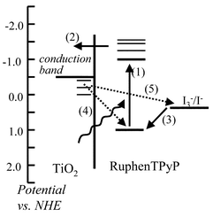 | ||
| Fig. 2 Schematic representation of the energy diagram of the dye sensitized photoelectrochemical cell (DSCC) and the most important kinetic processes after dye excitation. | ||
Typical AFM images of 4 µm thickness TiO2 (left) and TiO2/Ru2phenTPyP (right) nanoporous films deposited onto a F−-doped SnO2 substrate can be seen in Fig. 3. Both films show a rather uniform distribution of nanosized TiO2 pyramid-like crystals characteristic of the anatase structure. The TiO2 film (left) presents particles with diameters from 20 to 50 nm that are agglomerated forming typical “clusters”. After deposition of the supramolecule, the nanostructure of the TiO2/Ru2phenTPyP films is maintained, however, it appears more smooth and uniform showing particles with apparent increase in the average size up to 100 nm. This can be attributed to a coating effect, responsible for the disappearance of boundaries between the nanoparticles that constitute the “clusters”. Taking into account that the radii of the Ru2phenTPyP species are 23 and 30 Å for cis and trans isomers, respectively, and that the average pore dimensions is about 15 nm,3,31 the TiO2 matrix allows the supramolecules to be effectively incorporated within the pores.
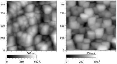 | ||
| Fig. 3 MAC Mode images of TiO2 (left) and TiO2/Ru2phenTPyP film (right). | ||
The UV-vis spectra of the supramolecular species in ethanol solution were similar to the sum of the ruthenium complex and corresponding porphyrin spectra, as shown in Fig. 4, together with emission spectra. For Ru2phenTPyP, the spectrum (Fig. 4(a)) has an intense Soret band at 415 nm and four Q bands at 510, 551, 589 and 648 nm in addition to the MLCT2 band of the peripheral ruthenium complexes (RuII (dπ) → phen (pπ*)) at 470 nm (shoulder). The Ru4phenTPyP spectrum (Fig. 4(b)) has a Soret band, MLCT2 and four Q bands at 417, 480 (sh), 516, 557, 592 and 650 nm, respectively. As expected, the ratio between the intensity of the phen (pπ) → phen (pπ*) (226 nm for both species) and the Soret band changes as more ruthenium complexes are coordinated to the porphyrin.25 Both species are fluorescent, exhibiting emission bands at 648 and 712 nm for Ru2phenTPyP and 656 and 712 nm for Ru4phenTPyP, corresponding to the decay from the low-lying Qx(0,0) and Qx(1,0) π–π* excited states of the porphyrin ring, whatever the exciting absorption band. The emission quantum yield decreased dramatically by coordination of the [Ru(phen)2Cl]+ fragment, but the emission profiles remained characteristic of the porphyrin center. Therefore, the luminescent state is expected to be centered on the lowest singlet excited state, S1 of the porphyrin. From the electronic and emission spectra it is possible to determine the E0–0 energy, equal to 617 (2.0 eV) and 588 nm (2.1 eV) for Ru2phenTPyP and Ru4phenTPyP, respectively.
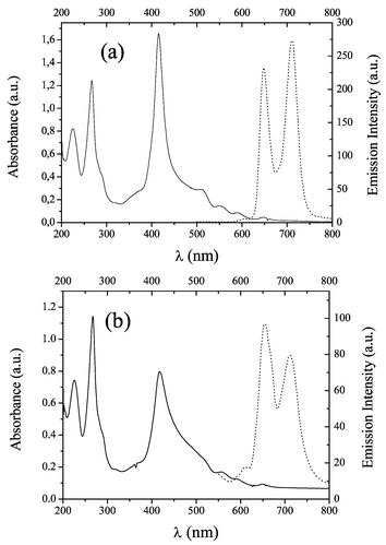 | ||
| Fig. 4 (a) Electronic and emission spectra (dashed line) for the supramolecular species Ru2phenTPyP. (b) Electronic and emission spectra (dashed line) for the supramolecular species Ru4phenTPyP. All spectra were obtained in ethanol solution at room temperature. | ||
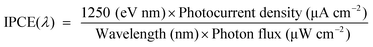
The adsorption of the RuphenTPyP species onto nanocrystalline TiO2 leads to active red–brown films. It is important to point out that even without the presence of an anchoring group such as hydroxyl, carboxyl or phosphonate, the dye remains strongly adsorbed onto the oxide films after the photoelectrochemical measurements. Dye desorption by solvent or electrolyte that could compromise cell integrity was not observed. Fig. 5 shows the monochromatic photon-to-electron conversion efficiency curve as a function of wavelength (IPCE) for the photoelectrochemical cells assembled with TiO2/RuphenTPyP films. IPCE(λ) is defined by eqn. (1).
 | (1) |
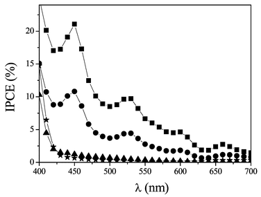 | ||
| Fig. 5 IPCE curves for the DSSC assembled with TiO2/RuphenTPyP films: (■) 4-µm TiO2/Ru2phenTPyP; (●) 8-µm TiO2/Ru2phenTPyP; (▲) 4-µm TiO2/Ru4phenTPyP; (★) bare TiO2. | ||
IPCE curves resemble the electronic absorption spectra of the supramolecular species, displaying the characteristic porphyrin and ruthenium complex bands. This is an indication that both components are active, participating in the electron transfer to the semiconductor conduction band.
The IPCE curves for the 4- and 8-µm-Ru2phenTPyP films are qualitatively similar, exhibiting Soret, MLCT and three Q bands at 450, 470 (sh), 530, 600 and 660 nm, respectively. It was not possible to observe the fourth Q band due to its low photocurrent response. The maximum IPCE was obtained with the photoelectrochemical cell assembled with the 4-µm TiO2/Ru2phenTPyP films and reached about 21% at 450 nm. This value is comparable with other porphyrin-related compounds10,11,17,32 but less than for Ru–phen complexes.33,34 The device assembled with the 8-µm TiO2/Ru2phenTPyP showed 11% IPCE at 450 nm, almost half of the value obtained with the thinner films. This result is expected for supramolecular species that give rise to better efficiencies when anchored to very thin films due to their low penetration into the deeper voids of the nanoporous film.7
In both cases only the porphyrin bands are red-shifted with respect to the species in solution (see Fig. 4), indicating that the supramolecular species interact with the TiO2 surface via the TPyP ring.
To our surprise, the supramolecular species Ru4phenTPyP showed negligible photocurrent, i.e., lower than 2. A possible explanation is the saturation of the pyridine groups in the tetrasubstituted porphyrin. It should be noticed that the meso-tetrapyridylporphyrin (TPyP) is strongly adsorbed onto TiO2 in contrast to the meso-tetraphenylporphyrins (TPP). Therefore, one can infer that the pyridine groups interact strongly with the TiO2 surface, due to their Lewis acid–base properties. Analogously, if the supramolecule also binds preferentially to the oxide surface through the pyridyl groups, the saturation with the ruthenium complexes would have a detrimental effect in the dye coverage and also in the efficiency of the DSSC charge transfer processes. This assumption was confirmed by measuring the optical density (OD) of the films. TiO2/Ru2phenTPyP and TiO2/Ru4phenTPyP films present OD at 590 nm equal to 1.0 and 0.3, respectively. Thus the supramolecular species containing two ruthenium complexes and two free pyridyl groups shows a ten-times better performance in comparison with the species with no free pyridyl groups.
The highest efficiency was achieved for the 4-µm TiO2/Ru2phenTPyP film of around 20% at excitation in the Soret band. Neglecting the reflective losses by the transparent electrode, the IPCE can be expressed by the product of three key parameters (eqn. (2)) where LHE is the light harvesting efficiency, ϕinj is the electron injection yield and η is the electron collecting efficiency in the external circuit.
 | (2) |
A detrimental effect of the LHE on the IPCE can be ruled out because of the strong absorption of these supramolecules in the visible region (high molar absortivity coefficients ranging from 104 to 106 M−1 cm−1). In addition, the optical density (OD) of the films is higher than 2.0 at the Soret band.
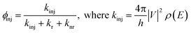
The electron collecting efficiency (η) is essentially controlled by two kinetics process: dye regeneration reaction by the iodide ions (kRR, step (3) in Fig. 2) and by the charge recombination reaction of the supramolecular species with the conduction/trap states electrons (kCR, step (4) in Fig. 2). The regeneration reaction is usually very fast (about 100 ns for the polypyridyl ruthenium complexes and zinc porphyrin)35,36 and thermodynamically favorable. The ground state oxidation potentials of the supramolecular species (0.98 V vs. NHE) are much more positive than the I−/I3− redox couple (0.44 V vs. NHE) giving rise to an electron transfer reaction with −ΔG > 0.6 eV. Recently Durrant and co-workers reported that the rate of the charge recombination between the conduction band/trap states electrons and the oxidized porphyrins dye is in the range of several milliseconds.37 In our case, preliminary transient absorption measurements (TAS) have revealed that there is a kinetic competition between regeneration and charge recombination processes, in the µs to ms timescale. This result can be explained by the lack of strong electronic connection between Ru2phenTPyP and TiO2. The charge recombination reaction is expected to be slow by the fact that the distance from the positive hole located at the ruthenium complexes and the conduction band electrons is larger for our supramolecular species than for the porphyrin alone. Thus, the parameter η can itself explain in part the low IPCE values. The last term controlling the IPCE is the efficiency of electron injection in the oxide conduction band (ϕinj) detailed in eqn. (3). The parameters kinj, kr and knr are rates of electron transfer to TiO2, radiative decay (determined by the fluorescence and phosphorescence lifetime) and non-radiative decay, respectively, V is the electronic coupling between the excited species and the semiconductor, and ρ(E) is the density of states of the conduction band.
 | (3) |
We believe that all factors controlling ϕinj contribute to the efficiency of electron injection. First, these supramolecular species fluoresces at ambient temperature as shown in Fig. 4. Due to their strong tendency to aggregate even in diluted solution, it is possible that the supramolecular species exist as monolayers and aggregate after deposition onto TiO2 substrate. These aggregates can occur isolated from the TiO2 nanoparticles precluding their direct participation in electron transfer, and consequently, after excitation, these species can undergo fast relaxation via radiative and non-radiative decays.
The rate of electron injection (kinj) from excited molecules adsorbed on the semiconductor surface to the conduction band is one the most important primary processes for the DSSC. A recent study showed that the rate of electron transfer for the free base 5,10,15,20-tetrakis(4-carboxyphenyl)porphyrin to a nanoporous TiO2 occurs within 10 ps.16 Assuming that the same is valid for our supramolecules and since the timescale for intersystem crossing from the lowest singlet excited state to the triplet state is several nanoseconds, injection via a triplet pathway can be ruled out, at least for the species in intimate contact with the TiO2 nanoparticles.38 As shown in eqn. (3), kinj involves the electronic coupling between the excited species and the semiconductor and the density of states of the conduction band. Argazzi et al. have suggested that the presence of an anchoring ligand attached to the chromophore is not required for an efficient electron transfer,39 however, it is plausible that a small electronic coupling between the supramolecular excited state and the 3d orbitals of the semiconductor, can decrease considerably the value of kinj. We believe that in the present study, IPCE is mainly limited by the last two terms in eqn. (2): electron injection yield and electron collecting efficiency.
The current–voltage characteristics of the photoelectrochemical cell assembled with the species Ru2phenTPyP is shown in Fig. 6. When irradiated at 100 mW cm−2 the cell exhibits short-circuit current (Isc), open circuit potential (Voc), fill factor (FF) and efficiency equal to 1.0 mA cm−2, 0.475 V, 0.62 and 0.3%, respectively.
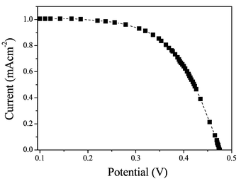 | ||
| Fig. 6 I–V Characteristics for the DSSC assembled with TiO2/Ru2phenTPyP film irradiated at 100 mW cm−2. | ||
The IPCE curves present contributions from the porphyrin and the ruthenium complexes as demonstrated in Fig. 5. Since we are dealing with two types of interconnected photosensitizers, their photoaction response can not be easily predicted on the basis of their isolated components, because of the expected cooperative supramolecular effects in the molecule. In order to evaluate what mechanisms are involved in the electron transfer process, and determine the extent of the participation from both components, we have performed some theoretical calculations for both species using the ZINDO/S method.
The relevant HOMO and LUMO levels and the fractional orbital mixture for the cis-Ru2phenTPyP supramolecule are shown in Table 1. A remarkable point is that the molecular orbitals in the HOMO levels (MO 258–259) and LUMO levels (MO 260–261) exhibit essentially porphyrin character (>75%). This result is the opposite found for the Ru4phenTPyP species (Table 2) where the HOMO levels are characterized by a mixing of the porphyrin and ruthenium molecular orbitals while the LUMO exhibits essentially porphyrin character.
| Mol. orbital | Energy/eV | RuII | Phen | Py | Py (bridge) | Porph |
|---|---|---|---|---|---|---|
| 266 | −4.41 | 1.04 | 98.69 | 0.00 | 0.26 | 0.01 |
| 265 | −4.48 | 4.43 | 92.07 | 0.12 | 1.46 | 1.83 |
| 264 | −4.48 | 4.23 | 92.72 | 0.02 | 2.05 | 0.91 |
| 263 | −4.65 | 2.20 | 95.23 | 0.08 | 0.98 | 1.44 |
| 262 | −4.65 | 2.24 | 95.00 | 0.04 | 1.23 | 1.42 |
| 261 | −4.95 | 0.12 | 1.72 | 3.77 | 5.37 | 89.02 |
| 260 (LUMO) | −5.08 | 0.36 | 2.53 | 2.76 | 13.33 | 81.01 |
| 259 (HOMO) | −9.62 | 7.08 | 2.68 | 7.04 | 6.77 | 76.28 |
| 258 | −10.10 | 8.91 | 3.88 | 2.00 | 2.04 | 82.78 |
| 257 | −10.20 | 52.07 | 23.32 | 0.24 | 6.91 | 14.39 |
| 256 | −10.20 | 56.84 | 24.99 | 0.89 | 6.35 | 7.26 |
| 255 | −10.50 | 77.77 | 15.27 | 0.00 | 3.20 | 0.45 |
| 254 | −10.50 | 75.81 | 15.88 | 0.27 | 2.60 | 1.77 |
| 253 | −10.50 | 73.09 | 21.23 | 0.00 | 1.74 | 0.38 |
| 252 | −10.50 | 72.24 | 20.39 | 0.27 | 2.67 | 1.62 |
| Mol. orbital | Energy/eV | RuII | Phen | Py (bridge) | Porph |
|---|---|---|---|---|---|
| 411 | −6.55 | 5.08 | 92.20 | 2.04 | 0.55 |
| 410 | −6.61 | 2.35 | 61.89 | 18.71 | 16.96 |
| 409 | −6.66 | 1.33 | 91.81 | 4.20 | 2.62 |
| 408 | −6.69 | 0.65 | 69.13 | 17.38 | 12.82 |
| 407 | −7.77 | 0.89 | 0.50 | 9.22 | 89.38 |
| 406 (LUMO) | −7.85 | 1.42 | 0.70 | 13.94 | 83.92 |
| 405 (HOMO) | −12.01 | 49.83 | 19.10 | 10.95 | 18.20 |
| 404 | −12.16 | 59.15 | 19.66 | 8.31 | 9.82 |
| 403 | −12.26 | 61.63 | 24.05 | 4.75 | 6.50 |
| 402 | −12.33 | 66.01 | 25.06 | 4.39 | 0.87 |
| 393 | −12.84 | 31.55 | 8.37 | 11.58 | 48.17 |
| 392 | −12.90 | 1.15 | 0.34 | 2.44 | 96.05 |
Below the HOMO level, there is a predominant ruthenium–phen character (MOs 252–257). Above the two degenerate LUMO levels (MOs 260–261) of porphyrin character, the molecular orbitals exhibit predominantly of π* phenanthroline character (MOs 262–266).
Some of the theoretical and experimental bands for cis-Ru2phenTPyP are collected in Table 3. The transitions 7 and 8 (MOs (258,259) → MOs (260,261)) are responsible for the vibronic Q(0–0) and Q(0–1) and the transitions 20, 32 and 51 (MOs (257,258,259) → MO(260,261)) are responsible for the Soret band. According to the theoretical calculations, the transitions in the [Ru(phen)2Cl]+ groups are expected to occur in the 400–540 nm range, contributing to the broad shoulder observed in this region (see electronic spectra in Fig. 4). In fact, the Ru (dπ) → phen (pπ) charge-transfer MLCT band can be found in the transitions 20, 32 and 51 [MOs (252,253,256,257) → MOs(262,263,264,265,267,267)].
| Assignment | ||||
|---|---|---|---|---|
| Transition # | λ/nm (calc.) | λ/nm (found) | MOi → MOf | Band type |
| 7 | 661.2 | 648 | 259 → 261; 258 → 260 | Porphyrin Q00 |
| 8 | 612.1 | 551 | 259 → 260; 258 → 261 | Porphyrin Q00 |
| 20 | 516.4 | 470 (sh) | 257 → 265; 256 → 264 | Ru → phen MLCT |
| 32 | 470.6 | 470 (sh) | 257 → 263; 256 → 262 | Ru → phen MLCT |
| 51 | 406.7 | Masked | 253 → 266; 252 → 267 | Ru → phen MLCT |
| 53 | 393.5 | Masked | 256 → 260 | Ru → Porph MLCT |
| 54 | 387.9 | 412 | 258 → 260 | Soret |
| 59 | 359.9 | Masked | 259 → 261; 257 → 260 | Soret + MLCT |
| 60 | 358.3 | Masked | 258 → 261; 256 → 260 | Soret + MLCT |
In the 350–400 nm range, the theoretical electronic transition 53 presents a distinct MO composition involving the corresponding HOMO levels with a predominant Ru(II) contribution (MO 256), while the LUMO ones have essentially porphyrin character (MO 260). This transition exhibits a large charge-transfer character, from the peripheral Ru(II) complexes to the porphyrin center. In this region, the Soret band is also observed, involving the porphyrin transitions from MO 259 and 258 to MO 260 and 261, as well as their combination with the Ru → Porph MLCT (from MO 257 and 256).
Thus, the presence of the peripheral groups changes dramatically the composition of the HOMO levels. In the Ru4phenTPyP we would expect a higher contribution from the peripheral groups due to a better connectivity between the two chromophores than in the cis-Ru2phenTPyP where the peripheral groups have low participation. However the IPCE spectra showed the opposite behavior. Therefore, the low IPCE obtained for the Ru4phenTPyP can only be ascribed to the lack of anchoring pyridyl groups, required for the effective binding of the supramolecular species onto the oxide surface. In agreement with this conclusion, it is important to note that in the cis-Ru2phenTPyP species, the bridging pyridine ligands contribute effectively to the ground and excited state properties (>13%), acting as a molecular wire in the photoinjection process.
Conclusion
In conclusion, supramolecules containing only two [Ru(phen)2Cl]+ groups are more efficient in transferring electrons to the conduction band of TiO2 than its analogue containing four peripheral groups. The free pyridyl groups play a fundamental role in the adsorption of the supramolecule in the oxide surface, being responsible for IPCE values of 21% for excitation in the Soret band. Experimental evidence and theoretical calculations suggest that the mechanism of TiO2 sensibilization by these supramolecules is through electron transfer from the excited singlet state of the porphyrin. This excited state can be populated directly by excitation at Soret and Q bands and also by a MLCT charge transfer electronic transition between the ruthenium complexes and the porphyrin.References
- B. O′Reagan and M. Grätzel, A low-cost, high-efficiency solar-cell based on dye-sensitized colloidal TiO2 films, Nature, 1991, 353, 737–740 CrossRef CAS.
- M. K. Nazeeruddin, A. Kay, R. Humphry-Baker, E. Muller, P. Liska, N. Vanchopoulos and M. Grätzel, Conversion of light to electricity by cis-X2Bis (2,2′-bipyridyl-4,4′dicarboxylate) ruthenium(II) charge transfer sensitizers (X = Cl−, Br−, I−, CN−,, and SCN−) on Nanocrystalline TiO2 electrodes, J. Am. Chem. Soc., 1993, 115, 6382–6390 CrossRef CAS.
- K. Kalyanasundaram and M. Grätzel, Applications of funcionalized transition metal complexes in photonic, and optoelectronic devices, Coord. Chem. Rev., 1998, 77, 347–414 CrossRef CAS.
- R. Argazzi, C. A. Bignozzi, T. A. Heimer, F. Castellano and G. J. Meyer, Enhanced spectral sensitivity from ruthenium(II) polypyridyl based photovoltaic devices, Inorg. Chem., 1994, 33, 5741 CrossRef CAS.
- O. Kohle, S. Ruile and M. Grätzel, Ruthenium(II) charge-transfer sensitizers containing 4,4′-dicarboxy-2,2′-bipyridine. Synthesis, properties, and bonding mode of coordinated thio-, and selenocyanates, Inorg. Chem., 1996, 35, 4779–4787 CrossRef CAS.
- C. A. Bignozzi, R. Argazzi, M. T. Indelli and F. Scandola, Design of supramolecular systems for spectral sensitization of semiconductors, Sol. Energy Mater. Sol. Cells, 1994, 32, 229–244 CrossRef CAS.
- C. A. Bignozzi, R. Argazzi and C. J. Kleverlaan, Molecular, and supramolecular sensitization of nanocrystalline wide band-gap semiconductors with mononuclear, and polynuclear metal complexes, Chem. Soc. Rev., 2000, 29, 87–96 RSC.
- A. Kay, R. Humphry-Baker and M. Grätzel, Artificial Photosynthesis. 2. Investigations on the mechanism of photosensitization of nanocrystalline TiO2 solar cells by chlorophyll derivatives, J. Phys. Chem., 1994, 98, 952–958 CrossRef CAS.
- K. Kalyanasundaram, N. Vanchopoulos, V. Krishnan, A. Monnier and M. Grätzel, Sensitization of TiO2 in the visible light region using zinc pophyrin, J. Phys. Chem., 1987, 91, 2342–2347 CrossRef CAS.
- T. Ma. K. Inoue, H. Noma, K. Yao and E. Abe, Effect of functional group on photochemical properties, and photosensitization of TiO2 electrode sensitized by porphyrin derivatives, J. Photochem. Photobiol. A, 2002, 152, 207–212 CrossRef.
- G. K. Boschloo and A. Goossens, Electron trapping in porphyrin-sensitized porous nanocrystalline TiO2 electrodes, J. Phys. Chem., 1996, 100, 19489–19494 CrossRef CAS.
- M. K. Nazeeruddin, R. Humphry-Baker, M. Grätzel, D. Wohrle, G. Schnurpfeil, G. Schneider, A. Hirth and N. Trombach, Efficient near-IR sensitisation of nanocrystalline TiO2 films by zinc, and aluminium phthalocyanines, J. Porphyrins Phthalocyanines, 1999, 3, 230–237 CrossRef CAS.
- S. Ferrere and B. A. Gregg, New perylenes for dye sensitization of TiO2, New J. Chem., 2002, 26, 1155–1160 RSC.
- K. Hara, T. Horiguchi, T. Kinoshita, K. Sayama, H. Sugihara and H. Arakawa, Highly efficient photon-to-electron conversion with mercurochrome-sensitized nanoporous oxide semiconductor solar cells, Sol. Energy Mater. Sol. Cells, 2000, 64, 115–134 CrossRef CAS.
- A. Hagfeldt and M. Gratzel, Light-induced redox reactions in nanocrystalline systems, Chem. Rev., 1995, 95, 49–68 CrossRef CAS.
- Y. Tachibana, S. A. Haque, I. P. Mercer, J. R. Durrant and D. R. Klug, Electron injection, and recombination in dye sensitized nanocrystalline titanium dioxide films: a comparison of ruthenium bipyridyl, and pophyrin sensitizer dyes, J. Phys. Chem. B, 2000, 104, 1198–1205 CrossRef CAS.
- F. Odobel, E. Blart, M. Lagrée, M. Villieras, H. Boujtita, N. E. Murr, S. Caramori and C. A. Bignozzi, Porphyrin dyes for TiO2 sensitization, J. Mater. Chem., 2003, 13, 502–510 RSC.
- M. J. Gunter and P. Turner, Metalloporphyrins as models for the cytochromes-P-450, Coord. Chem. Rev., 1991, 108, 115–161 CrossRef CAS.
- C. Shi and F. C. Anson, Cobalt meso-tetrakis(N-methyl-4-pyridiniumyl)-porphyrin becomes a catalyst for the electroreduction of O2 by four electrons when [(NH3)(5)Os](n+) (n = 2, 3) groups are coordinated to the porphyrin ring, Inorg. Chem., 1996, 35, 7928–7931 CrossRef CAS.
- D. Gust, T. A. Moore and A. L. Moore, Mimicking bacterial photosynthesis, Pure Appl. Chem., 1998, 70, 2189–2200 CAS.
- F. Bedioui, J. Devynck and C-J. Bied-Charreton, Electropolymerized manganese porphyrin films as catalytic electrode materials for biomimetic oxidations with molecular oxygen, J. Mol. Catal. A: Chem., 1996, 113, 3–11 CrossRef CAS.
- H. E. Toma and K. Araki, Supramolecular assemblies of ruthenium complexes, and porphyrins, Coord. Chem. Rev., 2000, 196, 307–329 CrossRef CAS.
- K. Araki and H. E. Toma, Syntehesis, and characterization of a multibriged porphyrin complex containing peripheral bis(bipyridine)-ruthenium(II) groups, J. Coord. Chem., 1993, 30, 9–17 CAS.
- K. Araki and H. E. Toma, Luminescence, spectroelectrochemistry, and photoelectrochemical properties of a tetraruthenated zinc porphyrin, J. Photochem. Photobiol. A, 1994, 83, 245–250 CrossRef CAS.
- F. M. Engelmann, P. Losco, H. Winnischofer, K. Araki and H. E. Toma, Synthesis, electrochemistry, spectroscopy, and photophysical properties of a series of meso-phenylpyridylporphyrins with one to four pyridyl rings coordinated to [Ru(bipy)2Cl]+ groups, J. Porphyrins Phthalocyanines, 2002, 6, 33–42 CAS.
- K. Araki, P. Losco, F. M. Engelmann, H. Winnischofer and H. E. Toma, Modulation of vectorial energy transfer in the tetrakis[tris(bipyridine)ruthenium(II)porphyrinate zinc complex, J. Photochem. Photobiol. A, 2001, 42, 25–30 CrossRef CAS.
- K. Araki, A. L. Araujo, M. M. Toyama, M. Franco, C. M. N. Azevedo, L. Agnes and H. E. Toma, Spectroscopic, and electrochemical study of a tetrapyridylporphyrin modified with four bis-(1,10-phenanthroline)chlororuthenium(II) complexes, J. Porphyrins Phthalocyanines, 1998, 2, 467–472 CrossRef CAS.
- N. L. Allinger, Conformational-analysis.130.MM2 – Hydrocarbon force-field utilizing V1, and V2 torsional terms., J. Am. Chem. Soc., 1977, 99, 8127–8134 CrossRef CAS.
- HyperChemTM version 6.01 for WindowsTM, Hypercube Inc. Gainesville, FL, USA, 2000.
- (a) M. C. Zerner, G. H. Loew, R. F. Kirchner and U. T. Mueller-Westerhoff, Intermediate neglect of differential-overlap technique for spectroscopy of transtion-metal compexes – ferrocene, J. Am. Chem. Soc., 1980, 102, 589–599 CrossRef CAS; (b) A. D. Bacon and M. C. Zerner, Intermediate neglect of differential-overlap technique for spectroscopy of transtion-metal compexes – Fe, Co, and Cu chlorides, Theor. Chim. Acta, 1979, 53, 21–54 CrossRef CAS; (c) J. E. Ridley and M. C. Zerner, Triplet-states via intermediate neglect of differential overlap – benzene, pyridine, and diazines, Theor. Chim. Acta, 1976, 42, 223–236 CrossRef CAS.
- C. J. Barbé, F. Arendse, P. Comte, M. Jirousek, F. Lenzmann, V. Shklover and M. Grätzel, Nanocrystalline titanium oxide electrodes for photovoltaic applications, J. Am. Ceram. Soc., 1997, 80, 3157–3171 CAS.
- R. Dabestani, A. J. Bard, A. Campion, M. A. Fox, T. E. Mallouk, S. E. Webber and J. M. White, Sensitization of titanium-dioxide, and strontium-titanate electrodes by ruthenium(II) tris(2,2′-bipirine-4,4′dicarboxylic acid), and zinc tetrakis(4-carboxyphenyl)porphyrin – an evaluation of sensitization efficiency for component photoelectrodes in a multipanel device, J. Phys. Chem., 1988, 92, 1872–1878 CrossRef CAS.
- H. Sugihara, L. P. Singh, K. Sayama, H. Arakawa, Md. K. Nazeeruddin and M. Gratzel, Efficient photosensitization of nanocrystalline TiO2 films by a new grass of sensitizer: cis-dithiocyanato bis(4,7-dicarboxy-1,10-phenanthroline)ruthenium(II), Chem. Lett., 1998, 1005–1006 CrossRef CAS.
- K. Hara, H. Horiuchi, R. Katoh, L. P. Singh, H. Sugihara, K. Sayama, S. Murata, M. Tachiya and H. Arakawa, Effect of the ligand structure on the efficiency of electron injection from excited Ru-phenanthroline complexes to nanocrystalline TiO2 films, J. Phys. Chem. B, 2002, 106, 374–379 CrossRef CAS.
- S. A. Haque, Y. Tachibana, D. Klug and J. R. Durrant, Charge recombination kinetics in dye-sensitized nanocrystalline titanium dioxide films under externally applied bias, J. Phys. Chem. B, 1998, 102, 1745–1749 CrossRef CAS.
- I. Montanari, J. Nelson and J. R. Durrant, Iodide electron transfer kinetics in dye-sensitized nanocrystalline TiO2 films, J. Phys. Chem. B, 2002, 106, 12203–12210 CrossRef CAS.
- J. N. Clifford, G. Yahioglu, L. R. Milgrom and J. R. Durrant, Molecular control of recombination dynamics in dye sensitised nanocrystalline TiO2 films, Chem. Commun., 2002, 1260–1261 RSC.
- J. E. Kroeze, T. J. Savenije and J. M. Warman, Contactless determination of the efficiency of photo-induced charge separation in a porphyrin-TiO2 bilayer, J. Photochem. Photobiol. A, 2002, 148, 49–55 CrossRef CAS.
- R. Argazzi, C. A. Bignozzi, T. A. Heimer and G. J. Meyer, Remote interfacial electron transfer from supramolecular sensitizers, Inorg. Chem., 1997, 36, 2–3 CrossRef CAS.
| This journal is © The Royal Society of Chemistry and Owner Societies 2004 |