Observation of fluidic behavior in a polymethylmethacrylate-fabricated microchannel by a simple spectroscopic analysis†
Hirofumi
Kawazumi
*a,
Asuka
Tashiro
a,
Kazuya
Ogino
b and
Hideaki
Maeda
c
aKyushu School of Engineering, Kinki University, Iizuka, 820-8555, Japan. E-mail: kawazumi@fuk.kindai.ac.jp; Fax: +81 948 225655
bFaculty of Engineering, Fukuoka University, Fukuoka, 814-0180, Japan
cThe National Institute of Advanced Industrial Science and Technology (AIST) Kyushu, Tosu, 841-0052, Japan
First published on 21st January 2002
Abstract
An easy-to-use and low cost microreactor made of polymethylmethacrylate was mechanically fabricated with a microchannel (200 µm × 200 µm). The laminar flow behavior was investigated by visualizing the flow of red and green aqueous solutions. Digitized color images from a CCD camera were analyzed by resolving the color in RGB mode. Numeric data from red and green color components in the images could reveal the fluidic behavior in the microchannel because the spatial spectroscopic information corresponds to the color solution flows. Effects of corner shapes in a turn, flow rate and surface roughness were observed on the mixing of the laminar flows. A right angle turn and unevenness of ±10% of the inner wall surface almost mixed the two color laminar flows.
Introduction
Miniaturized chemical analytical systems, that is, micro total analytical systems (μTAS), have been attracting interest owing to the advantages of fast analysis time, reduction of reagent consumption portability and disposability.1 Furthermore, miniaturized synthesis systems, that is, microreactors, have been demonstrated with better performance in the reaction time and yield.2–4 These profitable features mostly result from rapid mass transfer in micro scale and large specific interfacial area. Whitesides et al. demonstrated the patterning of reactions in multiphase laminar flows.5 Miyazaki et al.6 and Kitamori et al.7 reported the acceleration of enzyme-catalyzed hydrolysis and peroxidation, respectively, by using two laminar flows containing the enzyme and substrate in the microchannel with a few hundred micrometer dimensions in comparison with batch reactions. In these simple reactions, it is difficult to consider an increase of the intrinsic reaction rates. Therefore, effects of specific fluidic behavior in the microchannel might be taken into account such as disorder of the interface and mixing between the laminar flows.For practical applications of the microreactor for the chemical synthesis, the development of easy-to-fabricate and low cost microfluidic devices is very important. Polymethylmethacrylate (PMMA) is one of most attractive material because of hardness and transparency. Miyazaki et al.6 observed similar acceleration of an enzymatic reaction in a PMMA microreactor to that in a glass-fabricated one.7 However, effects of the PMMA surface still have to be evaluated with respect to its roughness and hydrophobicity. To study the microfluidic behavior the flow should be visualized and analyzed quantitatively. We observe color solutions in the microchannel with a color CCD camera and simply convert the color information into the fluidic behavior in the channel configuration of the PMMA microreactor.
Experimental
The microreactor with the microchannel (200 μm in width, 200 μm in depth, 40 cm in length) was mechanically fabricated on a PMMA plate (3 cm × 3 cm × 5 mm) using Robodrill equipped with a flat end mill (∅ 100 μm). The top plate assembly was applied by adding pressure (2 kg) and heat (120 °C and 4 h). Red and green pen inks were diluted and those color aqueous solutions were introduced through a Y-shape inlet using microsyringe pumps to visualize laminar flows, as shown in Fig. 1. Two colored flows were observed under a color CCD camera with a magnifying lens, HI-SCOPE DH-2700 (HIROX, Japan, 200× magnification). The microreactor was illuminated with a light source integrated in the lens head. Images from the CCD camera were stored digitally in a JPEG format with 24-bit color data and a resolution of 640 pixels × 480 pixels. The color data with spatial information were extracted from the image data files by using numerical analysis software, Igor Pro (WaveMetrics, OR), on a personal computer. The roughness was measured by a three dimensional profile microscope VK-8500 (Keyence, Japan) with a resolution of 0.03 μm.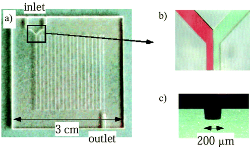 | ||
| Fig. 1 PMMA microreactor with a 200 μm width microchannel. (a) Picture of the whole microreactor plate, (b) Y-shape inlet and laminar flows of red (left) and green (right) solutions, (c) cross sectional view of the microchannel. | ||
Results and discussion
The color images are resolved into the major components of the colors, for example, red, green and blue in RGB mode, cyan, magenta, yellow and black in CMYK mode. Here, we analyzed the color data in RGB mode. Fig. 2 shows plots of the component color data along an intersectional line A–B in the Y-shape inlet. Each color has an 8-bit resolution and the 10-pixel data are accumulated along the perpendicular to the A–B direction for noise reduction. The plots are normalized in the peak intensities. The red and green plots clearly present the microchannel-shaped dips which correspond to the absorption of the red and green ink solutions in the two laminar flows, respectively. Therefore, even such a simple spectroscopic analysis could provide information on the fluidic behavior. Since the sensitivity of green is lower than those of red and blue, dark edges in the sides of the microchannel are enhanced in the green plot. The blue plot presents the absorption of both red and green inks. The red plot is used for the following analysis because it corresponds simply to the red laminar flow. | ||
| Fig. 2 Color resolving of CCD image in RGB mode along an intersectional line A–B. | ||
The red component plots along the flow across the microchannel reveal the mixing behavior of the laminar flows. Fig. 3 shows the effects of corner shapes in a turn and surface roughness of the inner wall. The flow rate is 20 μl min−1 for all results. The plots are the data around inlets (intersections C, E, and G in Fig. 3) and outlets (intersections at D, F, and H) at the first turn. The surface roughness is changed from 0.66 μm to 38.5 μm by speeding up the machining from 10 mm min−1 to 200 mm min−1; the roughness values are averages of variation of the depth profile in the microchannel. The numbers attached to the plots present the area ratios between the right and left halves; this number approaches unity as the mixing occurs. The mixing progresses in the rough channel even after a straight flow in 2 cm because the ratio at intersection C is larger than those at E and G. Very large mixing occurs in a right angle turn with a rough surface.
 | ||
| Fig. 3 Effects of corner shape in a turn and roughness of inner wall on the microfluidic behavior. The flow rate is 20 μl min−1. All plots correspond to the red component in the CCD image. Attached numbers are the area ratios between the right and left halves which correspond to the degree of mixing. | ||
An effect of the flow rate at the turn was found. Fig. 4 shows the pictures and plots at the first and second turn from the Y-shape inlet at the flow rate of 200 μl min−1. The plot at intersection I indicates that a degree of the mixing at the fast flow rate is similar to that at the low flow rate. Apparently, the two color flows mix with each other after the first turn. However, the two color solutions separate out after the second turn. This curious finding is explained as a result of rotation of the laminar flow along the flow axis in the microchannel owing to cornering inertia. This fluidic behavior especially would promote the mixing because the cross section of our microchannel is square.
 | ||
| Fig. 4 Rotation of the laminar flow along the flow axis at the turns. The flow rate is 200 μl min−1. | ||
A simple spectroscopic analysis has been applied for the observation of microfluidic behavior in the PMMA microreactor. The color solution injection could easily and quickly characterize the flow in the microchannel. This approach is very useful for designing a prototype of the microreactor.
References
- A. Manz, H. Becker, Microsystem Technology in Chemistry and Life Sciences, Springer, Berlin, 1998.
- N. G. Wilson and T. McCreedy, Chem Commun., 2000, 733 RSC.
- K. Kusakabe, D. Miyagawa, Y. Gu, H. Maeda and S. Morooka, J. Chem. Eng. Jpn., 2001, 34, 441 Search PubMed.
- H. Hisamoto, M. Tokeshi and T. Kitamori, High Yield Synthetic Chemical Reaction in Microchip toward High Throughput Synthesis, in 14th International Symposium on Microscale Separation and Analysis, 2001, Boston, USA Search PubMed.
- P. J. A. Kenis, R. F. Ismagilov and G. M. Whitesides, Science, 1999, 285, 83 CrossRef CAS.
- M. Miyazaki, H. Nakamura and M. Maeda, Chem. Lett., 2001, 442 CrossRef CAS.
- Y. Tanaka, M. N. Slyadnev, K. Sato, M. Tokeshi, H. B. Kim and T. Kitamori, Anal. Sci., 2001, 17, 809 CAS.
Footnote |
| † Presented at the International Symposium on Microchemistry and Microsystems (ISMM 2001), Kawasaki, Japan, September 16–18, 2001. |
| This journal is © The Royal Society of Chemistry 2002 |