Ultrasensitive, direct detection of a specific DNA sequence of Bacillus anthracis in solution
Alonso
Castro
* and
Richard T.
Okinaka
MS-D454, Los Alamos National Laboratory, Los Alamos, NM 87545, USA.. E-mail: acx@lanl.gov
First published on 7th January 2000
Abstract
A very fast and ultrasensitive method has been developed for the detection and quantitation of specific nucleic and sequences of bacterial origin in solution. The method is based on a two-color, single fluorescent molecule detection technique developed in our laboratory. The technique was applied to the detection of Bacillus anthracis DNA in solution.
Introduction
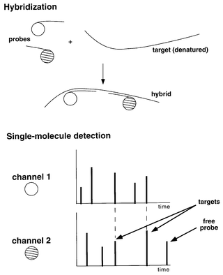
The ultrasensitive detection of viral and bacterial pathogens in air, water, and soil samples is of fundamental importance for maintaining public health, and for detecting environmental contamination due to biological warfare agents, among others reasons. Most techniques for environmental monitoring involve culturing the microorganisms present in a sample in order to multiply the number of target pathogens, and then using a gene probe for detecting specific nucleic acid sequences characteristic of a particular pathogen.1 These techniques are often problematic due to the time-consuming nature of the procedures involved, and to the lack of adequate sensitivity, which makes detection difficult. The sensitivity problem has been partially addressed by the development of the polymerase chain reaction (PCR) which, in this case, is used for selectively amplifying, or synthesizing, millions of copies of a short region of a DNA molecule from the target pathogen, so that it can be detected with a hybridization probe. PCR, however, often introduces ambiguities resulting from contamination and by other mechanisms not fully understood.2–6 Thus, its use is not suitable in situations when quantitation is critical or when false positives are not tolerable. Therefore, it is important to develop new techniques with the potential to succeed where the PCR fails.Here, we present our results on the development of a very fast and sensitive method for the detection and quantitation of specific nucleic acid sequences of bacterial origin in solution. The method is based on a two-color, single fluorescent molecule detection technique recently developed in our laboratory.7 The basis of our approach is to monitor for the presence of a specific nucleic acid base sequence of bacterial or viral origin in a sample. The nucleic acid sequences may be DNA or RNA sequences, and may be characteristic of a specific taxonomic group, or a specific physiological function. The detection scheme involves the use of peptide nucleic acid (PNA) probes, which possess much higher affinity and specificity for DNA targets than regular probes. Two fluorescent probes are used, each labeled with a different fluorescent tag, and whose sequences are complementary to the desired target (Fig. 1). When these probes are combined under controlled conditions with the sample containing the target nucleic acid, highly specific hybridization occurs. If the target sequence is present, our apparatus detects the two fluorescent colors simultaneously. Remaining free probes produce signals of one or the other color, but not both simultaneously. The high sensitivity and low background of our technique allows us to directly detect a specific sequence, thus avoiding the use of PCR amplification.
 | ||
| Fig. 1 Schematic representation of the double-label coincidence assay. Simultaneous detection of the two hybridized probes allow the detection of a specific sequence of the target at the single-molecule level of sensitivity. | ||
We demonstrate the use of this technique for the detection of Bacillus anthracis DNA in solution. B. anthracis is a gram-positive endospore-forming bacterium capable of producing fatal infections both in livestock and in humans.8 Virulent strains of B. anthracis are encapsulated and cause death in humans and animals by producing various toxins.9,10 The detection of a specific sequence of Bacillus anthracis in the presence of excess amounts of unrelated DNA from salmon sperm, and in the presence of large amounts of genomic DNA from a related bacillus, B. globigii (B. subtilis subsp. niger), are described in this paper.
Experimental
Sample preparation
The Bacillus anthracis DNA content consists of a 5.7 Mb genome, the 184-kb pXO1 virulent plasmid, and the 95-kb pXO2 capsule plasmid. Our target samples consisted of a 1∶1 mixture of the genomic and pXO2 plasmid components at a concentration of either 5 fM or 0.5 fM. In one series of experiments, salmon sperm DNA (Sigma, St. Louis, MO, USA) was added to the target sample at increasingly larger ratios (see Table 1), in order to simulate the large amounts of unrelated background DNA usually found in environmental samples. In another case, a 100∶1 genomic excess of B. globigii was added to a 5 fM B. anthracis sample in order to test the sensitivity and specificity of the technique for detecting the pathogen in the presence of another, closely related, bacillus. In all cases, the DNA target samples were denatured by heating at 95 °C, and then cooled to room temperature. Two fluorescently labeled, 12-base PNA probes were used (PerSeptive Biosystems, Inc., Framingham, MA, USA), one tagged with rhodamine 6G (Rho) and the other with bodipy-texas red (BTR). These probes have sequences that are complementary to a short region of the capB gene of the pXO2 plasmid, corresponding to nucleotides 475 to 486 and 1082 to 1093, respectively (H–Rho–O–CTGGTACATCTG–CONH2 and H–BTR–O–TGATCCCTCATC–CONH2). The PNA probes were added to the samples at a final concentration of 2 pM and allowed to hybridize to the target for 30 min. Immediately afterwards, the samples were loaded into the instrument and single-molecule data was collected as described below.| Experi ment 1 | Experi ment 2 | Experi ment 3 | Experi ment 4 | |
|---|---|---|---|---|
| a For an average sheared salmon sperm DNA fragment size of 700 base pairs. | ||||
| B. anthracis concentration/fM | 5 | 5 | 0.5 | 0 |
| B. anthracis/pg mL−1 | 5 | 5 | 0.5 | 0 |
| Salmon sperm/ng mL−1 | 0 | 100 | 100 | 100 |
| Mass ratio | — | 2 × 10⁴ | 2 × 10⁵ | — |
| Number of fragments ratioa | — | 4 × 10⁴ | 4 × 10⁵ | — |
Instrument
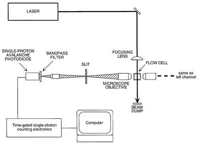
The system consists of a modified version of the apparatus used in previous single-molecule detection experiments (Fig. 2).11,12 The frequency-doubled output of a mode-locked Spectra-Physics Model 3800 Nd∶YAG laser producing 70 ps pulses at 532 nm was used as the excitation source. The laser output was attenuated to 2-5 mW and focused by a 6× microscope objective into the 0.8 × 0.8 mm id square cross-section capillary cell to yield a 10 μm spot (1/e2 value). The sample was pumped through the capillary cell at a rate of 200 μL h−1, which translates to a linear flow velocity of 87 μm s−1. As individual molecules move through the laser beam, repeated excitation–emission cycles produce a fluorescence photon burst. The apparatus incorporates two detection channels, which independently detect the photon bursts from each of the two dyes. Fluorescence in each channel is collected by a 40 × 0.85 numerical aperture microscope objective and spatially filtered by a 0.4 × 0.4 mm square slit, which defines a 10 × 10 μm detection area. In each channel, the light is then spectrally filtered by a 30 nm bandwidth eight-cavity interference filter which includes most of the emission spectral region of the corresponding dye, and detected by an EG&G Canada (Vaudreuil, Quebec) single-photon avalanche diode. Each detector output signal is analyzed by independent sets of time-correlated single-photon counting electronics under computer control. The detection electronics reject Raman and Rayleigh scattering by using a time-gate window set such that only delayed fluorescence photons are detected, thus increasing the signal-to-noise ratio of single-molecule detection.13 Fluorescence data from each channel was collected in 1 ms intervals for a total running time of 200 s. Cross-correlation analysis of the data reveals the amount of target present in the sample under investigation. | ||
| Fig. 2 Schematic diagram of the two-color single molecule detector. | ||
Results
(a) Detection of B. anthracis DNA in the presence of salmon sperm DNA
Table 1 summarizes the various experiments and controls performed in this case. Experiment 1 (Fig. 3a) corresponds to the detection of B. anthracis alone at a concentration of 5 fM. The large cross-correlation peak in Fig. 3a is an indication of the relative amount of B. anthracis DNA present in the sample. Experiments 2 and 3 (Fig. 3b and 3c) correspond to the detection of B. anthracis DNA in the presence of large amounts of salmon sperm background DNA. Experiment 4 was a control were no B. anthracis was added and only background DNA was present. No target signal is detected in this case (Fig. 3d). | ||
| Fig. 3 Cross-correlation results for the detection of Bacillus anthracis DNA. a, 5 fM B. anthracis alone; b, 5 fM B. anthracis DNA in the presence of 2 × 10⁴ times excess (by weight) salmon sperm DNA; c, 0.5 fM Bacillus anthracis DNA in the presence of 2 × 10⁵-times excess (by weight) salmon sperm DNA; (d) control: salmon sperm DNA alone. | ||
(b) Detection of B. anthracis DNA in the presence of B. globigii DNA
In this case, the complete genome of B. globigii was added to a 5 fM sample of B. anthracis DNA at a genomic ratio of 100:1. Fig. 4 shows the cross-correlation results (top trace). The large peak indicates the presence of the target. A control experiment was run under identical conditions, except that B. anthracis DNA was not added to the sample (Fig. 4, bottom trace). No cross-correlation peak was observed in this case, indicating that B. globigii does not contribute to the signal. | ||
| Fig. 4 Detection of B. anthracis in the presence of B. globigii. Top trace: 5 fM B. anthracis DNA and 100 X B. globigii genomic excess. Bottom trace: B. globigii alone. | ||
Discussion
In these experiments, we have demonstrated the rapid detection of a specific pathogen DNA sequence with high specificity and sensitivity. Excellent signal-to-noise ratios (SNR) were obtained in 200 s. For example, in the case of experiment 3 (Fig. 3c, B. anthracis DNA at 0.5 fM), a SNR of 15 was obtained. From this value, the limit of detection of B. anthracis DNA in the presence of excess salmon sperm DNA can be calculated to be 100 attomolar at a SNR of 3 in 200 s. The specificity of the technique is quite high as well. In the case of experiment 3, there was a single copy of the target sequence for every 3.3 × 10⁻¹³ g of salmon sperm DNA present in the sample. This is equivalent to detecting a single-copy gene in a 3 × 10⁸ Mbp genome. In previous experiments,7 we were able to detect a single-copy gene in a 3 × 10⁹ Mbp genome. The detection of B. anthracis DNA in the presence of an excess of B. globigii also demonstrates the high specificity of the technique, since these two bacterial species are closely related.14 The most widely used technique for the detection of low levels of specific DNA sequences of bacterial organisms is the polymerase chain reaction. However, the applicability of PCR methods is not universal. Sjostedt et al.,15 for example, have reported that no PCR products are obtained when attempting to amplify large quantities of B. anthracis bacteria present in various types of soils, probably due to polymerase activity inhibition by humic and phenolic compounds and metal ions. Also, the PCR is prone to yield false-positive results that are often impossible to predict. In a study by Reif et al.,16 it was shown that false-positive results were consistently obtained from Yersenia pestis samples, when PCR primers specific for the detection of the pXO2 plasmid of B. anthracis were used. We are in the process of conducting experiments for the detection of B. anthracis in soil samples, and in the presence of other pathogens such as Y. pestis. The results presented here are a first step towards the development of a powerful, new technique for the detection of specific pathogens with high sensitivity and specificity. This technique should allow the analysis of minute DNA samples directly, without the need for a target amplification step in most cases.The authors thank Brooks Shera for helpful discussions and Babs Marrone for providing the B. globigii samples.
References
- Standard methods for the examination of water and waste-water, ed. H. Greenberg, American Public Health Association, Washington DC, 1985. Search PubMed.
- J. Peccoud and C. Jacob, Biophys. J.,, 1996, 71, 101 Search PubMed.
- A. K. Bej, M. H. Mahbubani and R. M. Atlas, Crit. Rev. Biochem. Biophys., 1991, 26, 301 Search PubMed.
- J. Reiss, M. Krawczak, M. Schloesser, M. Wagner and D. Cooper, Nucleic Acids Res., 1990, 18, 973 CAS.
- A. M. Dunning, P. Talmud and S. E. Humphries, Nucleic Acids Res., 1988, 16, 10393 CAS.
- S. Kwoh and R. Higuchi, Nature, 1989, 339, 237 CrossRef.
- A. Castro and J. Williams, Anal. Chem., 1997, 69, 3915 CrossRef CAS.
- D. Lew, in Principles and Practice of Infectious Diseases, ed. G. L. Mandell, J. E. Bennett and R. Dolin, Churchill Livingstone, New York, 1995, pp. 1885–1889. Search PubMed.
- S. H. Leppla, Proc. Natl. Acad. Sci. USA, 1982, 79, 3162 CAS.
- J. L. Stanley, K. Sargeant and H. Smith, J. Gen. Microbiol., 1960, 22, 206 Search PubMed.
- A. Castro, E. Fairfield and E. B. Shera, Anal. Chem., 1993, 65, 849 CrossRef CAS.
- A. Castro and E. B. Shera, Anal. Chem., 1995, 67, 3181 CrossRef CAS.
- E. B. Shera, N. K. Seitzinger, L. M. Davis, R. A. Keller and S. A. Soper, Chem. Phys. Lett., 1990, 174, 553 CrossRef CAS.
- M. L. Nagpal, K. F. Fox and A. Fox, J. Microbiol. Meth., 1998, 33, 211 Search PubMed.
- A. Sjostedt, U. Eriksson, V. Ramisse and H. Garrigue, FEMS Microbiol. Ecol., 1997, 23, 159 CrossRef CAS.
- T. C. Reif, M. Johns, S. D. Pillai and M. Carl, Appl. Environ. Microbiol., 1994, 60, 1622 CAS.
| This journal is © The Royal Society of Chemistry 2000 |
