Discovery of a new class of 16-membered (2Z,11Z)-3,11-di(aryl/naphthyl)-1,13-dioxa-5,9-dithia-2,12-diazacyclohexadeca-2,11-dienes as anti-tumor agents†
Mohan Reddy Bodireddya,
Ranjeet Singh Mahlac,
P. Md. Khaja Mohinuddina,
G. Trivikram Reddya,
D. Vijaya Raghava Prasadb,
Himanshu Kumar*cd and
N. C. Gangi Reddy*a
aDepartment of Chemistry, School of Physical Sciences, Yogi Vemana University, Kadapa-516 003, Andhra Pradesh, India. E-mail: ncgreddy@yogivemanauniversity.ac.in
bDepartment of Microbiology, School of Life Sciences, Yogi Vemana University, Kadapa-516 003, Andhra Pradesh, India
cDepartment of Biological Sciences, Indian Institute of Science Education and Research (IISER), Bhopal-462023, Madhya Pradesh, India. E-mail: hkumar@iiserb.ac.in
dLaboratory of Host Defense, WPI Immunology Frontier Research Centre, Osaka University, Osaka, Japan
First published on 3rd August 2016
Abstract
A series of new 16-membered small macrocyclic compounds, (2Z,11Z)-3,11-di(aryl/naphthyl)-1,13-dioxa-5,9-dithia-2,12-diazacyclohexadeca-2,11-dienes (1a–k) were designed and developed by a simple and practical synthetic route from readily available substrates using simple organic transformations. Evaluation of in vitro anti-tumor activities on human triple negative breast cancer cells MDAMB-231 cell lines reveal that the macrocycles, 1a, 1f, 1g, 1i and 1k are promising anti-tumor compounds as evidenced from inhibition of cell migration and proliferation, upregulation of anti-tumor genes p53, MDA7 and TRAIL. The anti-proliferative effect of macrocycles is specific to cancer cells but no cytotoxic effect on normal breast epithelial cells has been observed (MCF10A). The developed synthetic route is free from metals, protecting groups and air-free techniques. The structure of macrocycle (1e) is confirmed by single crystal XRD studies.
Introduction
Design, synthesis and development of new anti-tumor agents has always been a challenging task for the scientific community as cancer is a major public health problem in many parts of the world1–4 particularly in developing countries increasingly, adopting a cancer-associated lifestyle options including smoking, westernized diets and lack of adequate exercise.5 Globally every 8th death is caused by cancer.6 Many of the available anti-tumor drugs have adverse effects on human health, therefore, there is a great need to develop new class of drugs for the treatment of cancer that are cost effective and chemically stable with low toxicity to normal cells.Macrocyclic molecules played a key role in drug discovery as evidenced from natural products.7 Among the macrocycles, the hetero-atoms incorporated macrocyclic molecules7 might be the best option as they possess unusual characteristics such as (i) structural complexity, regiogenicity, stereogenicity and rigidity, (ii) potential hydrogen bonding to interact with biomolecules such as RNA, DNA, proteins (enzymes),8,9 (iii) high degree of structural pre-organization which leads to the interaction of the key functional groups across extended protein's active site and (iv) ‘drug-like’ physico-chemical as well as pharmacokinetic properties.10,11 For instance, small macrocyclic molecules derived from natural products (I)12 and many synthetic macrocycles (II–IV)13–15 displayed apoptosis and 15-membered macrocycles (V)16 exhibited the highest levels of cytotoxicity (Fig. 1). Further, macrocyclic molecules exhibited various pharmacological properties which include anti-bacterial, anti-fungal, anti-tumor and anti-HIV activities.17
However, disinclination to investigate and develop macrocycles as new drug candidates in the academic and pharmaceutical industry was observed owing to poor yields during macrocyclization, difficulties in analog synthesis and unproductive side reactions during the joining of ends of a linear precursor molecule.18 Later, metal template methods were developed,19 but these methods were also associated with few disadvantages such as difficulties in choosing the correct template metal ion and its removal.20 Other methods were non-template, but required high dilution conditions21 which lead to huge organic waste generation along with low yields of desired product and also these methods require lengthy separations to remove oligomeric by-products. Later, ring closing metathesis,22 Glaser–Eglinton–Hay-type sp–sp coupling23 and head to tail macrocyclizations (build/couple/pair strategies)24 methods have emerged. But most of these methods suffer from one or more disadvantages as discussed above. Keeping in view of various pharmacological and biomedical applications and difficulties in the synthesis of macrocycles, we aimed to design and synthesize a new class of non-peptidic 16-membered macrocycles i.e. (2Z,11Z)-3,11-di(aryl/naphthyl)-1,13-dioxa-5,9-dithia-2,12-diazacyclohexadeca-2,11-dienes (1) from readily available substrates using simple organic transformations. The following factors formed the basis for the present design and synthesis: (i) high probability of cyclic oxime-ethers in the synthesis of biologically active synthetic molecules25 and (ii) accessibility of diverse topologies26 by the presence of sulfur atoms which were amiable to disrupt protein–protein interactions (PPIs).27
Retro-synthetic analysis
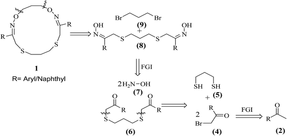
The retrosynthetic analysis of target 16-membered (2Z,11Z)-3,11-di(aryl/naphthyl)-1,13-dioxa-5,9-dithia-2,12-diazacyclohexadeca-2,11-dienes (1) is outlined in Scheme 1. | ||
| Scheme 1 Retrosynthetic analysis for the synthesis of (2Z,11Z)-3,11-di(aryl/naphthyl)-1,13-dioxa-5,9-dithia-2,12-diazacyclohexadeca-2,11-dienes (1). | ||
Herein, a simple, efficient and practical synthetic route is developed for the synthesis of structurally diverse non-peptidic 16-membered (2Z,11Z)-3,11-di(aryl/naphthyl)-1,13-dioxa-5,9-dithia-2,12-diazacyclohexadeca-2,11-dienes (1) (1a–k) from readily available substrates such as aralkyl ketones (2a–k), N-bromosuccinimide (3), 1,3-propanedithiol (5), NH2OH·HCl (7) and 1,3-dibromopropane (9) using simple organic transformations involving α-bromination, C–S bond construction, oxime formation followed by macrocyclization (C–O bond formation) as shown in Scheme 2. Further, the synthesized macrocyclic molecules (1a–k) are well characterized by 1H, 13C NMR, HRMS and single crystal XRD studies and evaluation of their anti-tumor activity is duly undertaken.
Results and discussion
Chemistry
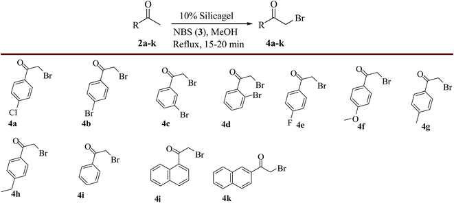
The synthetic route of title compounds i.e. (2Z,11Z)-3,11-di(aryl/naphthyl)-1,13-dioxa-5,9-dithia-2,12-diazacyclohexadeca-2,11-dienes (1) was accomplished via a four-step process as shown in Scheme 2. Accordingly, first 2-bromo-1-(4-chlorophenyl)ethanone (4a) was prepared from 4-chloroacetophenone (2a) by α-bromination using NBS (3) in MeOH in the presence of silica gel under reflux condition in high yield (91%) within a short period of time.28 The same procedure was applied for the preparation of other α-bromoaralkyl ketones (4b–k) from various acetophenones and acetonaphthones (2a–k) and the obtained results were presented in Scheme 3.Later, the reaction conditions for the synthesis of 2,2′-(propane-1,3-diylbis(sulfanediyl))bis(1-(4-chlorophenyl)ethanone) (6a) were optimized. Towards this direction, firstly, 1,3-propanedithiol was added drop wise to the α-bromo compound, 2-bromo-1-(4-chlorophenyl)ethanone (4a) in presence of Na2CO3 (3.0 equiv.) in ethanol at 20–25 °C and obtained 45% yield of the product (6a). To improve the yield further, the effect of various solvents such as dichloromethane, methanol, acetonitrile, THF, DMF and DMSO was investigated and 42%, 54%, 65%, 49%, 58% and 53% yields of product (6a) were obtained respectively. Preliminary screening of solvent study revealed that acetonitrile was the best option for maximum conversion of product (6a). To improve the yield of the product (6a) further, various bases such as K2CO3 (3.0 equiv.), Et3N (3.0 equiv.) and pyridine (3.0 equiv.) have been employed in acetonitrile which resulted in 85%, 65% and 55% yields of the product (6a) respectively. Finally, the effect of temperature on the course of C–S bond formation was also studied at various temperatures ranging from 5 to 25 °C. The study indicated that 15–20 °C was the optimum temperature for maximum yield of product 6a (98%). With the help of optimized reaction conditions, the substrate scope was tested for the synthesis of various 2,2′-(propane-1,3-diylbis(sulfanediyl))-bis(1-(aryl/naphthyl)ethanone) derivatives (6b–k) using α-bromoketones (4b–k) as substrates and the obtained results were presented in Scheme 4.
The third step was the oxime formation step which involves, the reaction between compound (6a) and NH2OH·HCl (7) in the presence of NaOAc in ethanol under reflux conditions29 to produce the precursor of the pharmacophore, (1Z,1′Z)-1-(4-chlorophenyl)-2-(3-((Z)-2-(4-chlorophenyl)-2-(hydroxyimino)-ethylthio)propylthio)ethanone oxime (8a) in excellent yield (98%). The same procedure was followed for the preparation of (1Z,1′Z)-1-(aryl/naphthyl)-2-(3-((Z)-2-(aryl/naphthyl)-2-(hydroxyimino)-ethylthio)propylthio)ethanone oximes (8b–k) using various substrates, 6b–k and the obtained results were presented in Scheme 5.
Then, the attention was focused on macrocyclization step to synthesize the title compound, (2Z,11Z)-3,11-bis(4-chlorophenyl)-1,13-dioxa-5,9-dithia-2,12-diazacyclohexadeca-2,11-diene (1a) by the reaction of pharmacophore, (1Z,1′Z)-1-(4-chlorophenyl)-2-(3-((Z)-2-(4-chlorophenyl)-2-(hydroxyimino)-ethylthio)propylthio)ethanoneoxime (8a) with linker i.e. 1,3-dibromopropane (9). Towards this direction, initially, a reaction was carried out by using the substrate, 8a and 1,3-dibromopropane (9) in MeOH in the presence of NaOH at 20–25 °C and lower yield (15%) of product, 1a was obtained. To improve the yield, the reaction conditions for the preparation of target 16-membered macrocycle 1a have been optimized using different solvents and bases. Initially, the effect of solvent or mixtures of solvents was studied for their impact on the course of macrocyclization. It was found that the solvents like MeOH, EtOH, acetonitrile, DMSO and DMF were provided 15%, 20%, 10%, 50% and 35% yields, respectively. From this study, it was found that DMSO provided considerable yield (50%) of the product 1a. For the improvement of the yield of the product 1a further, the effect of mixture of solvents was investigated. Towards this direction, a mixture of H2O and DMSO was selected and the study revealed that 1![[thin space (1/6-em)]](https://www.rsc.org/images/entities/char_2009.gif) :
:![[thin space (1/6-em)]](https://www.rsc.org/images/entities/char_2009.gif) 5 ratio of H2O and DMSO provided good yield (75%) of product 1a. Further, the effect of temperature was also studied and found that 5–10 °C was the optimum temperature for maximum yield of product 6a (86%). The effect of various bases such as Na2CO3, K2CO3, NaOH, KOH, Et3N and pyridine on the course of macrocyclization was studied. From this study, it was concluded that KOH was the best in obtaining maximum isolated yield (94%) of the desired product 1a in presence of 1
5 ratio of H2O and DMSO provided good yield (75%) of product 1a. Further, the effect of temperature was also studied and found that 5–10 °C was the optimum temperature for maximum yield of product 6a (86%). The effect of various bases such as Na2CO3, K2CO3, NaOH, KOH, Et3N and pyridine on the course of macrocyclization was studied. From this study, it was concluded that KOH was the best in obtaining maximum isolated yield (94%) of the desired product 1a in presence of 1![[thin space (1/6-em)]](https://www.rsc.org/images/entities/char_2009.gif) :
:![[thin space (1/6-em)]](https://www.rsc.org/images/entities/char_2009.gif) 5 ratio of H2O and DMSO at 5–10 °C. Encouraged by the productive results the same procedure was applied for the synthesis of a series of non-peptidic 16-membered (2Z,11Z)-3,11-di(aryl/dinaphthyl)-1,13-dioxa-5,9-dithia-2,12-diazacyclohexadeca-2,11-diene derivatives (1b–k) from various oximes (8b–k) and 1,3-dibromopropane (9) and the yields obtained were shown in Scheme 6.
5 ratio of H2O and DMSO at 5–10 °C. Encouraged by the productive results the same procedure was applied for the synthesis of a series of non-peptidic 16-membered (2Z,11Z)-3,11-di(aryl/dinaphthyl)-1,13-dioxa-5,9-dithia-2,12-diazacyclohexadeca-2,11-diene derivatives (1b–k) from various oximes (8b–k) and 1,3-dibromopropane (9) and the yields obtained were shown in Scheme 6.
The synthesized compounds were characterized by 1H-NMR, 13C-NMR and HRMS data and the structure of one of the macrocyclic compound, (2Z,11Z)-3,11-bis(4-fluorophenyl)-1,13-dioxa-5,9-dithia-2,12-diazacyclohexadeca-2,11-diene (1e) was confirmed by single crystal X-ray diffraction studies as shown in Fig. 2.
 | ||
| Fig. 2 ORTEP view of (2Z,11Z)-3,11-bis(4-fluorophenyl)-1,13-dioxa-5,9-dithia-2,12-diazacyclohexadeca-2,11-diene (1e). | ||
Anti-tumor activity studies
The anti-tumor activity of macrocyclic compounds (1a–k) was evaluated in vitro using triple negative breast cancer cells (TNBC) MDAMB-231. The anti-tumor effects of macrocyclic compounds were evaluated using standard anti-tumor assays, including wound healing assays (Fig. 3) and colonogenic assay (Fig. 4), depicting effect on cancer cells metastatic and cell division potential, respectively. For understanding the underlying molecular mechanism involved in anti-tumor effects of macrocyclic compounds (1a–k), transcription levels of key anti-tumor genes was measured by quantitative (q) RT-PCR (Fig. 5). Fragmentation of genomic DNA representing induction of apoptosis was analyzed by DNA laddering (Fig. 6) in agarose gel electrophoresis. The cytotoxic effects of these compounds (1a–k) were measured on normal breast epithelial cells (MCF10A) (Fig. 7).Conclusion
We designed and synthesized a new class of small macrocyclic molecules, (2Z,11Z)-3,11-di(aryl/naphthyl)-1,13-dioxa-5,9-dithia-2,12-diazacyclohexadeca-2,11-dienes (1a–k) from readily available substrates using simple organic transformations. These macrocycles were evaluated for their anti-tumor effect on triple negative breast cancer cell line, MDAMB-231. Compounds 1a, 1f, 1g, 1i and 1k restrict cell invasion, compounds 1e, 1f, 1g, and 1k restrict cell proliferation and colonogenic survival, compounds 1a, 1i and 1k induces TRAIL, MDA7 and p53 anti-tumor genes. None of the tested compound was found to be cytotoxic for normal breast epithelial cells. Further, it would be of great interest to evaluate these macrocyclic compounds in animal system, especially the compounds 1a and 1i to establish its effect under physiological condition. Other notable advantages are (i) accessibility of diverse topologies due to presence of two ‘S’ atoms that are amiable to disrupt protein–protein interactions (PPIs) and (ii) the presence of pharmacophore cyclic oxime-ether functionalities (C![[double bond, length as m-dash]](https://www.rsc.org/images/entities/char_e001.gif) N–O–) which are suitable for further ‘FG’ transformations holds the potential to access lead analogs other than the present reported macrocyclic molecules.
N–O–) which are suitable for further ‘FG’ transformations holds the potential to access lead analogs other than the present reported macrocyclic molecules.
Materials and methods
General information
All commercially available starting materials, reagents and solvents were purchased from Sigma Aldrich, Acros organics and Merck were used without further purification. Melting points of various obtained products were determined and uncorrected. NMR spectra were recorded on a Varian, Jeol/AL and Bruker 400 MHz. Chemical shifts were expressed in parts per million (ppm), coupling constants were expressed in Hertz (Hz). Splitting patterns describe apparent multiplicities and were designated as s (singlet), br s (broad singlet), d (doublet), t (triplet), q (quartet), quintet, m (multiplet). High-resolution mass spectra (MS) and compound purity data were acquired on a Waters LCT premier XE TOF HRMS and single quadrupole system equipped with electro spray ionization (ESI) source and Exactive orbitrap mass spectrometer (Thermo Scientific). Single crystal XRD analysis was carried out on a Bruker Apex ii diffractometer with CCD detector MoKα radiation (λ = 0.71069 Å) operating ω–2θ scanning mode at room temperature. Thin-layer chromatography was performed on 0.25 mm Merck silica gel plates and visualized with UV light. Column chromatography was performed on silica gel. The structures of isolated compounds were elucidated on the basis of spectroscopic data (1H & 13C-NMR, HRMS) and single crystal XRD analysis.Cell culture
Human triple negative breast cancer (TNBC) MDAMB231-cells (ATCC; cat no. HTB-26™) and neuroblastoma cell, IMR-32 were grown in 10% fetal bovine supplemented (FBS) (GIBCO, Life technologies, USA) complete Dulbecco's modified Eagles medium (cDMEM) (Sigma Aldrich, USA). The primary human breast epithelial cells (MCF10) were cultured in MEBM (Lonza/clonetics MEGM Kit cat# CC-3150), supplemented with 100 ng mL−1 cholera toxin, 2 mL per liter bovine pituitary extract (cat# CC4009G), 0.5 mL per liter gentamicin sulfate (cat# 4081G), 0.5 mL per liter recombinant growth factor human (cat# CC-4017G), recombinant insulin human (cat# CC-4021G), 0.5 mL per liter hypo cortisone (cat# CC-4031G). All the culture were maintained at 37 °C in humidified chamber (Thermo Fisher Scientific USA), supplied with supplied with 5% CO2. For subculture and seeding, TNBC MDAMB-231, IMR-32 cells were harvested with 0.01% trypsin-EDTA and MCF10A with 0.05% trypsin-EDTA, setting the cells density as per experimental requirements.Wound healing assay
For wound healing assay TNBC MDAMB-231 cells were seeded in six well plate, at density of 1 × 10⁶ cells per well in cDMEM. The 95% monolayer of cells carefully wounded using 20–200 μL micro tips. Cells were washed with DMEM for removing cell debris. Wounded monolayer of cells was treated with 1a, 1b, 1e, 1f, 1g, 1i, 1j and 1k (5 μM each) for 24 h 17AAG (5 μM) and solvent only treated cells were used as positive and negative control. Migration of cells into wounded area was captured by phase contrast microscope (Zeiss Axiovert model) at 0 h and 24 h time points.Colonogenic assay
The anti-proliferative effect of macrocycles (1a, 1b, 1e, 1f, 1g, 1i, 1j and 1k) were tested by this assay. TNBC MDAMB-231 cells were seeded in six well plates at density of 1 × 10³ cells per well and left in culture for 10 days for formation of colonies. Than cells were treated with test macrocyclic compounds 1a, 1b, 1e, 1f, 1g, 1i, 1j and 1k (5 μM each), positive control 17AAG (5 μM) or hydroxyl urea (1 mM) for 24 h. Followed by cells were washed with 1×-PBS and fixed using 70![[thin space (1/6-em)]](https://www.rsc.org/images/entities/char_2009.gif) :
:![[thin space (1/6-em)]](https://www.rsc.org/images/entities/char_2009.gif) 30 solution of ethanol acetic acid for 10 minutes. Fixed cells were stained with 1% methylene blue for 15 min. Extra stain was drained by washing with water. Dried plates were scanned for representation of effect of macrocycles on colonogenic survival of cells.
30 solution of ethanol acetic acid for 10 minutes. Fixed cells were stained with 1% methylene blue for 15 min. Extra stain was drained by washing with water. Dried plates were scanned for representation of effect of macrocycles on colonogenic survival of cells.
Quantitative-PCR analysis
The MDAMB-231 cells were seeded in six well plate at density of 0.2 × 10⁶. Next day, cells were treated with macrocycles 1a, 1b, 1e, 1f, 1g, 1i, 1j and 1k (5 μM each) for 24 h. The cells were washed with 1×-PBS and total RNA was extracted TRIzol reagent (Invitrogen USA) and first strand cDNA was synthesized for 1 μg RNA, using iScript-cDNA synthesis kit (BIORAD USA), according to manufacturers instruction. Relative quantification of anti-tumor genes (p53, MDA7, and TRAIL) and transcription factor STAT3 was performed with SYBR green master mix, using 18S as housekeeping control. Following, initial denaturation step at 95 °C for 10 min, 40 cycles of PCR amplification were performed with denaturation for 15 s and annealing at 60 °C for 1 min. In order to confirm amplification as single specific product, melt curve analysis was done as 95 °C for 15 s, 60 °C for 1 min, 95 °C for 15 s. The sequences of primer used in RNA quantification is given is Table 1.| Primer | Direction | Sequence | Size bp | Amplicon |
|---|---|---|---|---|
| S18 | Forward | ATCACCATTATGCAGAATCCACG | 23 | 93 bp |
| Reverse | GACCTGGCTGTATTTTCCATCC | 22 | ||
| P53 | Forward | GCCATCTACAAGCAGTCACAG | 21 | 143 bp |
| Reverse | TCATCCAAATACTCCACACGC | 21 | ||
| TRAIL | Forward | AGCAATGCCACTTTTGGAGT | 20 | 120 bp |
| Reverse | TTCACAGTGCTCCTGCAGTC | 20 | ||
| STAT3 | Forward | TTTGTCAGCGATGGAGTACG | 20 | 168 bp |
| Reverse | TGTTGACGGGTCTGAAGTTG | 20 | ||
| MDA7 | Forward | GAGGAACACGAGACTGAGAG | 20 | 116 bp |
| Reverse | TCCAGAGAAGCAGGGTAAAAC | 21 |
DNA fragmentation assay
Human TNBC MDAMB-231 and IMR32 cells were seeded in 6 well culture dish at density of 0.2 × 10⁶ cells per well. Next day cells were treated with macrocyclic compounds 1a, 1b, 1e, 1f, 1g, 1i, 1j and 1k. Followed to 48 h incubation, cells were harvested by trypsin-EDTA and pellet down at 200 g for 2 min. Cells washed with 1×-PBS and re-suspended lysis buffer (10 mM Tris-Cl: pH 8.0, 10 mM: EDTA, 0.5% Triton X-100) and nuclei pellet was collected at 13![[thin space (1/6-em)]](https://www.rsc.org/images/entities/char_2009.gif) 000 g for 20 min at 4 °C. Obtained nuclei pellet, re-suspended in lysis buffer and treated DNAse-free RNAse (0.1 mg mL−1) for 1 h at 37 °C. Then proteinase K (0.2 mg mL−1) and SDS (final concentration of 1%) were added and a resultant mixture was incubated further for 2 h at 50 °C. DNA from these samples was extracted using phenol chloroform isoamyl alcohol (25
000 g for 20 min at 4 °C. Obtained nuclei pellet, re-suspended in lysis buffer and treated DNAse-free RNAse (0.1 mg mL−1) for 1 h at 37 °C. Then proteinase K (0.2 mg mL−1) and SDS (final concentration of 1%) were added and a resultant mixture was incubated further for 2 h at 50 °C. DNA from these samples was extracted using phenol chloroform isoamyl alcohol (25![[thin space (1/6-em)]](https://www.rsc.org/images/entities/char_2009.gif) :
:![[thin space (1/6-em)]](https://www.rsc.org/images/entities/char_2009.gif) 24
24![[thin space (1/6-em)]](https://www.rsc.org/images/entities/char_2009.gif) :
:![[thin space (1/6-em)]](https://www.rsc.org/images/entities/char_2009.gif) 1). DNA precipitation was done using 3.5 volume of 100% ethanol and 0.1 volume of CH3COOK (pH 5.2) at −80 °C. The DNA pellet was resuspended in 40 μL of MQ and equal quantity of DNA was analyzed in 2% agarose gel.
1). DNA precipitation was done using 3.5 volume of 100% ethanol and 0.1 volume of CH3COOK (pH 5.2) at −80 °C. The DNA pellet was resuspended in 40 μL of MQ and equal quantity of DNA was analyzed in 2% agarose gel.
Cell cytotoxicity assay
The cytotoxic effect of newly synthesized macrocyclic compounds 1a, 1b, 1e, 1f, 1g, 1i, 1j and 1k was analyzed on human breast epithelial cells MCF10A. Cells were seeded in 24 well plates at density of 0.5 × 10⁶ cells per well, cultured till >90% cell density and treated with macrocyclic compounds 1a, 1b, 1e, 1f, 1g, 1i, 1j and 1k (200 μM each) for 24 h. Cells micrographs were taken before and after treatment at 0 h and 24 h time points using phase contrast microscopy.General experimental procedure
![[thin space (1/6-em)]](https://www.rsc.org/images/entities/char_2009.gif) :
:![[thin space (1/6-em)]](https://www.rsc.org/images/entities/char_2009.gif) 1 ratio of n-hexane and EtOAc) and pure product 3a was obtained in 91% yield. The same experimental procedure was adopted for the preparation of other α-bromoketones (4b–k).
1 ratio of n-hexane and EtOAc) and pure product 3a was obtained in 91% yield. The same experimental procedure was adopted for the preparation of other α-bromoketones (4b–k).![[thin space (1/6-em)]](https://www.rsc.org/images/entities/char_2009.gif) :
:![[thin space (1/6-em)]](https://www.rsc.org/images/entities/char_2009.gif) 1 ratio of n-hexane and ethyl acetate). The same experimental procedure was adopted for the preparation of other 2,2′-(propane-1,3-diylbis(sulfanediyl))bis(1-aryl/naphthyl)ethanone derivatives (6b–k).
1 ratio of n-hexane and ethyl acetate). The same experimental procedure was adopted for the preparation of other 2,2′-(propane-1,3-diylbis(sulfanediyl))bis(1-aryl/naphthyl)ethanone derivatives (6b–k).![[thin space (1/6-em)]](https://www.rsc.org/images/entities/char_2009.gif) :
:![[thin space (1/6-em)]](https://www.rsc.org/images/entities/char_2009.gif) 1 ratio of n-hexane and ethyl acetate). The same experimental procedure has been followed for the preparation of other macrocyclic compounds (1b–k). The synthesized compounds were characterized by 1H, 13C NMR and HRMS spectral data and the structure of compound 1e was confirmed by single crystal XRD data.
1 ratio of n-hexane and ethyl acetate). The same experimental procedure has been followed for the preparation of other macrocyclic compounds (1b–k). The synthesized compounds were characterized by 1H, 13C NMR and HRMS spectral data and the structure of compound 1e was confirmed by single crystal XRD data.Acknowledgements
The authors acknowledge the financial support for this work from the Council of Scientific and Industrial Research (CSIR), New Delhi, Government of India under a major research project (No. 01 (2391)/10/EMR-II) and Department of Atomic Energy-Board of Research in Nuclear Sciences (DAE-BRNS), Mumbai, Government of India through a major research project (No. 2011/37C/52/BRNS/2264). The study related to the analysis of biological activities is also supported by research grants number SR/S2/RJN-55/2009, BT/PR6009/GBD/27/382/2012 from Department of Science and Technology (DST) and Department of Biotechnology (DBT), Government of India respectively. This study was partly supported by the Intramural Research Grant of IISER, Bhopal, India and Grant for Joint Research Program of the Institute for Genetic Medicine, Hokkaido University, Japan to HK. Authors also thank Dr R.V.J. Kashyap, Dept. of English, Yogi Vemana University, for critical reading of the manuscript to check the linguistic corrections.Notes and references
- (a) R. Siegel, J. Ma, Z. Zou and A. Jemal, Cancer statistics, 2014, Ca-Cancer J. Clin., 2014, 64, 9–29 CrossRef PubMed; (b) C. E. DeSantis, C. C. Lin, A. B. Mariotto, R. L. Siegel, K. D. Stein, J. L. Kramer, R. Alteri, A. S. Robbins and A. Jemal, Cancer Treatment and Survivorship Statistics, 2014, Ca-Cancer J. Clin., 2014, 64, 252–271 CrossRef PubMed; (c) C. DeSantis, J. Ma, L. Bryan and A. Jemal, Breast Cancer Statistics, 2013, Ca-Cancer J. Clin., 2014, 64, 52–62 CrossRef PubMed.
- World Cancer Report 2014, World Health Organization, 2014, ch. 1.1., ISBN 9283204298 Search PubMed.
- The Top 10 Causes of Death Fact Sheet No 310, World Health Organization (http://www.who.int/mediacentre/factsheets/fs310/en/), May 2014 Search PubMed.
- World Cancer Report 2014, World Health Organization, 2014, ch. 1.3., ISBN 9283204298 Search PubMed.
- A. Jemal, F. Bray, M. M. Center, J. Ferlay, E. Ward and D. Forman, Global Cancer Statistics, Ca-Cancer J. Clin., 2011, 61, 69–90 CrossRef PubMed.
- R. L. Siegel, K. D. Miller and A. Jemal, Cancer statistics, 2015, Ca-Cancer J. Clin., 2015, 65, 5–29 CrossRef PubMed.
- (a) A. R. Shareef, D. H. Sherman and J. Montgomery, Chem. Sci., 2012, 3, 892–895 RSC; (b) R. M. Wilson and S. J. Danishefsky, Chem. Soc. Rev., 2007, 36, 1207–1226 RSC; (c) E. Marsault and M. L. Peterson, J. Med. Chem., 2011, 54, 1961–2004 CrossRef CAS PubMed.
- (a) T. E. Rose, K. V. Lawson and P. G. Harran, Chem. Sci., 2015, 6, 2219–2223 RSC; (b) D. F. Veber, S. R. Johnson, H. Y. Cheng, B. R. Smith, K. W. Ward and K. D. Kopple, J. Med. Chem., 2002, 45, 2615–2623 CrossRef CAS PubMed; (c) T. Rezai, B. Yu, G. L. Millhauser, M. P. Jacobsen and R. S. Lokey, J. Am. Chem. Soc., 2006, 128, 2510–2511 CrossRef CAS PubMed.
- (a) R. Tripier, O. Siri, F. Rabiet, F. Denat and R. Guilard, Tetrahedron Lett., 1999, 40, 79–82 CrossRef CAS; (b) C. Mcfarland, D. A. Vivic and A. V. Debnath, Synthesis, 2006, 5, 807–812 Search PubMed.
- E. M. Driggers, S. P. Hale, J. Lee and N. K. Terrett, Nat. Rev. Drug Discovery, 2008, 7, 608–624 CrossRef CAS PubMed.
- (a) A. K. Yudin, Chem. Sci., 2015, 6, 30–49 RSC; (b) J. Levin, Macrocycles in Drug Discovery, RSC Drug Discovery, The Royal Society of Chemistry, 2015 Search PubMed; (c) C. A. Lipinski, F. Lombardo, B. W. Dominy and P. J. Feeney, Adv. Drug Delivery Rev., 1997, 23, 3–25 CrossRef CAS.
- (a) V. Takiar, E. A. Strom, D. P. Baumann, F. Meric-Bernstam, R. H. Alvarez and A. M. Gonzalez-Angulo, Oncologist, 2013, 18, 265–270 CrossRef CAS PubMed; (b) C. C. Rohena and S. L. Mooberry, Nat. Prod. Rep., 2014, 31, 335–355 RSC.
- A. D. William, A. C. H. Lee, K. C. Goh, S. Blanchard, A. Poulsen, E. L. Teo, H. Nagaraj, C. P. Lee, H. Wang, M. Williams, E. T. Sun, C. Hu, R. Jayaraman, M. K. Pasha, K. Ethirajulu, J. M. Wood and B. W. Dymock, J. Med. Chem., 2012, 55, 169–196 CrossRef CAS PubMed.
- Z. F. Tao, L. Wang, K. D. Stewart, Z. Chen, W. Gu, M.-H. Bui, P. Merta, H. Zhang, P. Kovar, E. Johnson, C. Park, R. Judge, S. Rosenberg, T. Sowin and N. H. Lin, J. Med. Chem., 2007, 50, 1514–1527 CrossRef CAS PubMed.
- M. Marjanovic, M. Kralj, F. Supek, L. Frkanec, I. Piantanida, T. Sÿmuc and L. Tusek-Bozic, J. Med. Chem., 2007, 50, 1007–1018 CrossRef CAS PubMed.
- A. Muth, V. Pandey, N. Kaur, M. Wason, C. Baker, X. Han, T. R. Johnson, D. A. Altomare and O. Phanstiel, J. Med. Chem., 2014, 57, 4023–4034 CrossRef CAS PubMed.
- (a) P. Rajakumar, N. Venkatesan and G. Mohanraj, RSC Adv., 2014, 4, 21190–21194 RSC; (b) M. Altendorfer, A. Raja, F. Sasse, H. Irschik and D. Menche, Org. Biomol. Chem., 2013, 11, 2116–2139 RSC; (c) J. Choi, J. Kim, K. Kim, S. T. Yang, J. Il Kim and S. Jon, Chem. Commun., 2007, 1151–1153 RSC; (d) Y. X. Tan and F. E. Romesberg, Med. Chem. Commun., 2012, 3, 916–925 RSC; (e) J. M. Wilson, F. Giordani, L. J. Farrugia, M. P. Barrett, D. J. Robins and A. Sutherland, Org. Biomol. Chem., 2007, 5, 3651–3656 RSC; (f) E. L. Dodd and D. S. Bohle, Chem. Commun., 2014, 50, 13765–13768 RSC; (g) T. Takizawa, K. Watanabe, K. Narita, T. Oguchi, H. Abe and T. Katoh, Chem. Commun., 2008, 1677–1679 RSC; (h) T. L. Newkirk, A. A. Bowers and R. M. Williams, Nat. Prod. Rep., 2009, 26, 1293–1320 RSC; (i) W. M. Kazmierski, R. L. Jarvest, J. J. Plattner and X. Li, Linear and Macrocyclic Hepatitis C Virus Protease Inhibitors: Inhibitor Design and Macrocyclization Strategies for HCV Protease and Related Targets, Macrocycles in Drug Discovery, ch. 7, 2015 Search PubMedB. L. Dutton, R. R. A. Kitson, S. Parry-Morris, S. M. Roe, C. Prodromou and C. J. Moody, Org. Biomol. Chem., 2014, 12, 1328–1340 CAS; J. G. Rudick, M. M. Laakso, A. C. Schloss and W. F. DeGrado, Org. Biomol. Chem., 2013, 11, 7096–7100 Search PubMed.
- (a) D. Parker, Macrocycle Synthesis: A Practical Approach, Oxford University Press, Oxford, 1996 Search PubMed; (b) S. Vogel, K. Rohr, O. Dahl and J. Wengel, Chem. Commun., 2003, 1006–1007 RSC.
- (a) A. Noor, S. C. Moratti and J. D. Crowley, Chem. Sci., 2014, 5, 4283–4290 RSC; (b) Comprehensive Supramolecular Chemistry, ed. J. L. Atwood, J. E. D. Davies, D. D. MacNicol and F. Vögtle, Pregamon Press, NewYork, 1996, vol. 1–10 Search PubMed; (c) H. W. Gibson, H. Wang, K. Bonrad, J. W. Jones, C. Slebodnick, L. N. Zackharov, A. L. Rheingold, B. Habenicht, P. Lobue and A. E. Ratliff, Org. Biomol. Chem., 2005, 3, 2114–2121 CAS.
- K. E. Krakoviak, J. S. Bradshaw, W. Jiang, N. K. Dalley, G. Wu and R. M. Izatt, J. Org. Chem., 1991, 56, 2675–2680 CrossRef.
- F. Sancenon, R. Martinez-Manez and J. Soto, Angew. Chem., Int. Ed., 2002, 41, 1416–1419 CrossRef CAS.
- (a) S. Dasgupta and J. Wu, Org. Biomol. Chem., 2011, 9, 3504–3515 RSC; (b) S. A. Dietrich, L. Banfi, A. Basso, G. Damonte, G. Guanti and R. Riva, Org. Biomol. Chem., 2005, 3, 97–106 RSC.
- Naveen, S. A. Babu, G. Kaur, N. A. Aslam and M. Karanam, RSC Adv., 2014, 4, 18904–18916 RSC.
- M. E. Fitzgerald, C. A. Mulrooney, J. R. Duvall, J. Wei, B. C. Suh, L. B. Akella, A. Vrcic and L. A. Marcaurelle, ACS Comb. Sci., 2012, 14, 89–96 CrossRef PubMed.
- (a) A. Y. Sukhorukov and S. L. Ioffe, Chem. Rev., 2011, 111, 5004–5041 CrossRef CAS PubMed and references cited therein; (b) H.-U. Reissig and R. Zimmer, Science of Synthesis, ed. G. A. Molander, Thieme, Stuttgart, 2006, vol. 33, p. 371 Search PubMed.
- (a) J. M. Smith, N. C. Hill, P. J. Krasniak and R. Fasan, Org. Biomol. Chem., 2014, 12, 1135–1142 RSC; (b) J. Y. Lee, H. J. Kim, J. H. Jung, W. Sim and S. S. Lee, J. Am. Chem. Soc., 2008, 130, 13838–13839 CrossRef CAS PubMed.
- J. A. Robinson, ChemBioChem, 2009, 10, 971–973 CrossRef CAS PubMed.
- B. Mohan Reddy, V. V. Ramana Kumar, N. C. Gangi Reddy and S. Mahender Rao, Chin. Chem. Lett., 2014, 25, 179–182 CrossRef.
- B. S. Furniss, A. J. Hannaford, P. W. G. Smith and A. R. Tatchell, Vogel's text book of organic chemistry, Longmann Scientific & Technical and John Wiley & Sons, Inc., New York, 5th edn, 1989 Search PubMed.
- K. I. Hulkower and R. L. Herber, Pharmaceutics, 2011, 3, 107–124 CrossRef CAS PubMed.
- (a) U. Ramp, E. Caliskan, C. Mahotka, A. Krieg, S. Heikaus, H. E. Gabbert and C. D. Gerharz, Br. J. Cancer, 2003, 88, 1800–1807 CrossRef CAS PubMed; (b) S. Ekmekcioglu, J. B. Mumm, M. Udtha, S. Chada and E. A. Grimm, Cytokine, 2008, 43, 34–44 CrossRef CAS PubMed.
- A. Ashkenazi and V. M. Dixit, Science, 1998, 281, 1305–1308 CrossRef CAS PubMed.
- G. S. Wu, Cancer Lett., 2009, 285, 1–5 CrossRef CAS PubMed.
- A. Ventura, D. G. Kirsch, M. E. McLaughlin, D. A. Tuveson, J. Grimm, L. Lintault, J. Newman, E. E. Reczek, R. Weissleder and T. Jacks, Nature, 2007, 445, 661–665 CrossRef CAS PubMed.
- S. Chanda, A. M. Mhashilkar, Y. Liu, T. Nishikawa, D. Bocangel, M. Zheng, S. A. Vorburger, A. Pataer, S. G. Swisher, R. Ramesh, K. Kawase, R. E. Meyn and K. K. Hunt, Cancer Gene Ther., 2006, 13, 490–502 CrossRef PubMed.
- W. Zhu, L. Wei, H. Zhang, J. Chen and X. Qin, J. Exp. Clin. Cancer Res., 2012, 31, 51 CrossRef CAS PubMed.
- M. H. Mirzaei and A. Esmaeilzadeh, J. Med. Hypotheses Ideas, 2014, 8, 7–13 CrossRef.
- S. Huang and F. A. Sinicrope, Mol. Cancer Ther., 2010, 9, 742–750 CrossRef CAS PubMed.
Footnote |
| † Electronic supplementary information (ESI) available: Physical and spectral characterization data and single crystal XRD analysis data. CCDC 1046587. For ESI and crystallographic data in CIF or other electronic format see DOI: 10.1039/c6ra15140j |
| This journal is © The Royal Society of Chemistry 2016 |


