 Open Access Article
Optimization mechanism and applications of ultrafast laser machining towards highly designable 3D micro/nano structuring
Xiaomeng Yang†
a,
Ruiqi Song†a,
Liang He ab,
Leixin Wu
 a,
Xin Hea,
Xiaoyu Liu*a,
Hui Tang
 c,
Xiaolong Lua,
Zeyu Maa and
Peng Tian
 *a
*a
aSchool of Mechanical Engineering, Sichuan University, Chengdu 610065, China. E-mail: liuxiaoyu@scu.edu.cn; tianpeng@scu.edu.cn
bMed+X Center for Manufacturing, West China Hospital, Sichuan University, Chengdu 610041, China
cSchool of Materials and Energy, University of Electronic Science and Technology of China, Chengdu 611731, China
First published on 9th December 2022
Abstract
Three-dimensional (3D) micro/nano structures are significant in many applications because of their novel multi-functions and potential in high integration. As is known, the traditional methods for the processing of 3D micro/nano structures exhibit disadvantages in mass production and machining precision. Alternatively, ultrafast laser machining, as a rapid and high-power-density processing method, exhibits advantages in 3D micro/nano structuring due to its characteristics of extremely high peak power and ultra-short pulse. With the development of ultrafast laser processing for fine and complex structures, it is attracting significant interest and showing great potential in the manufacture of 3D micro/nano structures. In this review, we introduce the optimization mechanism of ultrafast laser machining in detail, such as the optimization of the repetition rate and pulse energy of the laser. Furthermore, the specific applications of 3D micro/nano structures by laser processing in the optical, electrochemical and biomedical fields are elaborated, and a valuable summary and perspective of 3D micro/nano manufacturing in these fields are provided.
1 Introduction
The emergence of lasers has accelerated the transformation of traditional manufacturing, providing a new means for modern industrial processing and expanding the strategies for processing modern precision devices.In various microfabrication technologies, laser processing, as an important type of processing method, is divided into long-pulse laser (nanosecond (10−9 s) and longer pulse width than nanosecond) and ultrafast laser (pulse width is shorter than a nanosecond, namely picosecond (10−12 s), femtosecond (10−15 s) and attosecond (10−18 s)).1 Compared with ultrafast laser processing, the heat-affected zone (HAZ) of long-pulse laser processing has a larger scale. Ultrafast laser processing has a smaller HAZ due to its higher peak power and shorter pulse width. Alternatively, in comparison with other non-contact processing, such as focused ion beam (FIB) and electron beam lithography (EBL), ultrafast laser is more economical and its procession speed is higher and thus is more promising for the fabrication of uniform micro/nano structures at a large scale.2 To the best of our knowledge, EBL is always applied to obtain ultrafine nanostructures without a mask (e.g., Cr photomask); however, large-area fabrication during a reasonable period is difficult to be achieved by this type of lithography.3 Ultrafast laser can flexibly produce a variety of patterns in one step, whereas FIB has limitations in the fabrication of periodic surface structures.4 Additionally, there is no development and rinsing in ultrafast laser processing; therefore, it has the advantages of few steps, no mask, fast processing and environmental friendliness.5–7 These merits lead to many applications in a variety of aspects, such as the use of ultrafast laser-induced microexplosion to create and recover high-density polymorphs,8 ultrafast laser pulse for building photonic lattices,9 and direct nanowriting in air.10 Also, these characteristics enable ultrafast laser to achieve ultrahigh precision in the processing of three-dimensional (3D) micro/nano structures. Therefore, ultrafast laser processing, as the forefront of laser processing technology, has attracted wide attention. Previous reviews focused on the principles of processing technologies, such as additive processing, subtractive processing, hybrid processing and other processing.11–13 Also, the different applications of ultrafast laser processing from a processing point of view were introduced.14–19 Alternatively, considering various structures, this review focuses on sorting the different 3D micro/nano structures processed by ultrafast laser and the micro/nano devices composed of these structures, as shown in Fig. 1.
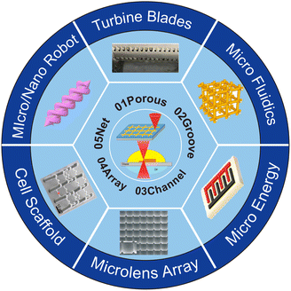 | ||
| Fig. 1 Schematic diagram of 3D micro/nano structures fabricated by ultrafast laser machining and their applications.20–26 Reproduced with permission from ref. 20. Copyright 2015, National Academy of Sciences. Reproduced with permission from ref. 21. Copyright 2018, Wiley. Reproduced with permission from ref. 22. Copyright 2021, American Association for the Advancement of Science. Reproduced with permission from ref. 23. Copyright 2018, Elsevier. Reproduced with permission from ref. 24. Copyright 2019, the American Chemical Society. Reproduced with permission from ref. 25. Copyright 2022, Springer Nature. Reproduced with permission from ref. 26. Copyright 2020, Elsevier. | ||
2 Methods and optimization of ultrafast laser
2.1 Principle of laser processing
Ultrafast laser, especially femtosecond laser, has ultra-short pulses of tens to hundreds of femtoseconds, and its energy absorption time is much shorter than the time required by thermal relaxation and other dynamic processes, and thus it can effectively minish the HAZ. The ultrafast laser has a very high instantaneous power, and due to this feature, it can not only process materials with high hardness, but also realize high quality and high precision in 3D processing. Furthermore, the ultrafast laser is capable of fabricating structures with ultra-high machining resolution and extremely small size. Therefore, it has been extensively applied, and the following description elaborates the two most common processing methods.Dry etching is often utilized to realize two types of functions. One is to modify various materials by ultrafast laser, and the other is to remove the laser-irradiated area by dry etching.33–35 Recently, some researchers applied femtosecond laser-induced plasma-assisted ablation to create high-aspect-ratio crack-free holes on sapphire substrates (Fig. 2(a)).36
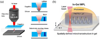 | ||
| Fig. 2 (a) Schematic of the experimental setup for the fabrication of high-aspect-ratio microstructures on sapphire.36 Reproduced with permission from ref. 36. Copyright 2020, ELSEVIER. (b) Schematic illustration of in-gel direct laser writing process.40 Reproduced with permission from ref. 40. Copyright 2018, Wiley. | ||
The uniformity and surface accuracy of the fabricated structure by wet etching will be affected to a certain extent via corrosion. Also, dry etching has limited application range and slow ablation efficiency under vacuum.
Picosecond laser etching and femtosecond laser etching employed in a laser generation system are different, but the steps and auxiliary mode are the same.37,38
In the case of laser etching, due to the Gaussian distribution of the laser beam, the temperature distribution of the irradiated point area is also approximately Gaussian distribution, and the temperature of the center area is much higher than the temperature of the edge, which will cause different reactions of substances in different areas. By choosing an appropriate power to match the temperature threshold of the object, the size of the etched hole is smaller than that of the laser spot, which is the reason for the machining resolutions of laser etching beyond the diffraction limit.39
TPP, a new approach, makes a big difference to 3D micro/nano structuring, owing to its characteristics of nonlinear absorption, high resolution and the universality of objects. The occurrence of TPP only proceeds under laser with high intensity, and also polymerization is triggered only at the focal point of the laser beam, without affecting other regions. This feature enables two-photon laser direct writing technology to achieve the high-precision processing of 3D micro/nano structures, but its point-by-point processing speed is low. Femtosecond projection two-photon lithography (FP-TPL) has been investigated for layer-by-layer printing, ensuring simultaneous temporal and spatial focusing of ultrafast light and leading to an increase in the writing speed.45
TPP can achieve spatial resolution below the diffraction limit of laser wavelength, which can be achieved by few-cycle laser pulse.42 In addition, TPP also has the universality of processing materials, which has gradually expanded from ordinary photoresist to polymethyl methacrylate (PMMA),46 polystyrene,47 polytetrafluoroethylene,48 hydrogels40,49–53 (Fig. 2(b)), etc. These characteristics are beneficial for the wide applications of TPP.
2.2 Optimization of processing efficiency
Ultrafast laser processing has significant advantages in micro/nano fabrication mainly due to the characteristics of ultra-short pulse and extremely high peak power. Compared with long pulse laser (such as nanosecond laser), the HAZ formed by ultrafast laser processing is ultra-small, and thus the processing quality is sharply improved. However, although ultrafast laser processing has excellent merits, it still faces the problem of low processing efficiency, resulting from the issues associated with the laser and the processing method. Important achievements have been made in optimizing ultrafast laser machining (Table 1).| PRF | Eout | Pout | Δτp | Ppeak | Ref. | |
|---|---|---|---|---|---|---|
| a PRF: pulse repetition frequency, Eout: pulse energy, Pout: average output power, Δτp: pulse duration, and Ppeak: peak power. | ||||||
| GHz amplified femtosecond laser source | 0.88, 1.76, 3.51 GHz (intraburst repetition rates) | — | 20 W | <550 fs | — | 55 |
| Inscribed thulium waveguide laser (Cr2+:ZnS saturable absorber) | 8 kHz | 6.9 μJ | 55 mW | 2.6 ns | 2.66 kW | 56 |
| Solid-state burst mode amplified laser source | 65 MHz (MHz burst mode), 2.5 GHz (GHz burst mode) | — | — | — | — | 57 |
| Thin-rod Yb:YAG amplifiers | — | 2.5 mJ | 28 W | — | — | 58 |
| Ultrafast fiber laser | — | 12 mJ | 700 W | 262 fs | — | 59 |
| Picosecond optical parametric oscillator | 10 kHz | 30.5 μJ | — | — | — | 60 |
Nowadays, ultrafast laser is receiving significant attention in industrial applications, where one of the key problems to be solved is its low processing speed, which should be improved, while its processing quality should be maintained. The processing speed is low because of the contradiction of a lower pulse energy and anabatic HAZ due to the increase in the repetition rate. The single pulse energy is relatively low, and thus the repetition rate needs to be improved to obtain an increased processing speed, but this will make the superposition of heat from a continuous single pulse, thereby increasing the formation of HAZ. Therefore, there are two methods to improve the processing speed, while ensuring the quality of ultrafast laser processing. One is to increase the single pulse energy of the laser and the other is to develop a laser that does not increase the HAZ even if it has a high repetition rate.
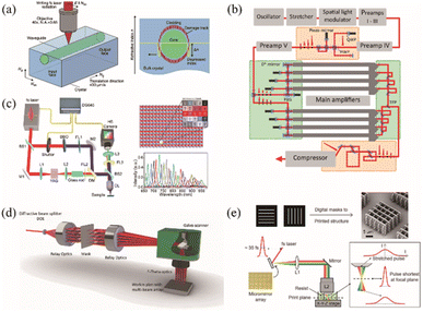 | ||
| Fig. 3 (a) Femtosecond direct laser writing of channel buried waveguides in a bulk Tm3+:KLu(WO4)2 crystal and illustration of the geometry of a depressed-index waveguide with a circular cladding (type III structure, end-facet view).56 Reproduced with permission from ref. 56. Copyright 2020, ELSEVIER. (b) Schematic setup of the eight-channel, four-pulse-replica FCPA system.59 Reproduced with permission from ref. 59. Copyright 2016, The Optical Society of America (OSA). (c) System configuration of CSMUP for femtosecond laser ablation measurement, filter arrangement on the CMOS sensor in the hyperspectral camera and spectral responses of 25 bands in the hyperspectral camera.77 Reproduced with permission from ref. 77. Copyright 2021, the American Chemical Society. (d) Principle of generating a static multi beam array.75 Reproduced with permission from ref. 75. Copyright 2021, IOP Publishing. (e) FP-TPL based on spatial and temporal focusing.45 Reproduced with permission from ref. 45. Copyright 2019, American Association for the Advancement of Science. | ||
The research on the GHz laser and burst mode is still in progress. Bonamis et al. emphasized the importance of experimental parameters to determine the optimal conditions of burst.55 Although the effect of the GHz laser on improving the processing speed is remarkable, due to the complexity of the heating/cooling balance ablation mechanism, these parameters are still being explored.55
An ultrafast laser with a high average power (>100 W) and a high pulse energy (>1 mJ) is required in many practical applications.58 This requires a reliable and compact laser source. Fiber lasers have remarkable advantages in terms of reliability; however, the peak power cannot be obtained due to the nonlinear effects in the fiber. Ultrafast fiber lasers usually emit ultra-short pulses with a pulse energy on the nJ level, and their average power is less than 100 mW.58 Many studies have been carried out to realize fiber lasers with a high average power and high pulse energy. Kienel et al. proposed an ultrafast, high-energy and high-power fiber-chirped-pulse-amplification (FCPA) system with a multi-dimensional amplification structure.59 Femtosecond pulses with an average power of 700 W and a maximum pulse energy of 12 mJ were obtained using four pulse replicas and eight amplifier channels.59 The performance demonstrated by the system reveals the great potential of the multi-dimensional amplification concept, which allows the full advantage of fiber optics, while mitigating its limitations (Fig. 3(b)).59 He and co-workers proposed a novel method for realizing a high-energy ultrafast optical parametric oscillator (OPO) using regenerative amplifier cavity pumping.60 It was proven that the pulse energy reaches 30 μJ when the pulse repetition rate is 10 kHz and the pulse width is 7.0 ps.60 This is the highest pulse energy of an ultrafast laser OPO to date.60 Kuznetsov et al. reported an ultrafast laser system with a high pulse energy (2.5 mJ) and high average power (28 W) based on a unique thin-rod gain module.58
Chirped pulse amplification (CPA) or optical parametric CPA (OPCPA) based on laser amplifiers has greatly increased the peak power of lasers by up to megawatts.61 Quasi-parametric CPA (QPCPA), as a new scheme, has the characteristics of high signal efficiency, wide bandwidth and high anti-phase mismatch ability. It achieves efficient amplification of the chirped signal pulse by exhausting the idle pulse, without the need for reverse conversion.62–64 Yin et al. demonstrated that QPCPA would break the average power barrier caused by thermal constraints.61 QPCPA has been shown to be robust to thermal degradation by blocking post-conversion effects.61 Numerical simulation demonstrated that the QPCPA based on an Sm:YCOB crystal supported a peak power of 3 TW, at 5 kHz and 13 kHz. It was 5 PW at the frequency of 1 Hz, and the average power exceeded 150 W in both cases.61
Femtosecond laser TPP (FL-TPP) is undoubtedly an important method to achieve 3D micro/nano machining, while near-threshold processing is considered as an indispensable strategy in FL-TPP, which can achieve ultra-high machining accuracy but has the problems of insufficient structural mechanical strength and long processing time. Several solutions have been proposed to overcome these problems. For example, Gross et al. proposed a post-processing method using oxygen plasma to improve the performance of TPP.65 They found that using octahedral micro lattices as a temporary support could prevent its movement during exposure, stabilize the beam during development, and most importantly, move it out after 25 min of exposure to oxygen plasma.65 They made a flawless flower using this method, which possessed a stem of 3.75 ± 0.12 μm and petal thickness of 1.5 ± 0.1 μm.65 It was proved that fine and complex structures can be produced by this method. To shorten the processing time, parallel multi-focus and focal field engineering (FFE) have also been proposed.65 Geng et al. proposed a novel laser nanomachinery process based on TPP and an ultrafast random-access digital micromirror device (DMD) scanner.66 This method expands single-focus scanning to parallel multi-focus scanning.66 The DMD scanner has unique advantages in precisely controlling the laser dose and focus position (∼100 nm).66 Therefore, it can be employed to design and manufacture complex 3D structures.66 Yang and co-workers firstly demonstrated 3D microstructures by a single-exposure TPP based on 3D FFE.67 Hu and co-workers proposed the DRS processing concept, where D refers to the use of low-power near-threshold femtosecond lasers and R refers to the high-power femtosecond laser enhanced scanning of the inner surface of structures.68 S represents irradiating internal unpolymerized materials by continuous ultraviolet (UV) light.68 Combining these three steps, the problems of low processing efficiency and mechanical strength can be effectively solved in near-threshold TPP.68
The spatial shaping of ultrafast lasers also has an important impact on the processing efficiency. The cross section of the laser beam emitted by the traditional laser is usually a Gaussian profile. When an object with a special shape is processed by this way, point scanning is needed, which puts forward high requirements for the quality and accuracy of the laser spot. Beam space shaping can directly complete the machining of microstructures with various shapes under the exposure of single or multiple pulses, thus improving the machining efficiency and resolution.69,70 In addition, it can be applied to single-step machining of special spatial contour microstructures,71 high-depth-aspect ratio structures,72,73 microarray structures and high-quality machining of various cross-scale hierarchical structures.74 Luo et al. proposed a method to process high-quality cylindrical microlens arrays (MLAs) on the surface of quartz glass through femtosecond laser shaping technology.71 They spatially fitted a Gaussian laser beam into a Bessel beam to obtain an anti-cylindrical laser intensity distribution in a specific section of the light field.71 The numerical simulation results showed that the envelope of light intensity distribution of this shape is consistent with the required morphology of cylindrical microlens, and the cylindrical MLA can be directly fabricated by line scanning of the shaped femtosecond laser on quartz glass.71 Furthermore, the radius and depth of the microlens element can be controlled by adjusting the laser power.71 Xie and co-workers used a femtosecond laser Bessel beam to control the local transient spatial electron density and performed single pulse processing of high-aspect-ratio and high-quality micropores in PMMA.72 The quality of the processed microholes at the entrance and side walls was higher than that of the microholes processed by a Gaussian beam.72 The aspect ratio of a microhole by femtosecond laser Bessel beams can be up to 330![[thin space (1/6-em)]](https://www.rsc.org/images/entities/char_2009.gif) :
:![[thin space (1/6-em)]](https://www.rsc.org/images/entities/char_2009.gif) 1.72 Roth et al. adopted adaptive beam shaping technology and used a femtosecond laser to process microchannels with almost no length limitation inside PMMA.73 They used a spatial light modulator (SLM) to compensate for the writing depth determined by spherical aberration, resulting in a precisely controllable and stable cylindrical shape.73 Hu and co-workers employed holographic FLDW technology to prepare aspheric microlens arrays (AMLAs) in parallel.74 In this process, they considered the inherent characteristics of SLM and compensated the defects of multi-focus mode to obtain higher intensity uniformity and optical efficiency.74
1.72 Roth et al. adopted adaptive beam shaping technology and used a femtosecond laser to process microchannels with almost no length limitation inside PMMA.73 They used a spatial light modulator (SLM) to compensate for the writing depth determined by spherical aberration, resulting in a precisely controllable and stable cylindrical shape.73 Hu and co-workers employed holographic FLDW technology to prepare aspheric microlens arrays (AMLAs) in parallel.74 In this process, they considered the inherent characteristics of SLM and compensated the defects of multi-focus mode to obtain higher intensity uniformity and optical efficiency.74
In laser ablation, Finger et al. reported a method for treating an arbitrary surface with high quality and efficiency, and they proposed a system that can use an average output power of 1 kW with fs pulse duration (Fig. 3(d)).75 Žemaitis and co-workers compared two methods for optimizing laser ablation efficiency, using optimal parameters for rapid ablation and 3D machining.76
In addition to the above-mentioned methods, Saha et al. developed FP-TPL technology to realize parallel printing of arbitrary complex 3D structures with sub-micron resolution (Fig. 3(e)).45 Yao and co-workers reported a chirped spectral mapping ultrafast photography (CSMUP) system for single real-time ultrafast imaging of femtosecond laser processing by combining femtosecond laser processing with visualization and imagination.77 This method proposed a new strategy for improving the accuracy and efficiency of femtosecond laser processing (Fig. 3(c)).77
3 3D micro/nano structures by ultrafast laser
Because it involves few steps, no mask, fast processing and is environmentally friendly, ultrafast laser is widely applied in many fields, resulting in the birth of a variety of ultrafast laser-processed structures. The size of the structures ranges from 100 μm to submicron.21,78–84 In this section, we review a variety of 3D micro/nano structures fabricated by ultrafast laser, including porous structures, grooves, channels, arrays and nets.3.1 Porous
As a common 3D structure, micro/nano porous structures are widely used in microsensors, aircraft turbine blades, nuclear fusion structures, microelectrodes, oil-water separation, etc.85,86 Although porous structures have been reported by many researches, and various porous structures studied, the processing on different materials, as well as the fabrication of pores with a high ratio of depth to diameter, and the blind holes with taper face great challenges.The quality, precision, and depth of ultrafast laser drilling rely on the material. Drilling in alloys is common.87–90 Zhai et al. drilled holes with an inner diameter of 162 μm, an aspect ratio of 15![[thin space (1/6-em)]](https://www.rsc.org/images/entities/char_2009.gif) :
:![[thin space (1/6-em)]](https://www.rsc.org/images/entities/char_2009.gif) 1 and almost zero taper on nickel-based alloy with thermal barrier coatings.89 Furthermore, they claimed that the depth and diameter of hole increased linearly with an increase in the pulse number.89 The self-focusing effect of femtosecond laser drilling has an important effect on the quality of deep holes. Wang and co-workers reported that the self-focusing effect has a regulating effect on enhancing the laser intensity in the cavity and a great influence on strengthening the intensity of microhole during the drilling process, which is not associated with the aperture.91,92
1 and almost zero taper on nickel-based alloy with thermal barrier coatings.89 Furthermore, they claimed that the depth and diameter of hole increased linearly with an increase in the pulse number.89 The self-focusing effect of femtosecond laser drilling has an important effect on the quality of deep holes. Wang and co-workers reported that the self-focusing effect has a regulating effect on enhancing the laser intensity in the cavity and a great influence on strengthening the intensity of microhole during the drilling process, which is not associated with the aperture.91,92
In addition to alloys, ultrafast laser drilling can be performed on polymers, ceramics, glass and other materials.82,93–96 Liu and co-workers fabricated an all-silicon uniform-aligned concave MLA through dry-etching-assisted femtosecond laser machining.34 Jiao et al. proposed a new method for applying a direct current to a silicon substrate during picosecond laser drilling.97 Because the voltage applied externally causes the free electrons to behave in a more orderly manner, it increases the current flow in the silicon substrate and enhances the energy absorption of silicon by laser. Besides the ultrafast laser drilling techniques adopted in different materials, some relatively general ultrafast laser drilling techniques also exist.98
Incredibly, Wang and co-workers processed holes on PMMA.94 In this study, an axial-hole lens with different cone angles was used to transform the incident Gaussian beam into a Bessel beam with different non-diffraction lengths and focus on the sample for processing. The aspect ratio of micropores was enhanced by increasing the pulse number and the cone angle of the axial cone lens. This method achieved the maximum ratio of depth to diameter of micropores (348![[thin space (1/6-em)]](https://www.rsc.org/images/entities/char_2009.gif) :
:![[thin space (1/6-em)]](https://www.rsc.org/images/entities/char_2009.gif) 1).94
1).94
In addition, ultrafast laser processing of porous has a self-cleaning effect, that is, the molten spatter after ultrafast laser etching is removed with continuous laser irradiation. Zhao et al. studied surface morphology and internal morphology based on the self-cleaning effect (Fig. 4(a)).78 Mishchik and co-workers indicated that when the focused numerical aperture is in the range of 0.08 to 0.15, the removal efficiency of dust is the highest when the holes are processed on sapphire.99
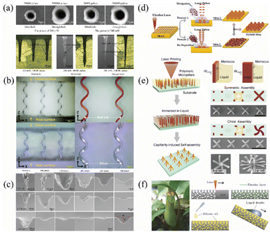 | ||
| Fig. 4 (a) SEM images of holes surfaces (wavelength: 1064 nm) and categories of the recast layers inside the holes based on the location (wavelength: 1064 nm).78 Reproduced with permission from ref. 78. Copyright 2018, Elsevier. (b) 3D helical microchannels.21 Reproduced with permission from ref. 21. Copyright 2018, Wiley. (c) Cross-section SEM microphotographs of the laser-machined grooves fabricated with different scan speeds at three certain energy fluences (1.14, 4.57, and 11.43 J cm−2). Inserted in the lower right corner: definition used herein for the width (W) and depth (d) of the grooves.79 Reproduced with permission from ref. 79. Copyright 2020, Elsevier. (d) Schematic illustration of the pulse injection controlled ultrafast laser direct writing strategy.80 Reproduced with permission from ref. 80. Copyright 2017, the American Chemical Society. (e) Preparation of deterministic chiral structures by combining laser printing and capillarity-induced self-assembly.81 Reproduced with permission from ref. 81. Copyright 2020, Wiley. (f) Process of preparing a slippery liquid-infused porous surface (SLIPS) by FLDW.82 Reproduced with permission from ref. 82. Copyright 2017, Wiley. | ||
3.2 Groove and channel
Groove structures are important common structures in micro/nano devices. The most representative ones are microfluidic channels applied in the biomedical field, micro/nano grating applied in the optical field, trench-like microelectrode applied in the electrochemical field, etc.100,101 Therefore, the machining of micro/nano grooves is of great significance. Laser engraving is currently the most common method of manufacturing.The working principle is similar to that of ultrafast laser drilling described in Section 3.1. Fig. 4(b) describes the glass micro channel fabricated by femtosecond laser combined with wet chemical etching.21 Also a silver film was placed on the wall of helical hollow microchannels. Moreover, different features were obtained in channel structures on different materials by ultrafast laser processing. For the grooves prepared on Al-doped silicon carbonitride (SiAlCN) ceramics, the number of femtosecond laser scans and the energy fluences will affect the quality of the grooves. Chen et al. pointed out that when the energy fluences increase, the ablation is serious and a HAZ is generated, resulting in low machining quality.79 However, increasing the scanning speed at the same energy fluences will decrease the removal rate and ablation degree. Also, the HAZ is associated with both the scanning speed and energy fluences. Laser parameters with a high scanning speed or low energy flux are helpful to avoid the formation of HAZ on SiAlCN ceramics.79 The results demonstrated that a scanning speed above 250 mm s−1 and energy flux below 4.57 J cm−2 are beneficial for the manufacture of high-quality grooves.79 Fig. 4(c) shows the influence of scan speed and energy on the laser-machined grooves. Grooving polyether ether ketone (PEEK) is widely applied to prevent bone formation.102 During this process, the femtosecond laser can form micro grooves and nanometer-scale pore clusters on the surface of the material, both of which have an obvious synergistic effect. The groove has a great correlation with the appearance of grooves and pore clusters. The results indicated that at the spacing of 2 μm, the surface of the processed material had a great amount of irregularly distributed pore clusters but no regular periodic grooves. When the widths of the surface groove of PEEK were 10 and 15 μm, the spine of the groove was also covered by nanopore clusters. In addition to making biomimetic bone, it also has an important role in the cluster and differentiation of osteoblasts.103
3.3 Array
Array structures enable structural or functional units to have special functions, such as anti-reflection,80,104–106 anti-icing,107,108 and electron emission.109 The array structure can also be used to obtain an increased specific surface area. However, the preparation of the array structures requires complicated steps, some of which cannot be achieved by traditional methods. Femtosecond laser processing can simplify the process and improve the preparation efficiency.For a surface with a special function, femtosecond laser processing can be applied to improve the preparation efficiency. For special functional surfaces that have anti-reflection performance, Wang et al. employed the femtosecond laser texturing technique to fabricate quasi-uniform cone-array microstructures on single-crystal silicon directly.104 The structure has a peak depth of up to 8 μm, which significantly reduces light reflection. Compared with planar silicon wafers, the relative reflectance of the cone array decreases to less than 9% in the measurement wavelength range (400–1000 nm). Fan and co-workers proposed and experimentally demonstrated a universal pulse injection-controlled ultrafast laser direct writing method for the fabrication of highly efficient antireflective structures on the surface of metals (Fig. 4(d)).80 By this method, the obtained minimum reflectance of the UV-NIR wideband spectral region on Cu, Ti, and W surfaces were 1.4%, 0.29%, and 2.5%, respectively.80 The proposed method is easy to access, suitable for different types of metals and large-area production, with double-scale characteristics. Li et al. fabricated an inverted pyramid and cone array with a pitch of about 2 μm and a total height of nearly 900 nm using an FLDW assisted by wet etching.105 The transmittance measurements between 3 μm and 5 μm were consistent with the simulation results using VirtualLab, and the transmittance reached a maximum of 92.5% at 4 μm.105 Liu and co-workers developed a collaborative 3D micro/nano manufacturing method using femtosecond laser and oxidation technology.106 They fabricated the conical structure of a uniformly loaded nanowire, namely, an urchin-like structure.106 This method was experimentally verified to be environmentally friendly and has no toxicity.106 In addition, the reflection test of the constructed sea urchin-like array demonstrated the excellent anti-reflection performance in the ultra-wide spectrum of UV, visible, infrared and far-infrared (FIR), which was fully realized on the surface of copper.106
For special functional surfaces that have anti-ice performance, Jiang et al. proposed a technique to prepare layered micro/nano structures on the surface of metals by high-temperature stamping with a mold by ultrafast laser ablation.107 The surface structure of the prepared copper was hydrophobic, which became superhydrophobic after chemical modification.107 This method combines the advantages of ultrafast laser ablation to fabricate a micro/nano mold with high-temperature lithography to obtain a micro/nano structure on the surface of the target metal.107 Pan and co-workers designed and fabricated a novel triple-scale micro/nanostructured superhydrophobic surface, with excellent ice-phobic and anti-icing performance.108 The surface was fabricated by chemical oxidation and ultrafast laser ablation.108 Also, it had excellent Cassie state stability and the critical Laplace pressure was up to 1450 Pa.108 Its ice adhesion strength had a low value of 1.7 kPa.108
For a special functional surface capable of electron emission, Xiao et al. fabricated a field emission array of single-crystal lanthanum hexaboride (LaB6) with uniform mountain emitter morphology using a UV femtosecond laser at a high speed.109 The height of the mountain emitter was 6 μm and the radius of the curvature was 100 nm.109
Array structures can also be employed to increase the surface area of active electrodes. Femtosecond laser processing can simplify this process and improve the preparation efficiency. Cui and co-workers developed a highly effective method to fabricate manganese oxide directly on the surface of a 3D manganese collector.110 In their process, a 3D conductive network on the surface of manganese was generated by a femtosecond laser and the network also acted as a current collector (named 3D Mn).110 Manganese oxides were then directly formed on the femtosecond laser-structured surface by chemical oxidation (named 3D Mn/MnOx).110 This structure had a large specific surface area, which shortened the transport path of electrons/ions, promoted electrolyte penetration, and reduced the contact resistance between 3D Mn and MnOx, which are conducive to long-term electrochemical cycling.110 Kwon et al. proposed a laser direct writing technique based on TPP for the fabrication of a photoresist-derived carbon cross finger electrode (PRC-IDE) with a large areal capacitance.111
In addition, femtosecond laser processing has extremely high preparation efficiency compared with other commonly used fabrication technologies. Hu et al. combined femtosecond laser printing with capillary-induced self-assembly to obtain a mesoscale chiral architecture with high designability and customizability (Fig. 4(e)).81 They took advantage of the flexibility of femtosecond laser printing to achieve a variety of chiral assemblies.81
3.4 Net
The net structure fabricated by femtosecond laser processing is a type of unique 3D structure, which also has some novel functions.Regular net structures such as cell scaffolds are processed by TPP. For example, Heitz et al. processed three-level pentaerythritol triacrylate (PETA):bisphenol a glycidyl methacrylate (BisGMA) cage structures by TPP on polyethylene terephthalate (PETG) substrates, which were used to fill bone-forming cells and generate 3D mineralized protein networks.112 Trautmann and co-workers adhered human primary fibroblasts to scaffolds prepared by TPP.113
Irregular net structures such as porous networks have been proposed. Yong et al. directly formed a 3D porous network structure on a polyamy-6 (PA6) substrate and tested a variety of liquids for their repellency (Fig. 4(f)).94 These liquids include water, cetane, lake water, ink, glycerin, coffee, milk, egg whites and egg yolks.94 In 2018, Yong and co-workers employed one-step FLDW to prepare porous network microstructures on the surface of various polymers.93 They also found that the growth of C6 glioma cells was promoted by the original laser-induced porous PET surface as a culture substrate, while the smooth surface of PET completely inhibited the growth of C6 glioma cells.93 As another type of irregular networking structure, the carbon nanoribbon network (CNRN) platform plays an important anti-cancer role. Chowdhury et al. developed a CNRN platform bottom-up by femtosecond laser ionization.83 They transformed an abiotically reactive graphite matrix into a bioactive interwoven CNRN platform.83 Their experiment proved two distinct simultaneous functions of the CNRN platform, i.e., the attractive properties of the extra cellular matrix (ECM) and the therapeutic properties without the need to bind any biomolecules/drugs.83 The CNRN platform contributes to enhancing the cellular attractiveness and selective functions, enabling fibroblasts to exhibit tissue-like behavior and HeLa cells to perform apoptosis-like functions.83 Huang and co-workers successfully prepared a precise 3D scaffold with living cell encapsulation using the TPP technique, and no obvious cell damage was observed during the manufacturing process.114 The advantages of laser processing for different structures are summarized, as shown in Table 2.
| Structural features | Ultrafast laser processing methods | Advantages of laser processing | Ref. | |
|---|---|---|---|---|
| Porous | Better surface topography and much deeper | Ablation | Self-cleaning effect, short pulse duration and high peak power | 78 and 89 |
| Groove and channel | High ratio of depth to diameter | Ablation and laser direct writing (LDW) | Excellent thermal stability, short pulse duration and high peak power | 99 |
| Array | Rational alignment | Ablation and LDW | High speed | — |
| Net | Complex and 3D | LDW | High resolution | 21 |
4 Micro/nano devices based on 3D structure
4.1 Turbine blades
Aircraft turbines can work at thousands of degrees (°C). Therefore, these high-temperature devices must have excellent cooling capacity. On the microscopic scale, aircraft turbine blades (including alloy structure, blade core and thermal barrier coating) always have the arrangement of a large number of micropores, which is a typical application of pore structures.For alloy structures on the outside of the blade, the treatment of high-quality hole structures on the turbine blade often utilizes ultrafast laser ablation. However, a more thorough understanding of the interaction between the laser and material during machining is required. The interaction can be based on the traditional two-temperature model.115 Besides, some studies analyzed the ground laser ablation of Ti6Al4V alloy using the two-temperature model and proposed the multi-pulse ablation method, which further improved the quality of processed holes.116 This two-temperature model is based on a set of coupled partial differential equations in the time and space domain.
In addition to the alloy blades, there are blade cores and thermal barrier coatings. The core of the turbine blade is typically porous alumina ceramic and its cavity plays an important role in the service life and performance of the turbine blade. Min and co-workers investigated the femtosecond laser treatment of porous alumina ceramics.117 They studied gas-assisted and water-assisted processing methods and determined the optimal working window of processing. For drilling a thermal barrier coating on turbine blades, Wang et al. performed drilling on the thermal barrier In718 coat using a femtosecond pulsed Ti:sapphire laser.118 Also, they analyzed the influence of the polarization of the laser beam on microhole profile and morphology of periodic structures on the sidewall of microhole.118 They found that with circular polarization, the periodic structure had a ball-like profile. Alternatively, under linear polarization, periodic structures of pine needle-like type were obtained.118
4.2 Micro energy devices
In recent years, there has been remarkable development in micro/nano electronic devices, and the emergence of micro/nano sensors, micro/nano robots, micro/nano actuators and flexible wearable devices also pose great challenges in the power supply.119,120 To meet the demand for power sources, micro energy devices must have a sufficient energy density. Thus, in the case of the manufacture of micro energy devices, the design, choice and treatment of materials with special structures become a critical issue, and therefore highly suitable and effective machining process is required.121–124 At present, most of the ultrafast laser processing of 3D structures applied in micro energy devices involves two approaches.One is the formation of active microelectrodes and composite microelectrodes by laser etching.125–127 Zheng et al. processed line and grid structures layer-by-layer on silicon/graphite (Si/C) electrodes.125 These two microstructures provided more free space for volume changes, and the mechanical stress of the electrode was reduced. Furthermore, the fabricated graphite electrode delivered an excellent high rate capability up to 3C beyond state-of-art cells. This processing is faster than those of other methods for the manufacture of microelectrodes. Kurra and co-workers described in detail the laser processing of 3D graphene electrodes.126 He et al. fabricated a high-performance in-plane Co–Zn alkaline micro-battery by laser dry etching. Fig. 5 shows the electrochemical performance of the fabricated Co–Zn microbattery.127
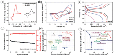 | ||
| Fig. 5 Electrochemical performances of Co–Zn micro-battery. (a) Cyclic voltammetry (CV) curves of Co(OH)2@NiCo-layered double hydroxide@Ni-coated textile and Zn@carbon clothes at 5 mV s−1. (b) CV curves, (c) galvanostatic charge–discharge (GCD) curves, (d) long-term cycling stability, and (e and f) Ragone plots of Co–Zn microbattery.127 Reproduced with permission from ref. 127. Copyright 2020, Wiley. | ||
The other approach is the use of LDW technology of TPP.128–130 Most processes are conducted for the treatment of polymers. For example, Staudinger et al. manufactured conductive microstructures on carbon nanofiller absorbers using this technology.128
4.3 Microfluidics
Microfluidic devices consist of a series of channels or chambers containing and manipulating fluid with ultra-small volume. They have been widely used in the biomedical field due to their high throughput, single-cell resolution and mixing efficiency.131 Since their appearance in the 1990s, microfluidic devices have been developing towards miniaturization and integration, which require high precision, high resolution, low cost and mass production for their manufacture. Ultrafast laser machining is emerging as a powerful process for the large-scale manufacturing of microfluidic devices, especially for devices with complex structures. For instance, laser wet etching technology was used in fused silica to obtain 3D microcoils,12 and Fig. 6(c) describes the process of filling 3D microcoils with metal gallium in fused silica.132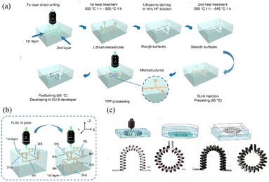 | ||
| Fig. 6 (a) Schematic illustration of the procedure for the fabrication of 3D multilayered microfluidic chips.134 Reproduced with permission from ref. 134. Copyright 2019, Springer Nature. (b) Schematic illustration of the process for the fabrication of eight-layered glass microchannels by femtosecond laser.134 Reproduced with permission from ref. 134. Copyright 2019, Springer Nature. (c) Schematic of the two-step fabrication process and different shapes of microchannels and inductances.132 Reproduced with permission from ref. 132. Copyright 2013, The Optical Society of America. | ||
TPP technology is used in processing polyethylene glycol-based hydrogels and protein-based biomolecules.133 To apply FLDW technology in the fabrication of 3D channels in glass, and then remove the glass matrix by HF, as similar as the procedure by Xu et al.,21 TPP and femtosecond-laser-assisted wet etching (FLWE) hybrid femtosecond laser processing can also be applied.
In the study by Wang and co-workers, “all-in-one” femtosecond laser processing was adopted to obtain multilayer microfluidic channels, and Fig. 6(a) shows their fabrication procedure.134 To create microchannels with high quality and uniformity, they optimized the power density of the laser. Fig. 6(b) demonstrates an eight-layered microfluidic chip integrated with the fabricated polymer microstructures. Wang et al. summarized numerous processing technologies for microfluidic devices.135
The slow progress in the treatment of malignant tumors, lack of tumor models and difficulty in simulating the narrow space of tissues and organs during their spread are important reasons that hinder pathophysiological research. Sontheimer-Phelps and co-workers, described in detail the important role of microfluidic devices in cancer treatment.136 Sima et al. processed submicron polymer channels of different lengths by TPP and chemically assisted femtosecond laser etching, which is a novel biochip with 3D polymer nanostructure.137 Also, they successfully observed the morphological change in cells during migration. In addition to pathological studies in cell cultures, Lao and co-workers produced a microchannel with a nanostructure for the local sensing of anti-cancer drugs (doxorubicin).138
Besides, microfluidic systems for applications in cell culture and observation, optical fluidic adaptive imaging and biomedical sensing are attracting attention.139,140
4.4 Microlens arrays
In MLAs, their aperture and relief depth are on the micrometer scale. They not only have the traditional lens focus and basic function such as imaging, but also have the characteristics of small cell size and high integration and can form various new optical systems. Nowadays, MLAs are important components in integrated micro-optics systems.Femtosecond laser processing and wet etching are used to fabricate MLAs. Yang et al. fabricated a double-sided concave MLA for generating novel and diverse imaging patterns by FLWE (Fig. 7(a)).23 Also, Zhang and co-workers employed this method to prepare a large-area concave MLA.141 They produced about 2 million quasi-periodic MLAs with an adjustable diameter and depression height within 30 min.141 Li et al. prepared a superhydrophobic polydimethylsiloxane (PDMS) MLA by combining FLDW and FLWE (Fig. 7(b)).142 The prepared MLA not only had ultra-low adhesion, ultra-high hydrophobicity and excellent imaging performance, but also had excellent water resistance and self-cleaning function compared with ordinary MLAs.142 Cao and co-workers prepared hard templates using a combination of femtosecond laser processing and wet etching, and prepared soft and thin PDMS MLAs through soft etching (Fig. 7(c)).143 Instead of making MLAs directly, they made silicon templates, which could then be used to obtain large-scale, highly homogeneous PDMS MLAs via soft lithography.143
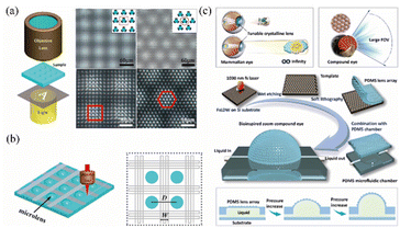 | ||
| Fig. 7 (a) Schematic illustration of the microscopic imaging system. The bottom side of the MLA observed by the top side of the MLA (insets respectively represent the arrangement of the rectangular-shaped and hexagonal-shaped MLA on each side) and imaging properties of the letter “A”.23 Reproduced with permission from ref. 23. Copyright 2018, Elsevier. (b) Schematic illustration of the fabrication of criss-cross reticular rough structures around the microlenses and top view of as-prepared MLA.142 Reproduced with permission from ref. 142. Copyright 2019, Wiley. (c) Schematic illustration of the design principles of a variable-focus compound eye, working principles and properties of a monocular eye, and working principles and properties of a compound eye. Schematic illustration of the fabrication process of the zoom compound eye. Regulation principle of the zoom compound eye.143 Reproduced with permission from ref. 143. Copyright 2020, the American Chemical Society. | ||
Furthermore, MLAs can also be prepared by femtosecond laser and ion beam etching. Liu et al. applied this method to prepare nanosmooth MLAs on rigid materials (such as molten quartz, gallium arsenide, silicon carbide and diamond) in a convenient and multifunctional way.144
4.5 Cell scaffold
Tissue engineering involves the use of cells and scaffolds/matrices to develop novel functional tissues for implantation into donors. With the great demand for tissues and organs, tissue engineering has developed to create living replacements for body. The need for tissue engineering is further proposed by the widening gap between demand and supply of organs or transplantable tissues. Cell scaffolds are an important component in tissue engineering. Scaffolds provide a favorable environment and support for the attachment and growth of tissue. They can also be applied as a template or carrier for tissue implantation and drug delivery.145 At present, the main method for the preparation of scaffolds is 3D printing based on femtosecond lasers.TPP 3D printing technology based on femtosecond lasers is widely employed. Thompson and co-workers proposed the preparation of a poly (caprolactone) (PCL) scaffold by TPP for human retinal progenitor cells.146 One month after the implantation of the cell-free scaffolds in pig models of retinitis pigmentosa, there was no infection, inflammation, local or systemic toxicity.146 Furthermore, comprehensive ISO 10993 tests of the photopolymerized scaffolds showed their high biocompatibility. Wang et al. prepared microcapsules via the TPP method based on structured laser beams and studied the culture of budding yeast using the microcapsules to demonstrate their application as a 3D cell culture scaffold.24 Richter et al. fabricated 3D scaffolds for cell culture via direct laser writing, where the scaffolds had a boxing-ring-like structure (Fig. 8).147 Song and co-workers reviewed hydrogel scaffolds prepared using TPP, provided an in-depth description on various biological and mechanical characterizations for evaluating the structures, and described the design requirements for cell and tissue-related applications.148 Three case studies of bone, cancer and heart tissue were also presented to elaborate the need for structural materials in the next generation of clinical applications.148
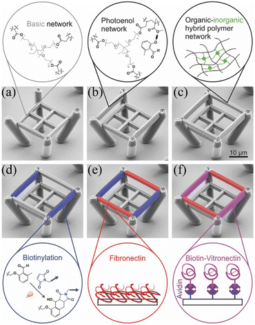 | ||
| Fig. 8 Pseudo-colored scanning electron microscopy (SEM) images illustrating the sample preparation process.147 (a) Scaffold produced in the first direct laser writing step using the basic resist. (b) In a second direct laser writing step, two beams of the functional resist (here, photoenol resist) were added to the scaffold. (c) In a third direct laser writing step, two additional OrmoComp beams were written. (d) Post-modification of the resulting microstructure via UV light leads to biotinylation of the functional resist (blue). (e) Fibronectin (red) exclusively binds to OrmoComp beams. (f) By subsequent coating steps with avidin and biotin–vitronectin (magenta), only the functional resist is addressed by this extracellular matrix protein. Reproduced with permission from ref. 147. Copyright 2016, Wiley. | ||
Cell scaffolds have also been prepared by femtosecond laser and 3D printing. Daskalova and co-workers achieved efficient production and post-processing of biomimetic and biodegradable PCL scaffolds using a combination of femtosecond laser processing and 3D printing.149 The results demonstrated that the hydrophilicity of PCL materials is improved by femtosecond laser processing.
4.6 Micro/nano robot
In addition to the above-mentioned microfluidic technology, micro/nano robots also have important applications in the treatment of some diseases due to their minimal invasiveness. Because of their micro scale and complex 3D structure, the accuracy and designability of the processing of micro/nano robots are necessary. Thus, ultrafast machining, especially FLDW technology in the fabrication of micro/nano robots is attracting great interest.Jeon et al. fabricated a micro-robot using FLDW, which is powered by a magnetic field and can provide precise positioning.150 The proposed micro robot was applied to metastasize colorectal cancer cells. Moreover, this FLDW was employed to create smart and deformable 3D micro actuators.151 Although micro robots show great potential in the medical field, they also face some challenges, especially biocompatibility.22
Because traditional laser processing is mostly focused on a single material, artificial organs and tissues cannot be precisely assembled from multiple materials. Ma and co-workers proposed an on-chip TPP strategy for the fabrication of artificial musculoskeletal systems, which also provides the possibility for fabricating various 3D structures.152 Furthermore, this strategy was preliminarily verified by producing a spider micro robot and 3D intelligent micro claw.
5 Conclusion and perspective
Ultrafast lasers are attractive because of their high power density, ultra-short pulse and extremely high peak power. Thus, due to these characteristics, ultrafast lasers are employed in various fields, ranging from micro/nano sensors, communication and consumer electronic-related components to aerospace. At present, ultrafast laser processing is gaining interest due to its high speed and high precision. The main ultrafast laser processing methods for 3D structures are picosecond laser etching, different auxiliary types of FLDW and femtosecond laser etching based on TPP. In this review, we introduced the principles of ultrafast laser fabrication and the optimization of its processing speed. Especially, the optimization of the repetition rate and pulse energy of laser is of interest. Furthermore, specific pulse intensities and fluences are required in suitable modifications, which are also very important parameters. For instance, the ablation pattern of protein crystals is determined by the pulse number and laser fluence.153 When the laser fluence is higher than 1 J cm−2, the protein crystal is directly removed to form the ablation hole.153 At a laser fluence below the threshold of single-pulse ablation, which was calculated to be ∼0.4 J cm−2 for a single pulse, the protein was denatured due to energy deposition. Through the accumulation of multiple pulses, the denatured protein could not maintain its crystal shape and formed a foaming form. Both the ablation pore and the foaming zone contributed to the surface topography of the ablation zone, resulting in unique physical and chemical performances. The nonlinear absorption of protein single crystals is proportional to the m order of the laser intensity, where m is the number of photons absorbed by an electron. The sharpening of the absorption region resulting from the multiphoton absorption is helpful for the ablation region exceeding the diffraction limit with high accuracy.153 For PMMA, the experimental method is to measure the average laser power delivered to the sample using a calibrated integrating sphere optometer after focusing the focal plane of the lens. The experimental results showed that the threshold fluence is predicted to be 0.97 J cm−2 when the number of pulses N is 20. Both the ablation depth and the average roughness of the ablation region increase with an increase in the laser fluence.154 Afterward, typical 3D structures machined by ultrafast laser, including porous structures, channels, arrays and net structures are elaborated. Also, the applications of these structures in turbine blades, microfluidics, micro energy devices, MLAs, cell scaffolds and micro/nano robots are described in detail. All these 3D structures applied in different devices are of great significance in medicine, energy and optics. The corresponding discussion and summary of these methods as well as optimization for 3D structures will also provide valuable strategies to researchers in future studies.To the best of our knowledge, ultrafast laser processing is limited by its high cost and immature control technology. Besides femtosecond and picosecond lasers, ultrafast lasers also include attosecond lasers. Attosecond lasers with a big leap in pulse width are expected to be an important driving force in microelectronic devices. At the current state, many studies have explored the interaction between metal micro/nano particles and attosecond laser pulses, achieving fascinating progress. This will promote the applications of attosecond lasers in many fields. Thus, it can be predicted that ultrafast lasers will become the main approach in the field of precision engineering.
Author contributions
P. Tian conceived the idea and supervised the whole study. X. Yang and R. Song wrote the manuscript. X. Liu, L. He, X. He, Z. Ma revised the manuscript. L. Wu, X. Liu, H. Tang, X. Lu helped to collect the information, and edited the figures and tables. All authors discussed the review and assisted during manuscript preparation.Conflicts of interest
There are no conflicts to declare.Acknowledgements
This work was supported by the Fundamental Research Funds for the Central Universities (20822041F4045), the Full-Time Postdoctoral Research and Development Fund of Sichuan University (2022SCU12129), and Science and Technology Cooperation Special Fund Project between Sichuan University and Zigong City (2020CDZG-12).References
- K. C. Phillips, H. H. Gandhi, E. Mazur and S. K. Sundaram, Adv. Opt. Photonics, 2015, 7, 684–712 CrossRef CAS.
- J. Huang, L. Jiang, X. Li, S. Zhou, S. Gao, P. Li, L. Huang, K. Wang and L. Qu, ACS Appl. Mater. Interfaces, 2021, 13, 43622–43631 CrossRef CAS PubMed.
- S. Bagheri, K. Weber, T. Gissibl, T. Weiss, F. Neubrech and H. Giessen, ACS Photonics, 2015, 2, 779–786 CrossRef CAS.
- Y. Liu, X. Li, J. Huang, Z. Wang, X. Zhao, B. Zhao and L. Jiang, ACS Appl. Mater. Interfaces, 2022, 14, 16911–16919 CrossRef CAS.
- J. Huang, K. Xu, J. Hu, D. Yuan, J. Li, J. Qiao and S. Xu, Nano Lett., 2022, 22, 6223–6228 CrossRef CAS.
- Y. Li, X. Zhang, T. Zou, Q. Mu and J. Yang, ACS Appl. Mater. Interfaces, 2022, 14, 21758–21767 CrossRef CAS PubMed.
- H. Liu, Z. Sun, Y. Chen, W. Zhang, X. Chen and C.-P. Wong, ACS Nano, 2022, 16, 10088–10129 Search PubMed.
- A. Vailionis, E. G. Gamaly, V. Mizeikis, W. Yang, A. V. Rode and S. Juodkazis, Nat. Commun., 2011, 2, 445 Search PubMed.
- H. B. Sun, Y. Xu, S. Juodkazis, K. Sun, M. Watanabe, S. Matsuo, H. Misawa and J. Nishii, Opt. Lett., 2001, 26, 325–327 CrossRef CAS PubMed.
- Z. Z. Li, L. Wang, H. Fan, Y. H. Yu, H. B. Sun, S. Juodkazis and Q. D. Chen, Light: Sci. Appl., 2020, 9, 41 CrossRef CAS PubMed.
- K. Sugioka, Int. J. Extreme Manuf., 2019, 1, 012003 Search PubMed.
- F. Sima, K. Sugioka, R. M. Vazquez, R. Osellame, L. Kelemen and P. Ormos, Nanophotonics, 2018, 7, 613–634 CAS.
- M. Malinauskas, A. Zukauskas, S. Hasegawa, Y. Hayasaki, V. Mizeikis, R. Buividas and S. Juodkazis, Light: Sci. Appl., 2016, 5, e16133 CrossRef CAS PubMed.
- J. B. Yu, M. T. Luo, Z. Y. Lv, S. M. Huang, H. H. Hsu, C. C. Kuo, S. T. Han and Y. Zhou, Nanoscale, 2020, 12, 23391–23423 RSC.
- M. Xi, J. L. Yong, F. Chen, Q. Yang and X. Hou, RSC Adv., 2019, 9, 6650–6657 Search PubMed.
- G. Wen, Z. G. Guo and W. M. Liu, Nanoscale, 2017, 9, 3338–3366 Search PubMed.
- Z. W. Mao, W. Cao, J. Hu, L. Jiang, A. D. Wang, X. Li, J. Cao and Y. F. Lu, RSC Adv., 2017, 7, 49649–49654 RSC.
- Y. Lu, L. D. Yu, Z. Zhang, S. Z. Wu, G. Q. Li, P. C. Wu, Y. L. Hu, J. W. Li, J. R. Chu and D. Wu, RSC Adv., 2017, 7, 11170–11179 RSC.
- M. S. S. Bharati, B. Chandu and S. V. Rao, RSC Adv., 2019, 9, 1517–1525 RSC.
- M. F. El-Kady, M. Ihns, M. Li, J. Y. Hwang, M. F. Mousavi, L. Chaney, A. T. Lech and R. B. Kaner, Proc. Natl. Acad. Sci. U. S. A., 2015, 112, 4233–4238 Search PubMed.
- J. Xu, X. L. Li, Y. Zhong, J. Qi, Z. H. Wang, Z. F. Chai, W. W. Li, C. B. Jing and Y. Cheng, Adv. Mater. Technol., 2018, 3, 1800372 Search PubMed.
- H. Ceylan, N. O. Dogan, I. C. Yasa, M. N. Musaoglu, Z. U. Kulali and M. Sitti, Sci. Adv., 2021, 7, eabh0273 CrossRef CAS PubMed.
- Y. Wei, Q. Yang, H. Bian, F. Chen, M. Li, Y. Dai and X. Hou, Appl. Surf. Sci., 2018, 457, 1202–1207 Search PubMed.
- C. Wang, L. Yang, Y. Hu, S. Rao, Y. Wang, D. Pan, S. Ji, C. Zhang, Y. Su, W. Zhu, J. Li, D. Wu and J. Chu, ACS Nano, 2019, 13, 4667–4676 Search PubMed.
- F. Jin, J. Liu, Y.-Y. Zhao, X.-Z. Dong, M.-L. Zheng and X.-M. Duan, Nat. Commun., 2022, 13, 1357 CrossRef CAS PubMed.
- Y. Z. Liu, Int. J. Mach. Tools Manuf., 2020, 150, 103510 CrossRef.
- X. Q. Liu, B. F. Bai, Q. D. Chen and H. B. Sun, Opto-Electron. Adv., 2019, 2, 190021 CAS.
- M. Chambonneau, D. Grojo, O. Tokel, F. O. Ilday, S. Tzortzakis and S. Nolte, Laser Photonics Rev., 2021, 15, 2100140 CrossRef.
- X. W. Li, Q. Xie, L. Jiang, W. N. Han, Q. S. Wang, A. D. Wang, J. Hu and Y. F. Lu, Appl. Phys. Lett., 2017, 110, 181907 CrossRef.
- Z. J. Lin, J. Xu, Y. P. Song, X. L. Li, P. Wang, W. Chu, Z. H. Wang and Y. Cheng, Adv. Mater. Technol., 2020, 5, 1900989 CrossRef CAS.
- A. Marcinkevicius, S. Juodkazis, M. Watanabe, M. Miwa, S. Matsuo, H. Misawa and J. Nishii, Opt. Lett., 2001, 26, 277–279 Search PubMed.
- X.-W. Cao, Q.-D. Chen, H. Fan, L. Zhang, S. Juodkazis and H.-B. Sun, Nanomaterials, 2018, 8, 287 CrossRef PubMed.
- X. Q. Liu, L. Yu, Q. D. Chen and H. B. Sun, Appl. Phys. Lett., 2017, 110, 091602 Search PubMed.
- X. Q. Liu, Q. D. Chen, K. M. Guan, Z. C. Ma, Y. H. Yu, Q. K. Li, Z. N. Tian and H. B. Sun, Laser Photonics Rev., 2017, 11, 1600115 Search PubMed.
- J. H. Noh, J. D. Fowlkes, R. Timilsina, M. G. Stanford, B. B. Lewis and P. D. Rack, ACS Appl. Mater. Interfaces, 2015, 7, 4179–4184 CrossRef CAS PubMed.
- H. G. Liu, Y. Li, W. X. Lin and M. H. Hong, Opt. Laser Technol., 2020, 132, 106472 Search PubMed.
- L. Chen and D. Q. Yu, J. Mater. Sci.: Mater. Electron., 2021, 32, 16481–16493 Search PubMed.
- X. L. Li, J. Xu, Z. J. Lin, J. Qi, P. Wang, W. Chu, Z. W. Fang, Z. H. Wang, Z. F. Chai and Y. Cheng, Appl. Surf. Sci., 2019, 485, 188–193 Search PubMed.
- K. Kurihara, T. Nakano, H. Ikeya, M. Ujiie and J. Tominaga, Microelectron. Eng., 2008, 85, 1197–1201 Search PubMed.
- A. Nishiguchi, A. Mourran, H. Zhang and M. Moller, Adv. Sci., 2018, 5, 1700038 Search PubMed.
- R. Batchelor, T. Messer, M. Hippler, M. Wegener, C. Barner-Kowollik and E. Blasco, Adv. Mater., 2019, 31, 1904085 CrossRef CAS PubMed.
- V. F. Paz, M. Emons, K. Obata, A. Ovsianikov, S. Peterhansel, K. Frenner, C. Reinhardt, B. Chichkov, U. Morgner and W. Osten, J. Laser Appl., 2012, 24, 042004 CrossRef.
- R. Infuehr, N. Pucher, C. Heller, H. Lichtenegger, R. Liska, V. Schmidt, L. Kuna, A. Haase and J. Stampfl, Appl. Surf. Sci., 2007, 254, 836–840 CrossRef CAS.
- K. Obata, A. El-Tamer, L. Koch, U. Hinze and B. N. Chichkov, Light: Sci. Appl., 2013, 2, e116 CrossRef CAS.
- S. K. Saha, D. Wang, V. H. Nguyen, Y. Chang, J. S. Oakdale and S.-C. Chen, Science, 2019, 366, 105–109 CrossRef CAS PubMed.
- G. L. Roth, C. Esen and R. Hellmann, Opt. Express, 2017, 25, 18442–18450 CrossRef CAS PubMed.
- O. I. Avila, N. B. Tomazio, A. J. G. Otuka, J. C. Stefanelo, M. B. Andrade, D. T. Balogh and C. R. Mendonca, J. Polym. Sci., Part B: Polym. Phys., 2018, 56, 479–483 CrossRef CAS.
- A. Zhizhchenko, A. Kuchmizhak, O. Vitrik, Y. Kulchin and S. Juodkazis, Nanoscale, 2018, 10, 21414–21424 Search PubMed.
- J. H. Li, C. T. Wu, P. K. Chu and M. Gelinsky, Mater. Sci. Eng., R, 2020, 140, 100543 CrossRef.
- J. F. Xing, M. L. Zheng and X. M. Duan, Chem. Soc. Rev., 2015, 44, 5031–5039 Search PubMed.
- M. Umar, K. Min and S. Kim, APL Photonics, 2019, 4, 120901 Search PubMed.
- P. Kunwar, Z. Xiong, Y. Zhu, H. Y. Li, A. Filip and P. Soman, Adv. Opt. Mater., 2019, 7, 1900656 CrossRef CAS.
- J. P. Aguilar, M. Lipka, G. A. Primo, E. E. Licon-Bernal, J. M. Fernandez-Pradas, A. Yaroshchuk, F. Albericio and A. Mata, Adv. Funct. Mater., 2018, 28, 1870158 CrossRef.
- C. Kerse, H. Kalaycioglu, P. Elahi, B. Cetin, D. K. Kesim, O. Akcaalan, S. Yavas, M. D. Asik, B. Oktem, H. Hoogland, R. Holzwarth and F. O. Ilday, Nature, 2016, 537, 84–88 CrossRef CAS PubMed.
- G. Bonamis, K. Mishchick, E. Audouard, C. Honninger, E. Mottay, J. Lopez and I. Manek-Honninger, J. Laser Appl., 2019, 31, 022205 CrossRef.
- E. Kifle, P. Loiko, C. Romero, J. R. V. d. Aldana, M. Aguilo, F. Diaz, P. Camy, U. Griebner, V. Petrov and X. Mateos, Prog. Quantum Electron., 2020, 72, 100266 Search PubMed.
- D. Metzner, P. Lickschat and S. Weissmantel, J. Laser Appl., 2021, 33, 012057 Search PubMed.
- I. Kuznetsov, I. Mukhin, O. Palashov and K.-I. Ueda, Opt. Lett., 2018, 43, 3941–3944 CrossRef CAS.
- M. Kienel, M. Mueller, A. Klenke, J. Limpert and A. Tuennermann, Opt. Lett., 2016, 41, 3343–3346 Search PubMed.
- L.-J. He, K. Liu, Y. Bo, Z. Liu, X.-J. Wang, F. Yang, L. Yuan, Q.-J. Peng, D.-F. Cui and Z.-Y. Xu, Opt. Lett., 2018, 43, 539–542 Search PubMed.
- Z. Yin, J. Ma, J. Wang, P. Yuan, G. Xie and L. Qian, IEEE Photonics J., 2019, 11, 1503612 CAS.
- J. G. Ma, J. Wang, P. Yuan, G. Q. Xie, K. N. Xiong, Y. F. Tu, X. N. Tu, E. W. Shi, Y. Q. Zheng and L. J. Qian, Optica, 2015, 2, 1006–1009 CrossRef CAS.
- J. G. Ma, J. Wang, P. Yuan, G. Q. Xie and L. J. Qian, Chin. Opt. Lett., 2017, 15, 021901 CrossRef.
- J. G. Ma, J. Wang, B. J. Zhou, P. Yuan, G. Q. Xie, K. N. Xiong, Y. Q. Zheng, H. Y. Zhu and L. J. Qian, Opt. Express, 2017, 25, 25149–25164 CrossRef CAS PubMed.
- A. J. Gross and K. Bertoldi, Small, 2019, 15, 1902370 CrossRef PubMed.
- Q. Geng, D. Wang, P. Chen and S.-C. Chen, Nat. Commun., 2019, 10, 2179 CrossRef PubMed.
- D. Yang, L. Liu, Q. Gong and Y. Li, Macromol. Rapid Commun., 2019, 40, 1900041 CrossRef PubMed.
- Z.-Y. Hu, H. Ren, H. Xia, Z.-N. Tian, J.-L. Qi, M. Wen, Q.-D. Chen and H.-B. Sun, J. Lightwave Technol., 2021, 39, 2091–2098 Search PubMed.
- C. Zhu, Y. Li, H. Yuan, Y. Wang, L. Liang, X. Sun and W. Wang, Optik, 2021, 242, 166991 Search PubMed.
- J. Li, Y. Tang, Z. Kuang, J. Schille, U. Loeschner, W. Perrie, D. Liu, G. Dearden and S. Edwardson, Opt. Lasers Eng., 2019, 112, 59–67 CrossRef.
- Z. Luo, J. a. Duan and C. Guo, Opt. Lett., 2017, 42, 2358–2361 Search PubMed.
- Q. Xie, X. Li, L. Jiang, B. Xia, X. Yan, W. Zhao and Y. Lu, Appl. Phys. A: Mater. Sci. Process., 2016, 122, 136 CrossRef.
- G.-L. Roth, S. Rung, C. Esen and R. Hellmann, Opt. Express, 2020, 28, 5801–5811 CrossRef PubMed.
- Y. Hu, Y. Chen, J. Ma, J. Li, W. Huang and J. Chu, Appl. Phys. Lett., 2013, 103, 141112 CrossRef.
- J. Finger and M. Hesker, J. Phys.: Photonics, 2021, 3, 021004 Search PubMed.
- A. Žemaitis, M. Gaidys, P. Gecys, G. Raciukaitis and M. Gedvilas, Opt. Lasers Eng., 2019, 114, 83–89 CrossRef.
- Y. Yao, Y. He, D. Qi, F. Cao, J. Yao, P. Ding, C. Jin, X. Wu, L. Deng, T. Jia, F. Huang, J. Liang, Z. Sun and S. Zhang, ACS Photonics, 2021, 8, 738–744 CrossRef CAS.
- W. Q. Zhao and Z. S. Yu, Opt. Lasers Eng., 2018, 105, 125–131 Search PubMed.
- Y. G. Chen, Y. J. Cao, Y. G. Wang, L. G. Zhang, G. Shao and J. F. Zi, Ceram. Int., 2020, 46, 11747–11761 Search PubMed.
- P. Fan, B. Bai, M. Zhong, H. Zhang, J. Long, J. Han, W. Wang and G. Jin, ACS Nano, 2017, 11, 7401–7408 Search PubMed.
- Y. Hu, H. Yuan, S. Liu, J. Ni, Z. Lao, C. Xin, D. Pan, Y. Zhang, W. Zhu, J. Li, D. Wu and J. Chu, Adv. Mater., 2020, 32, 2002356 CrossRef CAS PubMed.
- J. Yong, F. Chen, Q. Yang, Y. Fang, J. Huo, J. Zhang and X. Hou, Adv. Mater. Interfaces, 2017, 4, 1700552 CrossRef.
- A. K. M. R. H. Chowdhury, B. Tan and K. Venkatakrishnan, ACS Appl. Mater. Interfaces, 2017, 9, 19662–19676 CrossRef CAS PubMed.
- P. Dharmalingam, K. Venkatakrishnan and B. Tan, ACS Nano, 2021, 15, 9967–9986 CrossRef PubMed.
- J. L. Yong, Q. Yang, C. L. Guo, F. Chen and X. Hou, RSC Adv., 2019, 9, 12470–12495 Search PubMed.
- Z. Zhang, Y. H. Zhang, H. Fan, Y. L. Wang, C. Zhou, F. F. Ren, S. Z. Wu, G. Q. Li, Y. L. Hu, J. W. Li, D. Wu and J. R. Chu, Nanoscale, 2017, 9, 15796–15803 RSC.
- Z. Yu, J. Hu and K. M. Li, J. Mater. Process. Technol., 2019, 268, 10–17 Search PubMed.
- Q. Li, L. J. Yang, C. J. Hou, O. Adeyemi, C. Y. Chen and Y. Wang, Opt. Lasers Eng., 2019, 114, 22–30 Search PubMed.
- Z. Y. Zhai, W. J. Wang, X. S. Mei, M. Li and X. Li, Optik, 2019, 194, 163066 CrossRef CAS.
- T. V. Kononenko, C. Freitag, D. N. Sovyk, A. B. Lukhter, K. V. Skvortsov and V. I. Konov, Opt. Lasers Eng., 2018, 103, 65–70 CrossRef.
- R. J. Wang, X. Dong, K. D. Wang, X. M. Sun, Z. J. Fan and W. Q. Duan, Int. J. Adv. Manuf. Technol., 2021, 114, 857–869 CrossRef.
- R. J. Wang, X. Dong, K. D. Wang, X. M. Sun, Z. J. Fan and W. Q. Duan, Opt. Lasers Eng., 2019, 121, 406–415 CrossRef.
- J. Yong, J. Huo, Q. Yang, F. Chen, Y. Fang, X. Wu, L. Liu, X. Lu, J. Zhang and X. Hou, Adv. Mater. Interfaces, 2018, 5, 1701479 CrossRef.
- H. R. Wang, F. Zhang, K. W. Ding and J. Duan, Optik, 2021, 229, 166295 CrossRef CAS.
- J. L. Yong, F. Chen, J. L. Huo, Y. Fang, Q. Yang, J. Z. Zhang and X. Hou, Nanoscale, 2018, 10, 3688–3696 RSC.
- N. F. Ren, K. B. Xia, H. Y. Yang, F. Q. Gao and S. W. Song, Ceram. Int., 2021, 47, 11465–11473 CrossRef CAS.
- L. S. Jiao, H. Y. Zheng, Y. L. Zhang and E. Y. K. Ng, SN Appl. Sci., 2019, 1, 80 Search PubMed.
- Y. Fang, J. L. Yong, Y. Cheng, Q. Yang, X. Hou and F. Chen, Adv. Mater. Interfaces, 2021, 8, 2001334 CrossRef CAS.
- K. Mishchik, K. Gaudfrin and J. Lopez, J. Laser Micro/Nanoeng., 2017, 12, 321–324 CAS.
- G. Fu, L. L. Ma, F. F. Su and M. Shi, Optoelectron. Lett., 2018, 14, 212–215 CrossRef.
- J. F. Lu, Y. Dai, Q. Li, Y. L. Zhang, C. H. Wang, F. F. Pang, T. Y. Wang and X. L. Zeng, Nanoscale, 2019, 11, 908–914 RSC.
- H. Q. Xie, C. K. Zhang, R. Wang, H. Tang, M. D. Mu, H. S. Li, Y. P. Guo, L. Yang and K. L. Tang, Colloids Surf., B, 2021, 208, 112021 CrossRef CAS.
- P. Chen, M. Miyake, M. Tsukamoto, Y. Tsutsumi and T. Hanawa, J. Biomed. Mater. Res. A, 2017, 105, 3456–3464 Search PubMed.
- Q. Wang and W. Zhou, Opt. Mater., 2017, 72, 508–512 Search PubMed.
- Q.-K. Li, J.-J. Cao, Y.-H. Yu, L. Wang, Y.-L. Sun, Q.-D. Chen and H.-B. Sun, Opt. Lett., 2017, 42, 543–546 Search PubMed.
- H. Liu, J. Hu, L. Jiang, S. Zhan, Y. Ma, Z. Xu and Y. Lu, Appl. Surf. Sci., 2020, 528, 146804 CrossRef CAS.
- D. Jiang, P. Fan, D. Gong, J. Long, H. Zhang and M. Zhong, J. Mater. Process. Technol., 2016, 236, 56–63 CrossRef CAS.
- R. Pan, H. Zhang and M. Zhong, ACS Appl. Mater. Interfaces, 2021, 13, 1743–1753 CrossRef CAS.
- Y. Xiao, X. Zhang, R. Li, H. Liu, N. Zhou and J. Zhang, Vacuum, 2021, 184, 109987 Search PubMed.
- M. Cui, T. Huang, R. Xiao, X. Zhang, X. Qin, Q. Zhang, J. Xu and Q. Wu, ACS Sustainable Chem. Eng., 2019, 7, 14669–14676 CrossRef CAS.
- S. Kwon, G. Kim, H. Lim, J. Kim, K. B. Choi and J. Lee, Appl. Phys. Lett., 2018, 113, 243901 CrossRef.
- J. Heitz, C. Plamadeala, M. Wiesbauer, P. Freudenthaler, R. Wollhofen, J. Jacak, T. A. Klar, B. Magnus, D. Koestner, A. Weth, W. Baumgartner and R. Marksteiner, J. Biomed. Mater. Res. A, 2017, 105, 891–899 Search PubMed.
- A. Trautmann, M. Rueth, H.-D. Lemke, T. Walther and R. Hellmann, Opt. Laser Technol., 2018, 106, 474–480 Search PubMed.
- X. Huang, Y. Zhang, M. Shi, L.-P. Zhang, Y. Zhang and Y. Zhao, Eur. Polym. J., 2021, 153, 110505 CrossRef CAS.
- Z. N. Yang, P. F. Ji, Z. Zhang, Y. D. Ju, Z. Wang, Q. Zhang, C. C. Wang and W. Xu, Opt. Commun., 2020, 475, 126237 CrossRef CAS.
- K. K. Kumar, G. L. Samuel and M. S. Shunmugam, J. Mater. Process. Technol., 2019, 263, 266–275 CrossRef.
- C. Q. Min, X. B. Yang, M. Xue, Q. S. Li, W. J. Wang and X. S. Mei, Ceram. Int., 2021, 47, 461–469 Search PubMed.
- R. J. Wang, X. Dong, K. D. Wang, X. M. Sun, Z. J. Fan, W. Q. Duan and M. B. G. Jun, Appl. Surf. Sci., 2021, 537, 148001 Search PubMed.
- Y. X. Duan, G. C. You, K. E. Sun, Z. Zhu, X. Q. Liao, L. F. Lv, H. Tang, B. Xu and L. He, Nanoscale Adv., 2021, 3, 6271–6293 RSC.
- L. He, H. Liu, W. Luo, W. C. Zhang, X. B. Liao, Y. Q. Guo, T. J. Hong, H. Yuan and L. Mai, Appl. Phys. Lett., 2019, 114, 223903 CrossRef.
- Z. Liu, X. Yuan, S. Zhang, J. Wang, Q. Huang, N. Yu, Y. Zhu, L. Fu, F. Wang, Y. Chen and Y. Wu, NPG Asia Mater., 2019, 11, 12 CrossRef CAS.
- W. Yang, Y. X. Zhu, Z. F. Jia, L. He, L. Xu, J. S. Meng, M. Tahir, Z. X. Zhou, X. W. Wang and L. Q. Mai, Adv. Energy Mater., 2020, 10, 2001873 Search PubMed.
- L. He, T. J. Hong, X. F. Hong, X. B. Liao, Y. M. Chen, W. C. Zhang, H. Liu, W. Luo and L. Q. Mai, Energy Technol., 2019, 7, 1900144 Search PubMed.
- Z. Zhu, R. Y. Kan, P. J. Wu, Y. Ma, Z. Y. Wang, R. H. Yu, X. B. Liao, J. S. Wu, L. He, S. Hu and L. Q. Mai, Small, 2021, 17, 2103136 CrossRef CAS PubMed.
- Y. Zheng, H. J. Seifert, H. Shi, Y. Zhang, C. Kubel and W. Pfleging, Electrochim. Acta, 2019, 317, 502–508 CrossRef CAS.
- N. Kurra, Q. Jiang, P. Nayak and H. N. Alshareef, Nano Today, 2019, 24, 81–102 Search PubMed.
- Y. Wang, X. F. Hong, Y. Q. Guo, Y. L. Zhao, X. B. Liao, X. Liu, Q. Li, L. He and L. Q. Mai, Small, 2020, 16, 2000293 CrossRef CAS PubMed.
- U. Staudinger, G. Zyla, B. Krause, A. Janke, D. Fischer, C. Esen, B. Voit and A. Ostendorf, Microelectron. Eng., 2017, 179, 48–55 CrossRef CAS.
- S. T. Wang, Y. C. Yu, R. Z. Li, G. Y. Feng, Z. L. Wu, G. Compagnini, A. Gulino, Z. L. Feng and A. M. Hu, Electrochim. Acta, 2017, 241, 153–161 CrossRef CAS.
- B. Dorin, P. Parkinson and P. Scully, J. Mater. Chem. C, 2017, 5, 4923–4930 Search PubMed.
- S. Kim, J. Kim, Y. H. Joung, S. Ahn, C. Park, J. Choi and C. Koo, Lab Chip, 2020, 20, 4474–4485 Search PubMed.
- F. Chen, C. Shan, K. Liu, Q. Yang, X. Meng, S. He, J. Si, F. Yun and X. Hou, Opt. Lett., 2013, 38, 2911–2914 Search PubMed.
- C. Z. Liao, A. Wuethrich and M. Trau, Appl. Mater. Today, 2020, 19, 100635 Search PubMed.
- C. W. Wang, L. Yang, C. C. Zhang, S. L. Rao, Y. L. Wang, S. Z. Wu, J. W. Li, Y. L. Hu, D. Wu, J. R. Chu and K. Sugioka, Microsyst. Nanoeng., 2019, 5, 17 Search PubMed.
- H. Wang, Y. L. Zhang, W. Wang, H. Ding and H. B. Sun, Laser Photonics Rev., 2017, 11, 1600116 Search PubMed.
- A. Sontheimer-Phelps, B. A. Hassell and D. E. Ingber, Nat. Rev. Cancer, 2019, 19, 65–81 CrossRef CAS PubMed.
- F. Sima, H. Kawano, A. Miyawaki, L. Kelemen, P. Ormos, D. Wu, J. Xu, K. Midorikawa and K. Sugioka, ACS Appl. Bio Mater., 2018, 1, 1667–1676 Search PubMed.
- Z. X. Lao, Y. Y. Zheng, Y. C. Dai, Y. L. Hu, J. C. Ni, S. Y. Ji, Z. Cai, Z. J. Smith, J. W. Li, L. Zhang, D. Wu and J. R. Chu, Adv. Funct. Mater., 2020, 30, 1909467 Search PubMed.
- O. Kopach, K. Zheng, O. A. Sindeeva, M. Gai, G. B. Sukhorukov and D. A. Rusakov, Biomater. Sci., 2019, 7, 2358–2371 Search PubMed.
- Y. L. Hu, S. L. Rao, S. Z. Wu, P. F. Wei, W. X. Qiu, D. Wu, B. Xu, J. C. Ni, L. Yang, J. W. Li, J. R. Chu and K. Sugioka, Adv. Opt. Mater., 2018, 6, 1701299 CrossRef.
- F. Zhang, C. Wang, K. Yin, X. R. Dong, Y. X. Song, Y. X. Tian and J. A. Duan, Sci. Rep., 2018, 8, 2419 CrossRef CAS PubMed.
- M. Li, Q. Yang, F. Chen, J. Yong, H. Bian, Y. Wei, Y. Fang and X. Hou, Adv. Eng. Mater., 2019, 21, 1800994 CrossRef CAS.
- J.-J. Cao, Z.-S. Hou, Z.-N. Tian, J.-G. Hua, Y.-L. Zhang and Q.-D. Chen, ACS Appl. Mater. Interfaces, 2020, 12, 10107–10117 CrossRef CAS PubMed.
- X.-Q. Liu, L. Yu, S.-N. Yang, Q.-D. Chen, L. Wang, S. Juodkazis and H.-B. Sun, Laser Photonics Rev., 2019, 13, 1800272 Search PubMed.
- C. M. B. Ho, A. Mishra, K. Hu, J. An, Y.-J. Kim and Y.-J. Yoon, ACS Biomater. Sci. Eng., 2017, 3, 2198–2214 CrossRef CAS PubMed.
- J. R. Thompson, K. S. Worthington, B. J. Green, N. K. Mullin, C. Jiao, E. E. Kaalberg, L. A. Wiley, I. C. Han, S. R. Russell, E. H. Sohn, C. A. Guymon, R. F. Mullins, E. M. Stone and B. A. Tucker, Acta Biomater., 2019, 94, 204–218 Search PubMed.
- B. Richter, V. Hahn, S. Bertels, T. K. Claus, M. Wegener, G. Delaittre, C. Barner-Kowollik and M. Bastmeyer, Adv. Mater., 2017, 29, 1604342 CrossRef PubMed.
- J. Song, C. Michas, C. S. Chen, A. E. White and M. W. Grinstaff, Adv. Healthcare Mater., 2020, 9, 1901217 Search PubMed.
- A. Daskalova, B. Ostrowska, A. Zhelyazkova, W. Swieszkowski, A. Trifonov, H. Declercq, C. Nathala, K. Szlazak, M. Lojkowski, W. Husinsky and I. Buchvarov, Appl. Phys. A: Mater. Sci. Process., 2018, 124, 413 CrossRef.
- S. Jeon, S. Kim, S. Ha, S. Lee, E. Kim, S. Y. Kim, S. H. Park, J. H. Jeon, S. W. Kim, C. Moon, B. J. Nelson, J. Y. Kim, S. W. Yu and H. Choi, Sci. Robot., 2019, 4, eaav4317 CrossRef PubMed.
- Y. L. Zhang, Y. Tian, H. Wang, Z. C. Ma, D. D. Han, L. G. Niu, Q. D. Chen and H. B. Sun, ACS Nano, 2019, 13, 4041–4048 CrossRef CAS PubMed.
- Z. C. Ma, Y. L. Zhang, B. Han, X. Y. Hu, C. H. Li, Q. D. Chen and H. B. Sun, Nat. Commun., 2020, 11, 4536 CrossRef CAS.
- J. Yu, L. Jiang, J. Yan and W. Li, ACS Biomater. Sci. Eng., 2020, 6, 6445–6452 CrossRef CAS.
- C. De Marco, S. M. Eaton, R. Suriano, S. Turri, M. Levi, R. Ramponi, G. Cerullo and R. Osellame, ACS Appl. Mater. Interfaces, 2010, 2, 2377–2384 Search PubMed.
Footnote |
| † X. Yang and R. Song contributed equally to this work. |
| This journal is © The Royal Society of Chemistry 2022 |
