Temperature assisted acid catalyzed peptization of TiO2; facile sol–gel approach for thermally stable anatase phase
Vidya Kattoor,
Venu Sreekala Smitha,
A. Peer Mohamed,
Unnikrishnan Nair Saraswathy Hareesh and
Krishna Gopakumar Warrier*
Materials Science and Technology Division, National Institute for Interdisciplinary Science and Technology, (CSIR), Thiruvananthapuram, India. E-mail: warrier@niist.res.in; Fax: +91-471-2491712; Tel: +91-471-2515280
First published on 5th May 2014
Abstract
High temperature stable, phase pure anatase has been successfully synthesized by temperature assisted acid catalyzed peptization of hydrous titania through an aqueous sol–gel method. This facile method does not contain any metal or non metal dopants and is very effective in extending the anatase to rutile phase transformation by 200 °C. The temperature maintained during the peptization process has a significant effect on the particle size and morphology of titanium dioxide. The sol synthesized by peptization at 60 °C has the lowest particle size of 65.2 nm and after calcination at 800 °C, has a crystallite size of ∼53 nm. The relatively higher particle size of other samples is attributed to the aggregation of particles at respective peptization temperatures. An optimum temperature of 60 °C is effective in minimising the aggregation behaviour of titania sol which further increases the photocatalytic activity. Titanium dioxide peptized at 60 °C has higher photocatalytic activity (∼86%) than the one peptized at 30 °C (∼74%) under UV-A exposure for 100 minutes with apparent rate constants 0.02 and 0.01 min−1 respectively. An increased anatase to rutile transformation temperature of 800 °C will be very useful in increasing the annealing especially in ceramic substrates. Thus highly photoactive nanocrystalline titania containing thermally stable anatase that can be used for high temperature photocatalytic applications has been synthesized.
Introduction
Titanium dioxide exists in three different crystallographic forms anatase, rutile and brookite and the anatase phase has been widely exploited for photocatalytic applications.1–4 The crystal structure and size of TiO2 nanoparticles depend largely on the preparation method.5 Several synthetic methods such as sol–gel,6 hydrothermal,7 solvothermal,8 sonochemical,9 co-precipitation,10 chemical vapour deposition11 and physical vapour deposition12 have been widely reported for titanium dioxide. Sol–gel process appears to be the most convenient technique among them for obtaining TiO2 having high homogeneity and purity.13 An all aqueous sol–gel process which involves precipitation of hydrous oxides from metallic salt solution followed by peptization is more eco-friendly and less expensive.14In general, peptization is a de-agglomeration process carried out after precipitation by the addition of suitable electrolytes upon which the precipitate is converted into fine colloidal particles.15 The precipitate particles preferentially adsorb one particular type of ions of the electrolyte providing mutual repulsion between the particles having similar charge which results in the stable dispersion of colloidal particles and such electrostatic stability of sols can be attained by either acid or base.16 Larger average particle sizes and low surface area are usually observed with base catalysed peptization.16 Because of its high ionic potential, HNO3 is a powerful oxidant and hence is widely used as peptizing agent in aqueous sol–gel synthesis. Nanocrystalline titania with high surface area containing anatase–rutile mixture was synthesized by Mohammadi et al. by peptization using HNO3.17 Anatase, a meta stable phase structure, begins to transform irreversibly to the most stable rutile phase at ∼600 °C.18 A proper control to obtain phase pure anatase is of considerable significance since thermally stable anatase titania is highly useful for specific applications such as self cleaning coatings on high temperature substrates like ceramics and metals.
Many attempts were made by researchers to raise the anatase to rutile transformation temperature above 700 °C. Since anatase to rutile transformation is associated with growth of crystallite size beyond a critical size, the philosophy has been either to keep the initial particle size as minimum or to introduce a dopant/additive which will inhibit the growth while subjecting to heat treatment. Metal oxide doping19 and addition of complexing agents20 are successful to certain extent to raise the anatase to rutile transformation temperature. The formation of secondary impurity phases at high temperatures in such cases still remains a disadvantage. Yang et al. reported that peptization reaction when carried out in presence of heat and acid can influence the crystal structure of titania by structural rearrangement of amorphous TiO6 octahedral units for the formation of anatase or rutile nuclei.21 The effect of temperature assisted peptization on phase-transformation behaviour of sol–gel titania derived through alkoxide sources is reported, where titanium ethoxide is refluxed above 80 °C to get 100% anatase. Further increasing refluxing time leads to growth of anatase particles.22 Similar studies in an aqueous derived titanium dioxide prepared through colloidal route are not reported so far. Hence in the present work, a systematic investigation on acid catalyzed peptization of hydrous titanium oxide precipitate obtained by an aqueous sol–gel process was attempted to obtain phase pure anatase, stable at high temperatures. Peptization was carried out at different temperatures (30–75 °C) to study the effect of temperature during peptization in tuning the properties of nanocrystalline titania. The present investigation shows that it is possible to prepare anatase titania stable up to 800 °C which possesses excellent photoactivity.
Experimental
The nano titanium oxide sol was prepared from the titanyl sulphate, a non alkoxide precursor (TiOSO4, 0.2 M), obtained from Aldrich chemicals, USA. In a typical experiment, 156.7 mL of titanyl sulphate was dissolved in 1 L of distilled water to make 0.2 M solution and was hydrolysed by slow addition of ammonium hydroxide (10%, S.D. Fine Chemicals, India) solution under constant stirring at room temperature (32 °C), until pH 7.5. The precipitate obtained was separated by filtration and washed free of sulphate ions (as was confirmed by the BaCl2 test) with distilled water. The precipitate was then dispersed in 1 L distilled water and was peptized by addition of 20% HNO3 (Merck, India) solution at different temperatures (30, 45, 60 and 75 °C) in separate experiments, in beaker, until a stable pH of 1.8 was attained. A stable titania sol is thus formed over a period of 24 hours. The prepared sols were then dried and calcined at 600, 700 and 800 °C at a constant heating rate of 1 °C per minute.X-ray diffraction (XRD) patterns of the calcined powders were obtained using a Philips X'pert Pro Diffractometer in the 2θ range 20–80° using Cu Kα radiation (K = 1.5406 Å). The crystallite sizes of anatase titania were calculated using Scherrer eqn (1) provided below.
DXRD = 0.9λ/β![[thin space (1/6-em)]](https://www.rsc.org/images/entities/char_2009.gif) cosθ cosθ
| (1) |
| XR = 1/1 + 0.8[IA(101)/IR(110)] | (2) |
| 1/d2 = (h2 + k2)/a2 + l2/c2 | (3) |
|
ε = β/4tanθ
| (4) |
The photocatalytic activity of the samples was studied through methylene blue (MB, Analytical Grade, Qualigens, Fine chemicals, India) dye degradation experiments under UV irradiation. About 0.03 g of the sample was dispersed in an aqueous MB solution (10−5 M). The sample was stirred in the dark for half an hour prior to the experiment to eliminate any adsorption effect. UV irradiation was done in a photoreactor equipped with UV-A tubes, with a total intensity of 0.4 mW cm−2. The maximum intensity absorbance peak at 663.2 nm of MB solution was taken for measuring the degradation. The absorbance of MB solution after half hour dark stirring was taken as the initial absorbance (A0) and absorbance after UV exposure at 20 minutes time interval was taken as A. The degradation of MB was calculated by eqn (5) given below.
| C/C0 = (Atime=t/Atime=0) | (5) |
Results and discussion
The titania sol was peptized at pH 1.8 The hydrous titania particles get dispersed due to the high zeta potential created (56 mV) resulting in high surface charge repulsion. With increase in peptization temperature, it was observed that, the time and quantity of acid required to stabilize the sol were less and heating the sol during peptization was found to significantly enhance the rate of peptization as reported by Kurokawa et al.23 Similar observation regarding reduced peptization time was made by Colomer et al. and Kumar et al. in their study on determination of peptization time of alkoxide derived titania sol.24,25 Photograph of titanium dioxide sols peptized at different temperatures is provided in Fig. 1. Even though an increased turbidity was observed with respect to increase in the peptization temperature, destabilization of the prepared titania sol was not observed over a period of six months. | ||
| Fig. 1 Photograph of titania sol peptized at (a) 30 °C (b) 45 °C (c) 60 °C and (d) 75 °C. | ||
Particle size distribution of titania sol peptized at different temperatures is presented in Fig. 2. All the samples exhibited a monomodal distribution pattern. The mechanism of peptization appears to be rather independent of the range of temperature under present investigation. However, a closer examination reveals that there is appreciable influence of temperature on the size range. As the thermal energy is increased, brownian coagulation occurs in which the increased collision efficiency between the particles leads to particle–particle adherence resulting in the formation of large particles and hence an increased average particle size.26 Thus the energy absorbed by the suspended particles may influence the motion of particles in the sol resulting in specific distribution patterns and average particle size.24
 | ||
| Fig. 2 Particle size distribution of titania sol peptized at (a) 30 °C (b) 45 °C (c) 60 °C and (d) 75 °C. | ||
Room temperature (30 °C) peptized titania sol had a distribution range of 10–300 nm. As the temperature of peptization increases to 45 °C, the size distribution varied and positioned in the range 15–200 nm. For the sample peptized at 60 °C, the size distribution got shifted towards lower particle size region (10–200 nm) which is the reason for its lower average particle size. The size distribution increases again in the range 15–250 nm for the sample peptized at 75 °C. Therefore, average size varies within the range 10–300 nm, under present experimental conditions and the sol being quite stable in all cases as can be seen from Fig. 1. The zeta potential of titania sol is reported to have no significant change with respect to peptization temperature and it varies from 47.7 to 46.3 mV for peptization temperatures 50 and 70 °C respectively.17
Titania sol peptized at 60 °C shows the lowest average particle size. The variation of particle size with respect to temperature is shown in Fig. 3 which indicates titania sol is sensitive to the peptization temperature and exhibits a minimum (65.2 nm) at ∼60 °C. Particle size of room temperature (30 °C) peptized titania sol is higher (78 nm) and as the temperature increases, the size decreases and reaches a minimum at ∼60 °C, and further increase in temperature results in an increased particle size. Temperature is found to have a profound influence on the aggregation dynamics and on the resulting aggregate size distribution of the particles as reported by Jha et al.27 At low temperatures, particle–particle attraction is much higher than the thermal energy leading to the diffusion of particles that result in the formation of small aggregates.
 | ||
| Fig. 3 Variation in the average particle size of titania sol with respect to different peptization temperatures. | ||
As the temperature increases, particle–particle attraction becomes comparable to the thermal energy which prevents the aggregation behaviour. At high temperatures, thermal energy exceeds that of particle–particle interaction and the particles will try to relieve the excess energy absorbed by means of strong collision that ultimately results in aggregation. This proposed mechanism is supported by the reported literature by Yang et al. in which the crystal size of titania increases from 4.7 nm to 8.1 nm when the peptization temperature is increased from 20 to 70 °C.28
X-ray diffraction patterns of the titania powders peptized at different temperatures and further calcined at 600, 700 and 800 °C are presented in Fig. 4. All the samples calcined at 600 °C contain nearly 100% anatase with small amounts of brookite. In the case of room temperature (30 °C) peptized sample, anatase to rutile phase transformation starts at slightly above 600 °C with relative anatase and rutile percentages as 92.6% and 7.4% respectively for the sample heated at 700 °C.
 | ||
| Fig. 4 X-ray diffraction patterns of titania powders peptized at (a) 30 °C (b) 45 °C (c) 60 °C (d) 75 °C and calcined at (i) 600 °C, (ii) 700 °C (iii) 800 °C. | ||
When the calcination temperature is raised to 800 °C, the rutile percentage increased drastically to 95.3% and only 4.7% anatase was retained for the sample peptized at 30 °C. With increase in peptization temperature, the thermal stability of anatase was increased considerably and anatase phase was nearly 100% for the 75 °C peptized samples calcined at 800 °C. The relative percentages of anatase, brookite, rutile and anatase crystallite sizes of all samples are listed in Table 1. Anatase content increases with respect to peptization temperature and calcination temperature as evident from Table 1. This linear relation between anatase fraction and peptization/calcination temperature can be correlated with the initial particle size of titania sol. Winardi et al. reported that the initial size of anatase primary particles does not influence the phase-transformation behaviour in the case of well-packed titania, whereas loosely packed titania shows a strong initial anatase primary particle size dependence on the phase-transformation behaviour.29 Titanium dioxide synthesized by temperature assisted peptization in the present study has more loosely packed (mesoporous) structure. Fig. 5 shows the adsorption–desorption isotherm of room temperature (30 °C) peptized sample and the one peptized at 75 °C.
| Peptization temperature (°C) | Calcination temperature (°C) | Sample ID | Anatase (%) | Brookite (%) | Rutile (%) | Anatase crystallite size (nm) |
|---|---|---|---|---|---|---|
| 30 | 600 | T-30 (600) | 88.3 | 11.7 | 0 | 22.5 |
| 45 | T-45 (600) | 92.3 | 7.7 | 0 | 24.1 | |
| 60 | T-60 (600) | 83.8 | 16.3 | 0 | 24.2 | |
| 75 | T-75 (600) | 88.9 | 11.1 | 0 | 19.6 | |
| 30 | 700 | T-30 (700) | 92.6 | 0 | 7.4 | 30.8 |
| 45 | T-45 (700) | 96.5 | 0 | 3.5 | 35.2 | |
| 60 | T-60 (700) | 97.8 | 0 | 2.2 | 39.5 | |
| 75 | T-75 (700) | 100.0 | 0 | 0 | 39.0 | |
| 30 | 800 | T-30 (800) | 4.7 | 0 | 95.3 | 50.6 |
| 45 | T-45 (800) | 17.2 | 0 | 82.8 | 48.4 | |
| 60 | T-60 (800) | 75.7 | 0 | 24.3 | 55.6 | |
| 75 | T-75 (800) | 100.0 | 0 | 0 | 45.7 |
 | ||
| Fig. 5 Adsorption–desorption isotherms of titania peptized at (a) 30 °C and (b) 75 °C and annealed at 600 °C. | ||
The higher adsorption capability of titanium oxide peptized at high temperature compared to the one peptized at 30 °C indicates that temperature assisted peptization results in more loosely packed particles. BET surface area of titania peptized at 30 °C was 24.3 m2 g−1 while that peptized at 75 °C was 30.6 m2 g−1 after annealing at 600 °C. Thus the more loosely held titania particles in temperature assisted peptized samples show an inverse relation between the starting particle size and the final size of anatase crystallites (Table 1), following a trend reported by Winardi et al.29 The smaller particles in the case of 60 °C peptized sample have a larger driving force for growth resulting in the increased anatase crystallite size when compared with other samples.30 This observation is clear from Fig. 6 which provides the variation of anatase crystal size with respect to peptization and calcination temperatures.
 | ||
| Fig. 6 Variation of anatase crystallite size with respect to peptization temperature at different calcination temperatures of (a) 600 °C (b) 700 °C and (c) 800 °C. | ||
The anatase crystal size of titania gradually increased when the peptization temperature was raised from 45 to 60 °C and then decreased for the peptization temperature of 75 °C. The anatase crystallite size was higher for the samples peptized at 60 °C at all calcination temperatures due to the faster growth rate of smaller particles.
X-ray diffraction patterns (Fig. 4) indicate that an increase in calcination temperature induces a shift in the diffraction peak towards lower 2θ value for the sample peptized at 75 °C as shown in Fig. 7. Such a peak shift in XRD could be due to the morphological changes occurred to the sample while peptizing at higher temperature of 75 °C. Jan et al. has already reported a similar observation of peak shift in XRD pattern of ZnO nanostructure with respect to morphology change induced by Sn doping.31
 | ||
| Fig. 7 X-ray diffraction patterns of 75 °C peptized sample calcined at (a) 600 °C (b) 700 °C and (c) 800 °C. | ||
SEM and TEM images of room temperature peptized sample and 75 °C peptized sample, annealed at 800 °C are provided in Fig. 8. A higher peptization temperature 75 °C induces certain morphological changes to the sample as was observed from SEM images. The aggregated microstructure changes from nearly spherical to rod like morphology. Since the peptization reaction is carried out in an open vessel, evaporation of the aqueous media in which titania particles are dispersed is quite possible, resulting in an increased concentration of titania particles per unit volume inducing certain local aggregation behaviour which is attributed to the growth of titania aggregates having rod shape morphology.32 The morphological change occurring at higher peptization temperature is clearly revealed from the TEM images. For the room temperature peptized titania, a mixture of anatase and rutile crystallites with nearly spherical morphology can be seen while for the one peptized at 75 °C, small anatase crystallites tend to form rod shaped aggregates (Fig. 8d).
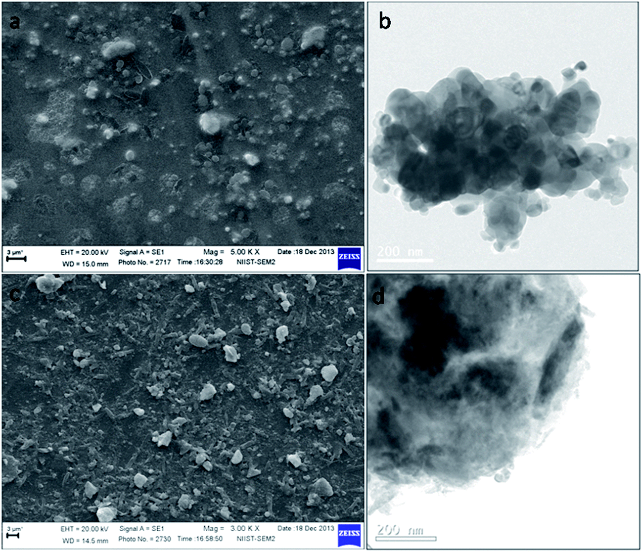 | ||
| Fig. 8 SEM and TEM images of (a and b) 30 °C and (c and d) 75 °C peptized sample, annealed at 800 °C. | ||
The anatase crystallite size was ∼50 nm for the room temperature peptized sample while it was ∼35 nm for the 75 °C peptized one. A shift in the X-ray diffraction peak towards lower 2θ value could be due to the variation in the lattice parameters caused by the morphology change.33 Further, the lattice parameters ‘a’ and ‘c’ for these samples were determined which is listed in Table 2. Lattice expansion was observed for the sample peptized at 75 °C with respect to increase in calcination temperature, which is attributed to the shift in the diffraction peak to a lower 2θ value.
| Sample ID | Lattice parameter ‘a’ | Lattice parameter ‘c’ | Lattice strain |
|---|---|---|---|
| T-75 (600 °C) | 3.7878 | 9.5153 | 0.30774 |
| T-75 (700 °C) | 3.8010 | 9.57604 | 0.23502 |
| T-75 (800 °C) | 3.8141 | 9.63692 | 0.17342 |
Lattice expansion of titania may be reflected in morphological changes.34 The calculated values for the lattice strains using Stokes–Wilson formula35 for the samples peptized at 75 °C (Table 2) were higher than that of the room temperature peptized samples with the values being 0.268, 0.193857 and 0.133975 for 600, 700 and 800 °C calcination. The crystal lattice is trying to relieve the strain induced by the higher peptization temperature by a lattice expansion. This increased lattice strain for higher peptization temperature (75 °C) may induce morphological changes. Even though peptization at 75 °C results in phase pure anatase at high temperatures of 800 °C, partial morphology changes happen to the aggregates as revealed by SEM, TEM and lattice expansion.
MB adsorption study of a few samples are carried out under dark condition for a period of 100 minutes prior to the photocatalytic experiments in order to confirm that the dye does not undergo any adsorption in presence of TiO2 after half an hour stirring under dark condition and the data are presented in Fig. 9.
 | ||
| Fig. 9 Methylene blue adsorption by (a) T-30 (800) (b) T-60 (700) and (c) T-75 (800) under dark condition for a period of 100 minutes. | ||
From the graph it is clear that the adsorption of MB on titanium dioxide gets saturated at 20–30 minutes of stirring under dark condition. Hence the initial half-an hour equilibration performed in dark is sufficient for eliminating any adsorption effect by the sample.
The photocatalytic activities of all the samples were then measured under UV-A light and the results are presented in Fig. 10. The photoactivity was comparatively higher for 60 °C peptized sample at all calcination temperatures. This may be due to the lower particle size of 60 °C peptized sample when compared with other samples. This lower particle size can provide an increased surface area which will enhance the photocatalytic activity.36 Among the three different calcination temperatures, the sample peptized at 60 °C after calcination at 700 and 800 °C has shown nearly same photoactivity (∼85%) which is the highest among all samples investigated.
 | ||
| Fig. 10 Methylene blue degradation by titania powders peptized at different temperatures (a) 30 °C (b) 45 °C (c) 60 °C (d) 75 °C and calcined at (i) 600 °C (ii) 700 °C (iii) 800 °C. | ||
This variation in photoactivity of the samples with respect to calcination temperature can be attributed to the phase changes of titania. All the samples contained small amounts of brookite in addition to anatase phase when calcined at 600 °C and hence exhibited lower activity than the ones calcined at higher temperatures. At 700 °C, the sample peptized at 60 °C contains 98% anatase and with increase in calcination temperature to 800 °C, the anatase phase decreased to 75% with 25% rutile. Even though the sample containing more anatase content (T-60-700) is expected to provide a better photoactivity, the photoactivities were nearly same for the two samples. The enhanced photoactivity of the latter sample (T-60-800) is attributed to the presence of anatase–rutile mixture (75:25) which enhances the photoactivity as reported in the case of Degussa titania containing 70% anatase and 30% rutile.37 The morphology of nano clusters in mixed-phase titania contains typically small rutile crystallites associated with anatase crystallites and this structural arrangement of similar TiO2 crystallites create catalytic hot-spots at the rutile–anatase interface38 which increases the electron transfer from rutile to anatase thereby increasing the photocatalytic activity. The rate of photocatalytic reactions was further calculated from the photocatalytic kinetic curves (Fig. 11) which followed a first order kinetics.39
 | ||
| Fig. 11 Photocatalytic kinetic curves for the titania powders peptized at different temperatures (a) 30 °C (b) 45 °C (c) 60 °C (d) 75 °C and calcined at (i) 600 °C (ii) 700 °C (iii) 800 °C. | ||
The variation in rate constant values (kapp) with respect to different peptization and calcination temperatures is presented in Fig. 12. The graph shows a hike for the peptization temperature of 60 °C at all calcination temperatures showing the influence of peptization temperature in photocatalytic activity of titania. There was a significant increase in the rate of 60 °C peptized sample annealed at 800 °C, possibly due to the presence of catalytic hot-spots at rutile–anatase interface.38
 | ||
| Fig. 12 Photocatalytic rate constants of TiO2 with respect to peptization temperatures and calcined at (a) 600 °C (b) 700 °C and (c) 800 °C. | ||
Even though 100% anatase phase was obtained for the samples peptized at 75 °C, at higher calcination temperatures of 700 and 800 °C, comparatively reduced photoactivity was observed which is attributed to the morphology changes occurring at high peptization temperature. Velasco et al. reported that spherically arranged titania would favour the fast motion of the charge carriers and minimize recombination processes40 resulting in enhanced photoactivity rather than a rod like arrangement.
Thus peptization temperature 60 °C was found to be effective in synthesising photoactive titanium dioxide which can meet high temperature applications.
Conclusion
Temperature assisted, acid catalyzed peptization of aqueous sol–gel titania can yield phase pure anatase stable up to 800 °C. Titanium dioxide sol prepared by peptization at 60 °C exhibited a lower particle size which is due to the lowering of electrostatic barrier against aggregation. The samples peptized at 60 °C showed enhanced photoactivity than those at other temperatures when calcined in the temperature range of 600–700 °C. The photocatalytic activities of the samples were found to depend on the phase content of titania. Even though the peptization temperature of 75 °C is effective in retaining 100% anatase phase after calcination at 800 °C, the induced morphology changes were found to reduce the photocatalytic activity. Thus a highly photoactive nanocrystalline titania containing phase pure anatase was synthesized by a temperature assisted peptization in an aqueous sol–gel process which can meet high temperature photocatalytic applications.Acknowledgements
The authors acknowledge Department of Science & Technology, and CSIR, Government of India for financial support. VSS and KGKW acknowledge CSIR, India for providing fellowship and support under CSIR Emeritus Scheme. The Authors acknowledge Mr M. Kiran and Mrs Lucy Paul for HR-TEM and SEM analysis.Notes and references
- K. Y. Jung and S. B. Park, J. Photochem. Photobiol., A, 1999, 127, 117 CrossRef CAS.
- V. S. Smitha, P. Francois, U. N. S. Hareesh and K. G. Warrier, J. Mater. Chem. A, 2013, 1, 12178 CAS.
- V. S. Smitha, C. K. Jyothi, A. P. Mohamed, S. Pillai and K. G. Warrier, Dalton Trans., 2013, 42, 4602 RSC.
- V. S. Smitha, K. B. Jaimy, P. Shajesh, J. K. Jeena and K. G. Warrier, J. Mater. Chem. A, 2013, 1, 12641 CAS.
- Y. Hwu, Y. D. Yao, N. F. Cheng, C. Y. Tung and H. M. Lin, Nanostruct. Mater., 1997, 9, 355 CrossRef CAS.
- V. S. Smitha, K. V. Baiju, P. Perumal, S. Ghosh and K. G. Warrier, Eur. J. Inorg. Chem., 2012, 2012, 226 CrossRef CAS.
- J. Thomas, K. P. Kumar and S. Mathew, Sci. Adv. Mater., 2010, 2, 481 CrossRef CAS PubMed.
- G. Melcarne, L. De Marco, E. Carlino, F. Martina, M. Manca, R. Cingolani, G. Gigli and G. Ciccarella, J. Mater. Chem., 2010, 20, 7248 RSC.
- K. Yang, J. M. Zhu, J. J. Zhu, S. S. Huang, X. H. Zhu and G. B. Ma, Mater. Lett., 2003, 57, 4639 CrossRef CAS.
- M. Toba, T. Sakuma, S. I. Niwa, H. Shimada and F. Mizukami, J. Sol-Gel Sci. Technol., 2000, 19, 695 CrossRef CAS.
- D. M. Schleich and B. Walter, Nanostruct. Mater., 1997, 8, 579 CrossRef CAS.
- H. Keskinen, J. M. Makela, M. Aromaa, J. Ristimaki, T. Kanerva, E. Levanen and T. Mantyla, J. Nanopart. Res., 2007, 9, 569 CrossRef CAS.
- U. Schubert, J. Chem. Soc., Dalton Trans., 1996, 3343 RSC.
- C. K. Jyothi, K. B. Jaimy, S. Ghosh, S. Sankar, V. S. Smitha and K. G. K. Warrier, J. Solid State Chem., 2011, 184, 1867 CrossRef CAS PubMed.
- C. J. Brinker and G. W. Scherer, Sol–Gel Science: The Physics and Chemistry of Sol–Gel Processing, Academic Press Inc, 1990 Search PubMed.
- S. Hore, E. Palomares, H. Smit, N. J. Bakker, P. Comte, P. Liska, K. R. Thampi, J. M. Kroon, A. Hinsch and J. R. Durrant, J. Mater. Chem., 2005, 15, 412 RSC.
- M. R. Mohammadi, M. C. Cordero-Cabrera, M. Ghorbani and D. J. Fray, J. Sol-Gel Sci. Technol., 2006, 40, 15 CrossRef CAS.
- D. A. H. Hanaor and C. C. Sorrell, J. Mater. Sci., 2011, 46, 855 CrossRef CAS.
- K. T. Ranjit, I. Willner, S. H. Bossmann and A. M. Braun, Environ. Sci. Technol., 2001, 35, 1544 CrossRef CAS.
- S. C. Pillai, P. Periyat, R. George, D. E. McCormack, M. K. Seery, H. Hayden, J. Colreavy, D. Corr and S. J. Hinder, J. Phys. Chem. C, 2007, 111, 1605 CAS.
- J. Yang, S. Mei and J. M. F. Ferreira, J. Am. Ceram. Soc., 2000, 83, 1361 CrossRef CAS.
- B. L. Bischoff and M. A. Anderson, Chem. Mater., 1995, 7, 1772 CrossRef CAS.
- Y. Kurokawa, T. Shirakawa, S. Saito and N. Yui, J. Mater. Sci. Lett., 1986, 5, 1070 CrossRef CAS.
- M. T. Colomer, J. Guzman and R. Moreno, Chem. Mater., 2008, 20, 4161 CrossRef CAS.
- K. N. P. Kumar, J. Kumar and K. Keizer, J. Am. Ceram. Soc., 1994, 77, 1396 CrossRef CAS.
- W. Yu-Ming, L. Jian-Zhong and C. Zhong-Li, Chin. Phys. Lett., 2011, 28, 14702 CrossRef.
- P. K. Jha, V. Kuzovkov, B. A. Grzybowski and M. O. de la Cruz, Soft Matter, 2012, 8, 227 RSC.
- S. Yang, Y. Liu, Y. Guo, J. Zhao, H. Xu and Z. Wang, Mater. Chem. Phys., 2003, 77, 501 CrossRef CAS.
- S. Winardi, R. R. Mukti, K. N. P. Kumar, J. Wang, W. Wunderlich and T. Okubo, Langmuir, 2010, 26, 4567 CrossRef CAS PubMed.
- F. F. Lange and B. J. Kellett, J. Am. Ceram. Soc., 1989, 72, 735 CrossRef CAS.
- T. Jan, J. Iqbal, M. Ismail, M. Zakaullah, S. H. Naqvi and N. Badshah, Int. J. Nanomed., 2013, 8, 3679 CrossRef PubMed.
- J. H. Park, C. S. Kim, D. H. Yoo, H. S. Yang, K. S. Hong, B. K. Moon, B. C. Choi and H. S. Lee, J. Korean Phys. Soc., 2005, 47, 884 CAS.
- K. Srinivasan, A. Cantoni and G. Bocelli, Cryst. Res. Technol., 2010, 45, 737 CrossRef CAS.
- M. Sahu and P. Biswas, Nanoscale Res. Lett., 2011, 6, 1 Search PubMed.
- A. Krishnan, T. S. Sreeremya, E. Murray and S. Ghosh, J. Colloid Interface Sci., 2013, 389, 16 CrossRef CAS PubMed.
- V. S. Smitha, K. A. Manjumol, K. V. Baiju, S. Ghosh, P. Perumal and K. G. K. Warrier, J. Sol-Gel Sci. Technol., 2010, 54, 203 CrossRef CAS.
- D. C. Hurum, A. G. Agrios, K. A. Gray, T. Rajh and M. C. Thurnauer, J. Phys. Chem. B, 2003, 107, 4545 CrossRef CAS.
- S. Bakardjieva, J. Subrt, V. Stengl, M. J. Dianez and M. J. Sayagues, Appl. Catal., B, 2005, 58, 193 CrossRef CAS PubMed.
- D. F. Ollis, E. Pellizzetti and N. Serpone, Environ. Sci. Technol., 1991, 25, 1523 CrossRef.
- L. F. Velasco, M. Haro, J. Parmentier, R. Gadiou, C. Vix-Guterl and C. O. Ania, J. Catal., 2012, 2013, 9 Search PubMed.
| This journal is © The Royal Society of Chemistry 2014 |