Photosensitivity of reflection notch tuning and broadening in polymer stabilized cholesteric liquid crystals
Kyung Min
Lee†
,
Vincent P.
Tondiglia‡
and
Timothy J.
White
*
Air Force Research Laboratory, Materials and Manufacturing Directorate, Wright-Patterson Air Force Base, Ohio 45433-7750, USA. E-mail: Timothy.White.24@us.af.mil; Tel: +1-937-255-9551
First published on 23rd November 2015
Abstract
The position or bandwidth of the selective reflection of polymer stabilized cholesteric liquid crystals (PSCLCs) prepared from negative dielectric anisotropy (“−Δε”) liquid crystalline hosts can be shifted by applying a DC voltage. The underlying mechanism of the tuning or broadening of the reflection of PSCLCs detailed in these recent efforts is ion-facilitated, electromechanical deformation of the structurally chiral, polymer stabilizing network in the presence of a DC bias. Here, we show that these electro-optic responses can also be photosensitive. The photosensitivity is most directly related to the presence of photoinitiator, which is a known ionic contaminant to liquid crystal devices. Measurement of the ion density of a series of control compositions before, during, and after irradiation with UV light confirms that the ion density in compositions that exhibit photosensitivity is increased by irradiation and correlates to not only the concentration of the photoinitiator but also the type. Thus, the magnitude of the electrically tuned or broadened reflection of PSCLC of certain compositions when subjected to DC field is further increased in the presence of UV light. While interesting and potentially useful in applications such as architectural windows, the effect may be deleterious to some device implementations. Accordingly, compositions in which photosensitivity is not observed are identified.
Introduction
Cholesteric liquid crystals (CLCs) are self-organized photonic crystals. Due to their helicoidal superstructure, these materials spontaneously can exhibit a circularly polarized and selective reflection.1,2 The reflection of a CLC is centered at the wavelength:| λ0 = navg × P0 | (1) |
| Δλ ≈ Δn × P0 | (2) |
Negative dielectric anisotropy liquid crystals have molecular structures within which the permittivity parallel to the long axis of the molecule (ε∥) is smaller than the permittivity on the short axis (ε⊥) (Δε = ε∥ − ε⊥ < 0). Accordingly, the orientation of liquid crystals with −Δε is perpendicular to the direction of the applied electric field. The molecular structures of these materials is largely proprietary, however the limited publications on the topic indicate that lateral electron-withdrawing substituents, such as cyano or fluorinated functional groups, induce the required dipole moment that is perpendicular to the principal molecular axis.2 In particular, superfluorinated liquid crystals are most widely used to create −Δε LCs due to their excellent resistivity, modest dipole moment, and low viscosity.3–5 The primary driver of the development of these materials is display modes in which that nematic liquid crystal is vertically aligned6–9 or in interdigitated devices.10–14
Our recent examinations have shown reflection notch broadening,15–17 red-shifting tuning18–20 and transmission control21 in PSCLCs formulated with −Δε nematic liquid crystals. As much as a seven-fold increase in bandwidth has been observed when the PSCLCs are subject to moderate DC fields (0–6 V μm−1).16 The bandwidth broadening is symmetric about the center of the reflection notch and returns to original bandwidth upon removal of the electric fields. Bandwidth broadening is only observed in samples which are composed of −Δε liquid crystal hosts, with polymer stabilization, and in the presence of a DC field. Large magnitude, high optical quality electrically induced reflection notch tuning in PSCLCs is recently reported.20 Repeatable tuning cycles from 100 nm to 400 nm range was observed. Removal of the applied DC field returns the PSCLCs to its original bandwidth and notch position. Recently, bistable switching of transmission was also reported21 where stable transparent or scattering states, including grayscale intermediates, were observed.
It is well known that liquid crystals contain a small amount of ionic impurities which are residual from the synthesis and purification (initiators, catalysts, salts, moisture). UV light exposure is known to degraded liquid crystal22–25 and alignment layers.26 Recently, our examinations have focused on the mechanism of the electro-optic effects we have observed in PSCLCs prepared with −Δε liquid crystals. These studies indicate that ions in the liquid crystal mixtures could be trapped on or within the polymer networks during the fast photopolymerization process. Subsequently, upon application of a DC field the ions distort the structurally chiral polymer stabilizing network. The movement of the structurally chiral polymer network distorts the local anchoring of the nematic liquid crystal recently directly observed with fluorescence confocal microscopy.17,20
Here, we report on the photosensitivity of the electro-optic effects observed in certain PSCLC formulations prepared with −Δε liquid crystal hosts. In PSCLC formulations exhibiting either tuning or broadening, if the samples are exposed to UV irradiation from a lamp or the sun while also subjected to a DC bias, the reflection notch is further tuned or broadened. Ion density measurements indicate the photosensitivity is most directly related to the presence of certain photoinitiators.
Results and discussion
The electro-optic response of the PSCLC compositions of our prior examinations is illustrated in Fig. 1.15–20 These compositions were prepared from a −Δε nematic liquid crystal host (ZLI-2079, Merck), two right-handed chiral dopants (5 wt% R1011 and 5 wt% R811, Merck), and contain 5 wt% polymer as a stabilizing network. Evident in Fig. 1(a), in certain preparation conditions and compositions (here with the achiral monomer RM82), applying a 110 V DC bias results in symmetrical bandwidth broadening of the selective reflection by as much as 7 times. In other conditions and compositions (here with the chiral monomer SLO4151), large red notch tuning (>400 nm) can be induced by subjecting the cell to 100 V DC (Fig. 1(b)). It should be noted that whether a PSCLC exhibits reflection dynamic bandwidth broadening or notch tuning is strongly related to the strength and extent of polymerization through the cell gap as well as secondarily influenced by factors such as polymer chirality. Further elucidating these dependencies are subjects of intense focus in our ongoing research activities. Notably for the work examined here, as the applied DC voltage is turned off, the displaced selective reflection or bandwidth returns to the initial notch position or original bandwidth. For simplicity, the PSCLC formulation that exhibits bandwidth broadening will be referred to as b-PSCLC and the PSCLC formulation in which reflection notch tuning is observed will be referred to as t-PSCLC.Motivated by a recent examination which demonstrated that the ion density of a LC mixture was sensitive to chemical additives similar to common photoinitiators,27–30 we examined the electro-optic response of some of the previously examined PSCLC formulations in the presence of UV light. The results for the b-PSCLC composition examined in Fig. 1(a) are presented in Fig. 2. Evident in the transmission spectra in Fig. 2(a-i), the bandwidth of the selective reflection is approximately 80 nm after preparation. Applying a 23 V DC field increases the reflection bandwidth to 150 nm (Δλ/Δλ0 ∼ 1.9) in Fig. 2(a-ii). Thereafter, exposing the PSCLC to 60 mW cm−2 UV light while maintaining 23 V DC further increases the bandwidth to 215 nm (Δλ/Δλ0 ∼ 2.7) in Fig. 2(a-iii). It should be noted that exposing this sample to 60 mW cm−2 UV light at 0 V induces no change in bandwidth. After removal of the UV light, the bandwidth nearly immediately returns to 150 nm (at 23 V DC, Fig. 2(a-iv)). Removing the field restores the bandwidth to the original 80 nm, as evident in Fig. 2(a-v). The influence of UV intensity on the photoinduced increase of bandwidth at 23 V DC is summarized in Fig. 2(b). Starting from the 150 nm bandwidth at 23 V DC, increasing UV intensity induces both larger (Δλ/Δλ0 ∼ 2.2, 2.7, and 3.1 for 20 mW cm−2, 60 mW cm−2, and 100 mW cm−2 UV intensities) and faster photoinduced bandwidth broadening.
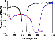
Similarly, UV light exposure can also affect the position of the selective reflection of t-PSCLC when subjected to a DC bias. This is illustrated in Fig. 3, for a sample that has an initial reflection notch position of 640 nm. Upon application of 50 V DC, the notch is displaced to 790 nm in Fig. 3(a-ii). In Fig. 3(a-iii), subsequent exposure to UV light while maintaining 50 V applied DC further shifts the position to 840 nm. Once again, if the UV light is removed but the 50 V DC bias is maintained, the reflection notch returns to 790 nm (Fig. 3(a-iv)). After removing the 50 V DC bias, the notch restores to the original 640 nm position in Fig. 3(a-v). The temporal response of the processes is illustrated in Fig. 3(b). The sample initially has a reflection centered at 640 nm that is displaced over 1–2 seconds to an equilibrium position of 790 nm. At approximately 10 seconds, the sample was exposed to 20 mW cm−2 of UV light which shifts the center of the reflection notch to 840 nm. Thereafter, removing the UV light the PSCLC reflection returns to 790 nm (Fig. 3(a-iv)) and subsequent removal of the 50 V DC field results in the center of the reflection notch returning to 640 nm (Fig. 3(a-v)). Here, it should be noted again that exposing this sample to 20 mW cm−2 UV light at 0 V induces no change in reflection notch position.
The reversibility and repeatability of the photosensitivity reported in Fig. 2 and 3 is presented in Fig. 4. In Fig. 4(a), the bandwidth of b-PSCLC is reversibly increased by exposing the sample to UV light. At the constant 23 V DC of this data set, 60 mW cm−2 UV exposure increases the bandwidth from 150 to 210 nm. The time constants of the relaxation of the bandwidth are identical, and the differences in the data set are simply due to differences in the time allowed for dark relaxation. Fig. 4(b) illustrates the photoinduced and reversible tuning at 40 V DC. Here, the initial notch position of 670 nm is shifted to 810 nm by the application of 40 V DC. Reversibly cycling 40 mW cm−2 of UV light on and off shifts the center of the selective reflection to and from 870 nm at the constant 40 V DC. After the DC voltage is removed, the center of the reflection notch returns to the original position.
 | ||
| Fig. 4 Photoinduced changes (on/off) with exposure to (a) 60 mW cm−2 of bandwidth broadening in b-PSCLC at 23 V DC and (b) 40 mW cm−2 of tuning of t-PSCLC at 40 V DC. | ||
To elucidate the mechanism of the photoinduced changes, the ion density of the mixtures was measured at 1 Vpeak and 3 Hz. The ion density values are the average of five measurements. The applied AC field of 1 V is below the threshold voltage. The ion density was calculated from current measurements with the triangle method. The −Δε liquid crystal ZLI-2079 examined here (Fig. 5(a-i)) is contrasted to the commonly used +Δε LC E7 (Fig. 5(a-ii)). Comparatively, the ion density of ZLI-2079 (2.3 × 10¹³ ions per cm3) is an order of magnitude less than that of E7 (2.1 × 10¹⁴ ions per cm3). As soon as the UV light is exposed to the nematic hosts, the ion density increases significantly over 5–10 min and then plateaus. After 2 h of UV exposure, the ion density increases an order of magnitude to 5 × 10¹⁴ ions per cm3 for ZLI-2079 and 7.5 × 10¹⁴ ions per cm3 for E7. The photoinduced change in ion density has been previously reported25,27–30 and in part attributed to the photoionization of associated ions. Fig. 5(b) illustrates the average ion densities of a variety of formulations prepared from the materials examined here. From these values, it is clear that mixtures composed with the photoinitiators I-369 or I-907 have a considerably larger ion density. Comparatively, the common photoinitiator I-651 contributes minimally to the average ion density when mixed with ZLI-2079.
While the nematic liquid crystal host employed here (ZLI-2079) does exhibit some change in ion density to UV exposure, that change is irreversible, at least in the timescale examined in Fig. 5(a) and in the experiments reported here. From the data in Fig. 5(b), the photoinitiators I-369 and I-907 with polar morpholine heterocycles strongly contribute to the ion density of the mixtures. To further elucidate the contribution of photoinitiator to the photosensitivity of the electro-optic effects examined here, Fig. 6 examines the response of t-PSCLCs prepared with a range of I-369 concentrations. As summarized in Fig. 6, a PSCLC prepared a mixture with 0.1 wt% I-369 exhibits 150 nm tuning upon application of 40 V DC (Fig. 6(a-ii)). Subsequent exposure to UV light induces an additional 25 nm red shift as evident in Fig. 6(a-iii). Interestingly, a t-PSCLC prepared with higher I-369 concentration (0.5 wt%) shows less tuning (100 nm) when subjected to 40 V DC – likely due to the influence of the photoinitiator on the robustness of the polymer stabilizing network. However, comparatively larger UV induced red tuning (55 nm) is observed as can be seen in Fig. 6(b). A t-PSCLC prepared with 1 wt% I-369 exhibits less tuning range when subjected to 40 V but larger photosensitivity (70 nm) in Fig. 6(c). Tuning range and ion density as a function of initiator concentration are summarized in Fig. 6(d). The ion density increases with increasing I-369 concentration which is concurrently accompanied by an increase in photosensitivity. The sensitivity of the photoinduced changes in notch position are indirect evidence that the photoinitiator is the dominant source of the photosensitivity of the electro-optic effects reported here with minor contributions attributed to the photosensitivity of the nematic host and potentially the alignment layers.
To confirm that the effect is not isolated to samples prepared with I-369, Fig. 7 examines the photosensitivity of t-PSCLC prepared with the related photoinitiator I-907. In Fig. 7(ii) the reflection notch is shifted 100 nm by the application of 40 V DC. Irradiation of t-PSCLC with 50 mW cm−2 UV while maintaining 40 V DC shifts the reflection notch an additional 50 nm, similar to the results obtained for the t-PSCLC formulations prepared with I-369 (Fig. 7(iii)).
Fig. 8 confirms that t-PSCLC can be prepared absent of the photosensitivity discussed hereto by preparing the sample with the photoinitiator I-651. As can be seen in the data presented in Fig. 5(b-vii), I-651 does not seem to affect the ion density, potentially due to differences in the chemical structure. The application of 45 V DC moves the reflection notch 150 nm (Fig. 8(ii)), however, irradiation of t-PSCLC with 70 mW cm−2 shifts the reflection notch only very slightly (Fig. 8(iii)). This is further confirmation that the photosensitivity of the nematic liquid crystal host and alignment layer are minor contributors to the photosensitivity reported in Fig. 2–7.
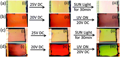
Fig. 9 visually illustrates the change in the optical properties of the t-PSCLC and b-PSCLC samples in the presence of sunlight31,32 or in laboratory conditions upon irradiation with a UV lamp. The t-PSCLC shows color change from light orange (Fig. 9(a-i), 0 V) to reddish orange color (Fig. 9(a-ii), 25 V DC). As the sample is exposed to sunlight while the 25 V DC field is maintained, further color change is observed (Fig. 9(a-iii)). UV light induces fast color changes from red (Fig. 9(b-ii), 20 V DC) to red/infrared (Fig. 9(b-iii)) at a constantly applied DC voltage. Similar photosensitivity in b-PSCLCs are observed in Fig. 9(c) and (d). The reflection color in b-PSCLC sample changes from red-orange (Fig. 9(c-i), 0 V) to yellowish orange (Fig. 9(c-ii), 20 V DC) and then greenish color (Fig. 9(c-iii), 50 mW cm−2 of UV light at the constant 20 V DC). Upon UV exposure by lamp, the color of the PSCLC is a bright green.
 | ||
| Fig. 9 Reflection images of t-PSCLC and b-PSCLC upon exposure to (a and c) sunlight (30 min exposure, 25 V DC) and (b and d) UV lamp (20 V DC). | ||
From the data reported here, it is clear that UV light can increase the ion density of the PSCLC samples. Indirect evidence presented here indicates that while the nematic liquid crystal host can exhibit changes in ion density when exposed to UV light, these changes seem to be irreversible. The dominant factor of the photosensitivity of the reflection notch broadening or tuning reported here is the photoinitiator. Most clearly, the increase in photoinduced broadening or tuning upon UV light exposure for samples prepared with increasing photoinitiator concentrations is the clearest indicator of this dependence. Further, the fact that certain formulations (for example PSCLCs prepared with I-651) do not exhibit photoinduced broadening or tuning are yet more indirect evidence that the photoinduced responses do not seem to be attributable to the nematic liquid crystal host, alignment layer, or any other component of the optical system.
The question remains as to how does the concentration and type of photoinitiator affect the optical properties of the PSCLCs in the presence of UV irradiation? We postulate two potential explanations that we are unable to delineate at this time. On one hand, the results reported here could be attributed to the photoinitiator molecules, covalently attached or dissolved within the polymer stabilizing network, in improving the “trapping” efficiency of the system,33 thereby subjecting the polymer stabilizing network to an increase in mechanical force. Further, there is literature preference that the polar heterocycle morpholine unit in I-369 or I-907 can attract ions.3–5 UV light might induce partial polarization of ion sources interacted with polar group in I-369 or I-907 and could be recombined as the UV light is removed. Such ion sources are trapped in the semisolid polymer networks and complete dissociation might be difficult. Another potential explanation is that these photoinitiators simply increase the ion density upon UV exposure and correspondingly, the increase in the number of ions increases the ions available to trap on and within the polymer network thereby increasing the sensitivity to an applied DC bias.
Materials and methods
Preparation of −Δε PSCLCs
Alignment cells were self prepared from ITO-coated glass slides (Colorado Concepts). The glass slides were coated with a polyimide alignment layer. The alignment layers were rubbed with a cloth, and the cell was constructed with planar alignment conditions. The cell gap was controlled by mixing 15 μm thick glass rod spacers into an optical adhesive. Samples were prepared by mixing 1 wt% of the photoinitiator Irgacure 369, two right-handed chiral dopants (5 wt% R1011 and 5 wt% R811, Merck), 5 wt% of an achiral liquid crystal monomer (RM82, Merck) or a chiral liquid crystal monomer (SLO4151, Alpha Micron), and 84 wt% of a −Δε achiral nematic liquid crystal (ZLI-2079, Merck). The polymer stabilizing network was formed within the samples by photoinitiated polymerization with 50–80 mW cm−2 of 365 nm light (Exfo) for 3 min. To ensure homogeneous curing conditions, the cell was rotated at an angular velocity of 200 Hz during polymerization. While samples prepared with more traditional, one-sided curing exhibit similar electro-optic responses to that reported here, the consistency and sample-to-sample repeatability are improved by rotating the sample during curing. All materials were used as received without any purification.Experimental setup and measurements
Transmission spectra were collected with a fiber optic spectrometer. Unless otherwise mentioned, the white light probe was unpolarized. Transmission spectra were collected before, during, and after application of DC fields. The ion density of the mixtures was measured with a commercial instrument from LC Vision in homeotropic alignment cells. During the experiment, the samples were subjected to a 1 Vp bias at a frequency of 3 Hz. The ion density values reported here in the Fig. 5 are the average of five measurements. There is no switching response of liquid crystals because the applied alternating voltage (1 Vp) is below the threshold voltage. The ion density is calculated from current measurements with the triangle method. Optical and polarized optical microscopy was employed to characterize the PSCLCs.Conclusions
In this contribution, the tuning range and bandwidth broadening of polymer stabilized cholesteric liquid crystals (PSCLCs) is shown to increase with UV light exposure when an applied DC bias is applied to the cell. The photoresponsivity of the samples is primarily related to the presence and type of photoinitiator. The ion density in the mixtures examined here increases with photoinitiator concentration. Irradiation of the samples with sunlight in the presence of an applied DC bias is shown to further increase the electro-optic response of the PSCLCs.Acknowledgements
The authors acknowledge funding from the Materials and Manufacturing Directorate of the Air Force Research Laboratory.Notes and references
- H.-S. Kitzerow and C. Bahr, Chirality in Liquid Crystals, Springer-Verlag, New York, 2001 Search PubMed.
- S.-T. Wu and D.-K. Yang, Reflective Liquid Crystal Displays, Wiley, West Sussex, UK, 2001 Search PubMed.
- P. Kirsch, et al. , Eur. J. Org. Chem., 2008, 3479–3487 CrossRef CAS.
- P. Kirsch and K. Tarumi, Angew. Chem., Int. Ed., 1998, 37, 484–489 CrossRef CAS.
- S. N. Naemura, Y. Nakazono, K. Nishikawa, A. Sawada, P. Kirsch, M. Bremer and K. Tarumi, Mater. Res. Soc. Symp. Proc., 1998, 508, 235–240 CrossRef CAS.
- M. F. Schiekel and K. Fahrenschon, Appl. Phys. Lett., 1971, 19, 391–393 CrossRef CAS.
- F. J. Kahn, Appl. Phys. Lett., 1972, 20, 199–201 CrossRef CAS.
- A. Takeda, S. Kataoka, T. Sasaki, H. Chida, H. Tsuda, K. Ohmuro, T. Sasabayashi, Y. Koike and K. Okamoto, SID Int. Symp. Dig. Tech. Pap., 1998, 29, 1077–1080 CrossRef.
- K. Ohmuro, S. Kataoka, T. Sasaki and Y. Koike, SID Int. Symp. Dig. Tech. Pap., 1997, 28, 845–850 Search PubMed.
- Z. Ge, X. Zhu, T. X. Wu and S. T. Wu, J. Disp. Technol., 2006, 2, 114–120 CrossRef.
- M. Oh-e and K. Kondo, Appl. Phys. Lett., 1995, 67, 3895–3897 CrossRef CAS.
- S. H. Lee, S. L. Lee and H. Y. Kim, Appl. Phys. Lett., 1998, 73, 2881–2883 CrossRef CAS.
- Y. Chen, Z. Luo, F. Peng and S. T. Wu, J. Disp. Technol, 2013, 9, 74–77 CrossRef.
- H. J. Yun, M. H. Jo, I. W. Jang, S. H. Lee, S. H. Ahn and H. J. Hur, Liq. Cryst., 2012, 39, 1141–1148 CrossRef CAS.
- V. P. Tondiglia, L. V. Natarajan, C. A. Bailey, M. M. Duning, R. L. Sutherland, D.-K. Yang, A. Voevodin, T. J. White and T. J. Bunning, J. Appl. Phys., 2011, 110, 053109 CrossRef.
- K. M. Lee, V. P. Tondiglia, M. E. McConney, L. V. Natarajan, T. J. Bunning and T. J. White, ACS Photonics, 2014, 1, 1033–1041 CrossRef CAS.
- H. Nemati, S. Liu, R. S. Zola, V. P. Tondiglia, K. M. Lee, T. J. White, T. J. Bunning and D.-K. Yang, Soft Matter, 2015, 11, 1208 RSC.
- M. E. McConney, V. P. Tondiglia, L. V. Natarajan, K. M. Lee, T. J. White and T. J. Bunning, Adv. Opt. Mater., 2013, 1, 417–421 CrossRef.
- T. J. White, K. M. Lee, M. E. McConney, V. P. Tondiglia, L. V. Natarajan and T. J. Bunning, SID Int. Symp. Dig. Tech. Pap., 2014, 45, 555–558 CrossRef.
- K. M. Lee, V. P. Tondiglia, T. Lee, I. I. Smalyukh and T. J. White, J. Mater. Chem. C, 2015, 3, 8788–8793 RSC.
- K. M. Lee, V. P. Tondiglia and T. J. White, MRS Commun, 2015, 5, 223–227 CrossRef CAS.
- W. Lee, C.-T. Wang and C.-H. Lin, Displays, 2010, 31, 160–163 CrossRef CAS.
- B. Gosse and J. P. Gosse, J. Appl. Electrochem., 1976, 6, 515–519 CrossRef CAS.
- C.-H. Wen, S. Gauza and S. T. Wu, Liq. Cryst., 2004, 31, 1479–1485 CrossRef CAS.
- P.-T. Lin, S. T. Wu, C. Y. Shang and C. S. Hsu, Mol. Cryst. Liq. Cryst., 2004, 411, 243–253 CrossRef.
- L. Lu, V. Sergan and P. J. Bos, Phys. Rev. E: Stat., Nonlinear, Soft Matter Phys., 2012, 86, 051706 CrossRef PubMed.
- C.-H. Wen, S. Gauza and S.-T. Wu, Liq. Cryst., 2004, 31, 1479–1485 CrossRef CAS.
- C.-H. Wen, S. Gauza and S.-T. Wu, J. Soc. Inf. Disp., 2005, 13, 805–811 CrossRef CAS.
- J.-H. Son, S. B. Park, W.-C. Zin and J.-K. Song, Liq. Cryst., 2013, 40, 458–467 CrossRef CAS.
- C.-Y. Tanga, Y.-C. Lin and W. Lee, Photonics Global Conf., 2010, 1–4 Search PubMed.
- L. B. N. Laboratory, Cool Colors Project: Improved Materials for Cooler Roofs, in Newsletter, edited, Berkeley, CA, 2004.
- K. Biernat, A. Malinowski and M. Gnat, The Possibility of Future Biofuels Production Using Waste Carbon Dioxide and Solar Energy, in Biofuels – Economy, Environment and Sustainability, ed. Z. Fang, InTech, 2013, pp. 123–172 DOI:10.5772/53831.
- R.-Q. Ma and D.-K. Yang, Phys. Rev. E: Stat. Phys., Plasmas, Fluids, Relat. Interdiscip. Top, 2000, 61, 1567–1573 CrossRef CAS.
Footnotes |
| † Also with Azimuth Corporation, 4027 Colonel Glenn Hwy, Beavercreek, Ohio 45431, USA. |
| ‡ Also with Leidos, 3745 Pentagon Boulevard, Beavercreek, Ohio 45431, USA. |
| This journal is © The Royal Society of Chemistry 2016 |


![[thick line, graph caption]](https://www.rsc.org/images/entities/char_e117.gif) ), (ii) during application of 50 V DC (
), (ii) during application of 50 V DC (


