
A nano-scale probing system with a gold nano-dot array for measurement of a single biomolecular interaction force†
Sung Joo Kim‡
a,
Jeong Hyun Seo‡bd,
Jin Woo Lee‡ac,
Dong-Woo Cho*a,
Hyung Joon Cha*d and
Wonkyu Moon*a
aDepartment of Mechanical Engineering, Pohang University of Science and Technology, Pohang 790-784, Korea. E-mail: wkmoon@postech.ac.kr; dwcho@postech.ac.kr
bSchool of Chemical Engineering, Yeungnam University, Gyeongsan 712-749, Korea
cDepartment of Molecular Medicine, College of Medicine, Gachon University, Inchon 406-840, Korea
dDepartment of Chemical Engineering, Pohang University of Science and Technology, Pohang 790-784, Korea. E-mail: hjcha@postech.ac.kr
First published on 9th December 2015
Abstract
A new nano-scale probing system was proposed and developed to measure and analyze the interaction force between biomolecules at the single molecular level. A probing instrument adopting the working principle of atomic force microscopy (AFM) was designed to measure a single biomolecular interaction force with a patterned gold nano-dot array and functionalized AFM probe. The constructed probing system was evaluated through measurement of the unbinding force between a protein (Vibrio cholerae toxin B subunit) and a carbohydrate (GM1 pentasaccharide) as a model single biomolecular interaction. Thus, the developed probing system might be a potential platform for the effective measurement of single biomolecular level interactions.
Life phenomena such as cellular metabolism are driven by biomolecular interactions including cell adhesion, cell differentiation, cell communication, and immunological responses. Therefore, scientists have paid attention to the study of biomolecular interactions and their applications. The development of nano-scale probing systems, including the surface forces apparatus (SFA)1–3 and atomic force microscopy (AFM),4–6 has made it possible to detect and analyze molecular interactions. These instruments have been used as powerful tools for measuring the unbinding forces between biomolecules at the single molecule level and consequently accelerated research in the area of single-molecule force spectroscopy (SMFS).7,8
Particularly, AFM has been known as a good tool for measuring force due to its relatively facile measurement process. In general, the AFM operation method is classified by three modes according to the motion of the tip: contact mode, non-contact mode, and tapping mode.4,9–17 The contact mode drags the tip across the surface of a specimen and measures the surface topograph using the deflection of the cantilever or the feedback signal for keeping the cantilever position in a constant state. In tapping mode, the cantilever is oscillated with an amplitude of 100 to 200 nm by a piezoelectric component that is mounted on the cantilever holder like in non-contact mode. When the tip comes close to the sample surface, van der Waals forces, which cause an attractive force between the tip end and the surface, decrease this oscillation amplitude. Consequently, the adjusting signals of the actuator are monitored and visualized by a surface image.
In the AFM system, the interaction force can typically be measured by drawing a force–distance (F–D) curve from the force exerted on the functionalized probe and the distance to the substrate, where the samples are uniformly applied.1–4,6–8,18 The magnitude of the molecular interaction force is believed to be obtained by extracting the smallest common measurement from the forces shown in the F–D curves by confirming the repeated appearance of adhesive forces or the presence of the linker material (Scheme 1).2,8,10 The treatment of the sample for fixing the single molecule sparsely on the substrate (or on the probe) would be a tedious method to find the most effective measurement.
 | ||
| Scheme 1 Schematic representation of single biomolecular unbinding force measurement using an Au nano-dot. | ||
In traditional AFM analysis, the actual behavior of the individual molecular interaction has been deduced from collective behaviors (i.e., measurement of the ensemble average of many molecules in parallel at the same time). Although the nano-scale probing system has been used to detect and analyze interactions at the molecular level, it is necessary to improve the method to more easily measure and guarantee a single molecule interaction.
Carbohydrate–protein interactions play critical roles in living organisms, such as by initiating the infection of host cells by viruses and bacterial toxin proteins.19–22 Therefore, studies related to the characterization of these interactions have become increasingly important because they provide useful information on biological processes in living organisms and can help in the development of protein biomedical agents. Particularly, because the interaction between the GM1 pentasaccharide and the Vibrio cholerae toxin B subunit has been recognized as a standard carbohydrate–protein interaction and a well-known pathogenic mechanism, it has received attention in the research realm of the unbinding force.23 Although several results describing the interaction have been reported,24–26 there have been few reports on the exact unbinding force of the GM1 pentasaccharide–V. cholerae toxin B subunit at the single molecular level. Thus, a report on the exact interaction mechanism through the measurement of the unbinding force would be very meaningful to current research.
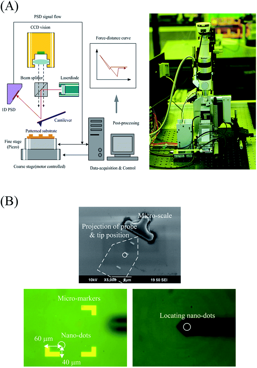
In the present work, a new method that possesses an ultra-precise position control system with a long working range was proposed for the reliable binding force measurement of biomolecules by employing the basic principle of AFM and micro-machined gold (Au) nano-dots (Fig. 2). For the effective measurement of the unbinding force of a single biomolecular interaction, unlike in the conventional technique, the array was treated by a crosslinking chemical to covalently attach a single molecule onto a nano-dot (Fig. 2A). Then, by drawing the F–D curves, the unbinding forces can be measured while probing around the nano-dot. Additionally, an ultra-precise probing system with a long-range positioner was developed to find the locations of the nano-dots, which cannot easily be detected by an optical microscope due to their extremely small size, and to locate the initial position of the measurement (Fig. 2B). The unbinding force between a single protein (V. cholerae toxin B subunit) and a carbohydrate (GM1 pentasaccharide) was selected as a target measurement.
Au nano-dots were successfully fabricated by a micromachining process in most conditions (Fig. 1B), but electron-beam lithography and Cr/Au evaporation processes were not successful for nano-dots of 20 nm diameter. Therefore, we conducted experiments for measuring the interaction force on Au nano-dot array patterns with 30–70 nm diameters. After fabrication, the linker and protein were attached on the Au nano-dots in series.27 After attaching the carbohydrate onto the AFM cantilever probe, the probe was mounted onto the probing system.
 | ||
| Fig. 1 (A) Design and MEMS process of Au nano-dots of various sizes on a silicon wafer. (B) Optical and SEM images of fabricated Au nano-dots and macro-markers. | ||
 | ||
| Fig. 2 (A) A laboratory-constructed nano-scale probing instrument. (B) Defining the position of the cantilever and locating the probe around the Au nano-dots. | ||
To confirm the fabricated nano-dots in advance to measure the unbinding forces between the biomaterials at the single molecular level, the surface topograph was imaged using a conventional AFM (SPA400; Seiko Instrument, Tokyo, Japan). Fig. 3A shows the topography of Au nano-dots of 70 nm diameter, which were measured in tapping mode before and after protein attachment. The topography was measured immediately after the substrate was rinsed with the buffer solution and slightly dried in air. The height was elevated by approximately 20 nm after protein attachment onto the nano-dots of 40–70 nm diameters. Considering the combined size of the V. cholerae toxin B subunit protein and the linker material for attachment, it is reasonable that a single protein molecule is attached to the Au nano-dots of 40–70 nm diameters. To verify the performance of the developed probing system, we scanned the fabricated Au nano-dot patterns of 50 nm diameter to which the attachment of a single V. cholerae toxin B subunit molecule had already been confirmed (left, Fig. 3B). The scanned patterns were shown to have the topography of 50 nm diameter Au nano-dots in a 25 × 25 array with 200 nm central spacing. The scanning area was 1 μm2, and the starting position was successfully located from the macro-marker at once using the positioning system. The repeatability of the positioning system was shown to the sub-micron level. Using raw data gathered by just one scanning step, we could obtain the surface image (right, Fig. 3B). This result indicates that the surface profile can be acquired very quickly using our probing system, and it might be able to acquire a high-quality image like a conventional AFM system by removing noise and scanning repeatedly.
 | ||
| Fig. 3 (A) Elevation change of 70 nm diameter Au nano-dots before (above image) and after (below image) protein attachment. (B) 50 nm diameter Au nano-dots in 25 × 25 array with 200 nm central spacing and the topography of a 1 μm2 area from the first trial of locating the probe around the nano-dots. | ||
Based on the successful measurement of the binding force of a single protein on Au nano-dot pattern using the conventional AFM and the fast surface reconstruction performance of the developed high-speed scanning system, we measured the unbinding force between the carbohydrate and protein on the fabricated Au nano-dots. The F–D curves were obtained from the unbinding force measurement using the constructed probing system (Fig. 4). The unbinding forces appeared on certain adjacent spots but not on the whole surface of the measured area. The unbinding force was approximately 400 pN and appeared after a delay of approximately 50–100 nm (left, Fig. 4A). The magnitude of the unbinding force was close to the value expected from our preliminary research.28 The delay in the detection of the attraction signal demonstrates the existence of the linker material, which guarantees that the result was the unbinding force between the target materials.8 Successive adhesive forces appeared in the F–D curves in some locations, which indicates that there was more than one protein molecule on the Au nano-dot, because the fabricated size (40–70 nm) of the nano-dot was larger than that needed for the attachment of a single molecule (∼5 nm) (right, Fig. 4A). The use of smaller nano-dots (e.g., 30–40 nm diameter) might reduce this phenomenon and consequently increase the potential for measuring single molecular interactions. Finally, we visualized the force map for the GM1 pentasaccharide–V. cholerae toxin B subunit interaction based on the obtained force values (Fig. 4B).
 | ||
| Fig. 4 (A) Representative F–D curves of the unbinding process between GM1 pentasaccharide and V. cholerae toxin B subunit. (B) Adhesive force map around Au nano-dots. | ||
In summary, a new nano-scale probing system was proposed and developed to measure and analyze single biomolecular level interactions. An Au nano-dot array was fabricated to measure the unbinding force between the GM1 pentasaccharide and V. cholerae toxin B subunit as a model carbohydrate–protein interaction. By using the probing system and nano-dot array, we successfully measured the unbinding force for a single carbohydrate–protein interaction. The newly proposed measurement method and the developed nano-scale probing system provide good potential for the effective measurement of the biomolecular interaction force at the single molecular level. Determination and analysis of the single biomolecular interaction can provide useful information on biological processes in living organisms and help in the development of biomedical agents. However, more elaborate trials of other biomolecules with various sizes and interaction force levels (∼pN–nN) are needed to evaluate the repeatability and reliability of the measurements using the developed nano-scale probing system.
Acknowledgements
This work was supported by National Research Foundation, Korea (2010-0018294; to J. W. L.), Priority Research Centers Program (2014R1A6A1031189; to J. H. S.) funded by the Ministry of Education, Korea, Marine Biotechnology Program (Marine BioMaterials Research Center; to H. J. C.) funded by the Ministry of Oceans and Fisheries, Korea, and Basic Core Technology Development Program for the Oceans and the Polar Regions of the National Research Foundation (NRF-2015M1A5A1037055; to H. J. C.) funded by the Ministry of Science, ICT & Future Planning, Korea. The authors declare no competing financial interests.Notes and references
- D. E. Leckband, F.-J. Schmitt, J. N. Israelachvili and W. Knoll, Biochemistry, 1994, 33(15), 4611 CrossRef CAS PubMed.
- J. Wong, A. Chilkoti and V. T. Moy, Biomol. Eng., 1999, 16, 45 CrossRef CAS PubMed.
- D. X. Oh, S. Shin, H. Y. Yoo, C. Lim and D. S. Hwang, Korean J. Chem. Eng., 2014, 31, 1306 CrossRef CAS.
- G. Binnig, C. F. Quate and C. Gerber, Phys. Rev. Lett., 1986, 56, 930 CrossRef PubMed.
- E. L. Florin, V. T. Moy and H. E. Gaub, Science, 1994, 264, 415 CAS.
- N. C. Santos and M. A. R. B. Castanho, Biophys. Chem., 2004, 107, 133 CrossRef CAS PubMed.
- M. Carrion-Vazquez, A. F. Oberhauser, T. E. Fisher, P. E. Marszalek, H. B. Li and J. M. Fernandez, Prog. Biophys. Mol. Biol., 2000, 74, 63 CrossRef CAS PubMed.
- J. Zlatanova, S. M. Lindsay and S. H. Leuba, Prog. Biophys. Mol. Biol., 2000, 74, 37 CrossRef CAS PubMed.
- M. Benoit, D. Gabriel, G. Gerisch and H. E. Gaub, Nat. Cell Biol., 2000, 2, 313 CrossRef CAS.
- H. Clausen-Schaumann, M. Seitz, R. Krautbauer and H. E. Gaub, Curr. Opin. Chem. Biol., 2000, 4, 524 CrossRef CAS PubMed.
- A. Engel and D. J. Muller, Nat. Struct. Biol., 2000, 7, 715 CrossRef CAS PubMed.
- T. E. Fisher, P. E. Marszalek and J. M. Fernandez, Nat. Struct. Biol., 2000, 7, 719 CrossRef CAS PubMed.
- P. Hinterdorfer, W. Baumgartner, H. J. Gruber, K. Schilcher and H. P. Schindler, Proc. Natl. Acad. Sci. U. S. A., 1996, 93, 3477 CrossRef CAS.
- G. U. Lee, L. A. Chrisey and R. J. Colton, Science, 1994, 266, 771 CAS.
- A. F. Oberhauser, P. E. Marszalek, H. P. Erickson and J. M. Fernandez, Nature, 1998, 393, 181 CrossRef CAS PubMed.
- M. Rief, H. Clausen-Schaumann and H. E. Gaub, Nat. Struct. Biol., 1999, 6, 346 CrossRef CAS PubMed.
- M. Rief, M. Gautel, F. Oesterhelt, J. M. Fernandez and H. E. Gaub, Science, 1997, 276, 1109 CrossRef CAS.
- B. Cappella and G. Dietler, Surf. Sci. Rep., 1999, 34, 1 CrossRef CAS.
- C. R. Bertozzi and L. L. Kiessling, Science, 2001, 291, 2357 CrossRef CAS.
- G. E. Ritchie, B. E. Moffatt, R. B. Sim, B. P. Morgan, R. A. Dwek and P. M. Rudd, Chem. Rev., 2002, 102, 305 CrossRef CAS PubMed.
- J. Roth, Chem. Rev., 2002, 102, 285 CrossRef CAS PubMed.
- N. E. Zachara and G. W. Hart, Chem. Rev., 2002, 102, 431 CrossRef CAS.
- W. B Turnbull, B. L. Precious and S. W. Homans, J. Am. Chem. Soc., 2004, 126, 1047 CrossRef.
- G. M. Kuziemko, M. Stroh and R. C. Stevens, Biochemistry, 1996, 35, 6375 CrossRef CAS PubMed.
- S. Lauer, B. Goldstein, R. L. Nolan and J. P. Nolan, Biochemistry, 2002, 41, 1742 CrossRef CAS PubMed.
- C. R. MacKenzie, T. Hirama, K. K. Lee, E. Altman and N. M. Young, J. Biol. Chem., 1997, 272, 5533 CrossRef CAS PubMed.
- J. H. Seo, K. Adachi, B. K. Lee, D. G. Kang, Y. K. Kim, K. R. Kim, H. Y. Lee, T. Kawai and H. J. Cha, Bioconjugate Chem., 2007, 18, 2197 CrossRef CAS PubMed.
- J. H. Seo, C. S. Kim, H. Y. Lee, T. Kawai and H. J. Cha, Anal. Chem., 2011, 83, 6011 CrossRef CAS PubMed.
Footnotes |
| † Electronic supplementary information (ESI) available. See DOI: 10.1039/c5ra23186h |
| ‡ Equally contributed to this work. |
| This journal is © The Royal Society of Chemistry 2015 |