
An activatable fluorescent probe with an ultrafast response and large Stokes shift for live cell bioimaging of hypochlorous acid†
Feng Liu,
Ying Tang,
Yongqing Kuang,
Dan Pan,
Xianjun Liu,
Ru-Qin Yu* and
Jian-Hui Jiang*
State Key Laboratory of Chemo/Biosensing and Chemometrics, College of Chemistry and Chemical Engineering, Hunan University, Changsha 410082, P. R. China. E-mail: rqyu@hnu.edu.cn; jianhuijiang@hnu.edu.cn; Fax: +86-731-88822872; Tel: +86-731-88822577
First published on 14th October 2016
Abstract
A novel activatable fluorescent probe for hypochlorous acid (HOCl) imaging, has been developed based on HOCl-triggered aldehyde recovery reaction. This probe features with a long emission wavelength because of the use of intramolecular charge transfer (ICT) effect in the fluorophore. Moreover, this probe displayed ultrafast response (within 1 s), good selectivity and high sensitivity (detection limit of 50 nm) toward HOCl with excellent independency upon pH of the assay system. Live cells imaging studies demonstrate that the probe can be applied successfully to detect exogenous and endogenous HOCl.
Introduction
Hypochlorous acid (HOCl), an important reactive oxygen species (ROS) in living organisms, has emerged as a critical signaling molecule for both physiological and pathological processes.1 Endogenous HOCl is mainly produced by the enzyme myeloperoxidase (MPO)-catalyzed peroxidation of chloride ions2 in leukocytes,3 which is closely linked to the immune defense systems.4,5 Under the physiological environments, HOCl is highly reactive and short-lived.6 However, an excess amount of HOCl can lead to tissue damage and diseases, such as atherosclerosis, osteoarthritis, rheumatoid arthritis and cancers.7 Therefore, it is crucial to investigate the distribution and functions of HOCl for biological research and clinical diagnoses.To date, lots of analytical techniques, such as colorimetric, fluorometric, electrochemical, and chromatographic methods,8 have been exploited for the detection of HOCl in living systems. Among these methods, fluorescence imaging possesses unique advantages for its excellent temporal and spatial resolution capability in living cells,9,10 which make it a promising method to monitor the level, localization and dynamics of HOCl at subcellular levels.11–15 So far, a number of small-molecule fluorescent probes with different responsive moieties for specific detection of HOCl have been designed and developed for applications in vitro and in vivo.16 However, most of them still suffer some drawbacks, such as interferences from other ROS and RNS, relatively slow activation kinetics and strong pH dependency, which prevents the applications of these probes for bioimaging of rapid and multi-parameter ROS signaling. As a result, it is highly desired to develop novel fluorescent probes that can overcome these limitations in detecting HOCl.
It is known that 2,1,3-benzoxadiazole (BD) derivatives with an electron-donating group at the 4-position and an electron-withdrawing group at the 7-position exhibit strong intramolecular charge transfer (ICT) effect.17 Such fluorophores can deliver strong fluorescence with a large Stokes shift, which makes them a useful probe for high contrast imaging in biological systems. In this study, we develop a novel BD derivative fluorophore, 7-(dimethylamino)-benzo-[c]-[1,2,5]-oxadiazole-4-carbaldehyde (DBDC), with dimethyl-amine as the electron donating group at the 4-position and carbaldehyde as the electron withdrawing group at the 7-position, as shown in Scheme 1. This “donor–acceptor” structure is able to induce a strong ICT effect and a relatively large Stokes shift. Based on this design, we reason that protection of the carbaldehyde moiety will decrease the electron-withdrawing ability, thereby quenching the fluorescence of DBDC. Motivated by this hypothesis, we design a novel “turn-on” fluorescent probe for high selectivity, rapid detection and imaging of HOCl based on the protection of carbaldehyde with 2-mercaptoethanol.18 The resulting BD derivative with oxathiolane, HPBD, is supposed to be non-fluorescent due to low electron-withdrawing ability of the oxathiolane group. When reacting with HOCl, this probe is able to be recovered to DBDC, which shows intense fluorescence as the indicator for the concentration of HOCl. The results revealed that this probe indeed exhibits a color change and substantial fluorescence enhancement with a large Stokes shift over 100 nm in response to HOCl. Moreover, this fluorescence activation process is very rapid, high sensitive with a detection limit of 50 nM, and highly selective to HOCl with little dependency to other ROS or RNS as well as pH variations. The excellent sensing properties of the HPBD probe enables its use in living cells for real-time and high-contrast imaging of HOCl in HeLa cells and RAW264.7 macrophage cells.
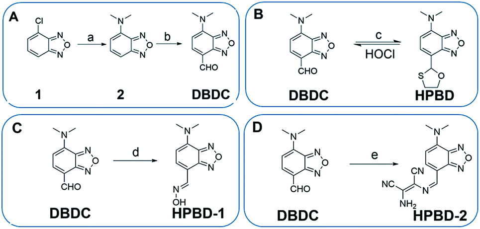 | ||
| Scheme 1 Design and synthesis of the fluorescent probes for HOCl. Reaction conditions: (a) Me2NH·HCl, EtOH, 150 °C, 2 d; (b) POCl3, DMF, 0–20 °C, 6 h; (c) 2-mercaptoethanol, 40 °C, 3 h; (d) hydroxylamine hydrochloride, 60 °C, 3 h; (e) diaminomaleonitrile, room temperature, 3 h. | ||
Experimental
Reagents and materials
Myeloperoxidase (MPO) inhibitor (4-aminibenzohydrazide, 4-ABH) and phorbol myristate acetate (PMA) were purchased from Sigma-Aldrich (USA). All other reagents were commercially available from Sinopharm Chemical Reagent Co., Ltd. (Shanghai, China) and used as received without further purification. RAW 264.7 cells and HeLa cells were obtained from the cell bank of Central Laboratory at Xiangya Hospital (Changsha, China). Thin-layer chromatography (TLC) was performed on precoated silical gel 60 F254 plates, and column chromatography was conducted over silica gel (mesh 200–300), both of which were obtained from Qingdao Ocean Chemicals (Qingdao, China). All solvents used were purified by standard methods prior to use. Ultrapure water used was obtained through a Millipore Milli-Q water purification system (Billerica, MA) and had an electric resistance >18.25 MΩ.Cell cytotoxicity assay and cell culture
The cytotoxic effects of the probe were assessed using a methyl thiazolyl tetrazolium (MTT) assay. HeLa cells were grown in RPMI-1640 medium (Thermo Scientific HyClone) supplemented with 10% fetal bovine serum (Invitrogen), 100 U mL−1 penicillin, and 100 U mL−1 streptomycin at 37 °C in a humidified atmosphere incubator containing 5 wt%/vol CO2. RAW 264.7 macrophage cells were cultured in Dulbecco's modified Eagle's medium (DMEM) containing 10% fetal bovine serum (Invitrogen) and antibiotics (100 U mL−1 penicillin and 100 U mL−1 streptomycin), maintaining at 37 °C in a humidified atmosphere incubator containing 5 wt%/vol CO2. The cell density was determined using a TC20 automated cell counter (Bio-Rad).Fluorescence imaging of living cells
For the detection of exogenous HOCl, HeLa cells was seeded in glass bottom dishes in culture medium with a density of 1 × 10⁵ cells per dish, and incubated for 24 h. Then, the cells were incubated with 10 μM HPBD for 1 h at 37 °C. After washing with phosphate-buffered saline (PBS) (pH 7.4) for three times, the cells were treated with HOCl (40 μM) for 10 min at 37 °C. Prior to imaging, the cells were washed three times with PBS (pH 7.4). For the detection of endogenous produced HOCl, RAW 264.7 macrophage cells was seeded into a glass bottom dish with a density of 1 × 10⁵ cells per dish, and incubated for 24 h. Subsequently, the cells were exposed to 10 μM HPBD solution for 1 h at 37 °C. The solution was then removed, and the cells were washed with PBS (2 mL × 3) to clear HPBD molecules attached to the surface of cells. The cells incubated with 10 μM HPBD in culture medium for 1 h at 37 °C, then washed with PBS. The washed cells were separated and incubated with 500 ng mL−1 phorbol myristate acetate (PMA) for imaging of endogenous production of HOCl. The culture medium was removed, and the treated cells were washed three times with PBS (2 mL × 3) before observation. We could watch the fluorescent change through fluorescence confocal microscopy. The confocal cell images were collected confocal laser scanning microscopy (Olympus, FV-1000) with a 100× oil objective. The samples were excited with 488 nm, observation at 560–650 nm for the green channel.Preparation of ROS and RNS
Various ROS and RNS including HOCl, ˙OH, H2O2, 1O2, NO2−, NO3−, NO, ONOO−, O2− and t-BuOOH were prepared according to the following methods. HOCl was prepared from the source of NaClO at room temperature in PBS buffer (pH 7.4, 20 mM). The concentration of HOCl was determined by titration with Na2S2O3. Hydroxyl radical (˙OH) was generated by Fenton reaction. To prepare ˙OH solution, ferrous chloride was added in the presence of 10 equiv. of H2O2. Singlet oxygen (1O2) was generated by the addition of NaClO and H2O2 according to the literature.19 The source of NO2−, NO3− from NaNO2 and NaNO3. Nitric oxide (NO) was generated from sodium dihydrate. Peroxynitrite (ONOO−) was prepared as the reported method.20 Superoxide O2− is prepared from KO2. t-BuOOH was obtained commercially from J&K.Synthetic procedures
Compound 2 and DBDC was synthesized according to previously reported methods.21![[double bond, length as m-dash]](https://www.rsc.org/images/entities/char_e001.gif) ), 11.24 (s, 1H, –OH). 13C NMR (100 MHz, d6-DMSO) δ (ppm): 42.32, 55.38, 105.03, 107.58, 135.38, 140.26, 145.73, 147.99. HRMS (EI): calcd. For C9H10N4O2 206.0804; found 206.0806.). 13C NMR (100 MHz, d6-DMSO) δ (ppm): 42.84, 104.78, 104.86, 108.75, 114.10, 115.16, 125.09, 137.09, 142.53, 145.41, 148.98, 150.13. HRMS (EI): calcd for C13H11N7O 281.1025; found 281.1014.
), 11.24 (s, 1H, –OH). 13C NMR (100 MHz, d6-DMSO) δ (ppm): 42.32, 55.38, 105.03, 107.58, 135.38, 140.26, 145.73, 147.99. HRMS (EI): calcd. For C9H10N4O2 206.0804; found 206.0806.). 13C NMR (100 MHz, d6-DMSO) δ (ppm): 42.84, 104.78, 104.86, 108.75, 114.10, 115.16, 125.09, 137.09, 142.53, 145.41, 148.98, 150.13. HRMS (EI): calcd for C13H11N7O 281.1025; found 281.1014.Results and discussion
Design and synthesis of HOCl probes
We designed an ICT-based BD derivative fluorophore, 7-(dimethylamino) benzo-[c]-[1,2,5]-oxadiazole-4-carbaldehyde (DBDC), as the fluorescence reporting group. The synthetic route of DBDC is shown in Scheme 1A. The HPBD probe for HOCl was prepared via a simple one-step reaction (Scheme 1B): DBDC reacted with 2-mercaptoethanol in dry dichloromethane in the presence of a right amount of methanesulfonic acid to synthesize HPBD (59.6% yield). Several functional groups, such as oxime and diaminomaleonitrile, have also been identified as HOCl-reactive moieties.22–26 For comparison, hydroxylamine and diaminomaleonitrile were also synthesized (HPBD-1 and HPBD-2). The synthetic procedures of HPBD-1 and HPBD-2 were shown in Scheme 1C and D, respectively. The synthetic intermediates and HOCl probes were characterized by 1H NMR, 13C NMR and high resolution mass spectrometry (HRMS) (Fig. S1–S12 in the ESI†).Comparing the fluorescent response of HPBD, HPBD-1, HPBD-2 to HOCl
To compare the fluorescent responses of HPBD, HPBD-1, and HPBD-2 to HOCl, three probes were titrated with HOCl from 0 to 40 μM. It was found that the fluorescence intensity of HPBD enhanced very significantly at low concentration of HOCl (Fig. 1), indicating its high sensitivity for HOCl detection. In contrast, oxime based probe HPBD-1 showed only slight activation to HOCl, which might be attributed to the fact that effective oxidative deprotection of oxime by HOCl only occurred under basic conditions.27 The probe HPBD-2 almost exhibited no fluorescence activation in the concentration range of HOCl, which seemed ascribed to the low efficiency of HOCl-induced de-diaminomaleonitrile reaction under the condition of low concentration of HOCl. Therefore, HPBD was chosen as the probe for HOCl detection and its basic photophysical properties were investigated (Table S1†). | ||
| Fig. 1 Plot of fluorescence intensity changes of three probes as a function of HOCl concentrations in a PBS buffer–CH3CN (v/v = 4/1, 20 mM PBS, pH 7.4). | ||
Photophysical properties and response of HPBD
The fluorescence titration of HPBD (10 μM) was performed in a PBS buffer–CH3CN (v/v = 1/4, 20 mM, pH 7.4). As depicted in Fig. 2, the HPBD probe (10 μM) showed a very weak fluorescence signal due to low electron-withdrawing ability of the oxathiolane group. The fluorescence titration of the probe with HOCl led to an increase of the fluorescence emission intensity (over 187-fold) at 585 nm. This result indicated the anticipated HOCl-promoted carbaldehyde recovery from 7-oxathiane with a concomitant recovered ICT effect. The Stokes shift was as large as 130 nm, suggesting the potential of the developed probe for minimizing autofluorescence and self-absorption in biological analysis. Moreover, the probe displays a linear response to HOCl in the range from 0 to 40 μM with a limit of detection of 50 nM (Fig. S13 and S14 in the ESI†), which indicated the high sensitivity of the HPBD probe to HOCl and its ability to monitor amounts of intracellular HOCl. | ||
| Fig. 2 (a) Fluorescence emission spectra of HPBD (10 μM) in response to HOCl of varying concentrations. λex = 455 nm. (b) Absorption spectra of HPBD (50 μM) in response to HOCl of varying concentrations. | ||
The absorption spectroscopic analysis, as shown in Fig. 2b, showed that the HPBD probe only gave an absorption peak at 448 nm. Upon addition of HOCl, the peak increased with increasing concentrations of HOCl with a bathochromic shift to 458 nm as well as a new absorption band appearing at 316 nm. We also observed that the solution turned from pale yellow to bright yellow. The increased red-shifted peak implied that the reaction of the probe with HOCl caused an enhanced ICT effect.
A salient advantage of the HPBD probe is the very rapid response rate, as shown in Fig. 3. Time-dependent measurements of the fluorescence intensities at 585 nm revealed that, on reacting with HOCl, the fluorescence signal gave a sudden increase and the plateau was reached almost within 1 s. These findings implied the extremely rapid activation kinetics of the probe in response to HOCl, which indicated the potential of this probe for real-time monitoring of the fast HOCl signaling in biological systems.
 | ||
| Fig. 3 The time course of fluorescence intensity of HPBD (10 μM) at 585 nm in a PBS buffer–CH3CN (v/v = 4/1, 20 mM PBS, pH 7.4) before and after adding 10 and 20 μM HOCl. λex = 455 nm. | ||
Next, the response capability of the HPBD probe was examined towards various ROS and RNS, including HOCl, ˙OH, H2O2, O2, NO2−, NO3−, NO, ONOO−, O2−, and t-BuOOH. In the solution of HPBD (10 μM) in a PBS–CH3CN buffer (v/v = 1/4, 20 mM PBS, pH 7.4), a given species of ROS or RNS was added. As shown in Fig. 4a, it was found that a strong fluorescence emission at 585 nm was only obtained for the addition of HOCl (40 μM). Moreover, the fluorescence enhancement induced by HOCl was almost unchanged even in the presence of large amounts of other competitive ROS and RNS. All these results confirm that HPBD is high selective to sense HOCl even in the presence of large excesses of other ROS and RNS. The absorption spectra of HPBD in the presence of 10 equiv. other ROS and RNS were almost the same as that of free HPBD, confirming its colorimetric sensing ability for HOCl (Fig. S15 in the ESI†). Investigation of the effects of pH on the response of the probe to HOCl manifested that the probe was chemically stable under conditions of varying pH and its fluorescence response toward HOCl was significant across a wide range of pH values (4–9), suggesting the utility of this probe for live cell imaging application, and the optimal activity was achieved at physiological conditions of pH 7.4 (Fig. 4b).
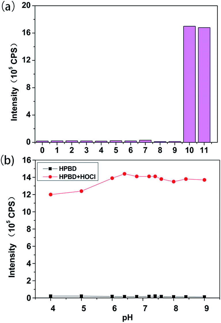 | ||
| Fig. 4 (a) Fluorescence intensity of HPBD (10 μM) at 585 nm before and after addition of various ROS and RNS in a PBS buffer–CH3CN (v/v = 4/1, 20 mM PBS, pH 7.4). (0: blank, 1: ˙OH, 2: H2O2, 3: 1O2, 4: NO2−, 5: NO3−, 6: NO, 7: ONOO−, 8: O2−, 9: t-BuOOH, 10: HOCl, 11: a mixture of 1–10). [HOCl] = 40 μM, [˙OH] = [ONOO−] = [1O2] = [t-BuOOH] = [H2O2] = [NO] = [NO2−] = [NO3−] = [O2−] = 400 μM. λex = 455 nm. (b) The effect of pH values on the fluorescence intensity of HPBD (10 μM) in the absence or presence of HOCl (30 μM). λex/em = 455/585 nm. | ||
Further experiments were performed to interrogate the mechanism and performance for the HPBD probe. MS analysis showed that reaction mixture yielded two new peaks with m/z of 192.1 and 214.1, which were assigned as [HPBD + H]+ and [HPBD + Na]+, respectively (Fig. S16 in the ESI†). This result revealed that HPBD (m/z of 251.1 for [M + H]+) was converted into DBDC in the presence of HOCl. High performance liquid chromatography (HPLC) was also used to confirm the response mechanism (Fig. S17 in the ESI†). We found that the chromatographic peak at 24.26 min, which was ascribed to the HPBD probe, decreased remarkably after reacting with HOCl, while a new peak was observed at 16.24 min, which was attributed to the reaction product DBDC. All these results confirmed the aldehyde recovery mechanism proposed in Scheme 1B, and suggested that the HOCl-induced emission enhancement arose from the formation of the fluorophore DBDC with strong ICT effect. Cytotoxicity assay using RAW264.7 cells revealed that the probe only generated marginal toxicity (cell viability decreased by ∼7% after 24 h incubation) at a concentration up to 25 μM (Fig. S18 in ESI†), implying the utility for this probe in live cell imaging at a working concentration below 25 μM.
Fluorescence imaging of HOCl in live cells
The desirable properties of HPBD for HOCl detection motivated us to utilize it for imaging of intracellular HOCl in live cells. First, we explored the possibility of using HPBD for imaging exogenous HOCl in HeLa cells (Fig. 5). When HeLa cells were incubated with HPBD (10 μM) for 1 h, no appreciable fluorescence signal was obtained. In contrast, after the cells were incubated with 10 μM probe followed by treatment with 40 μM HOCl for 10 min, bright fluorescence was observed in the cells. The merged picture confirmed that the fluorescence signals were only localized in the cytoplasmic areas, suggested effective internalization of the probe in cells. The above experiments proved that the HPBD probe was well cell-permeated and had the ability to detect HOCl in live cells. | ||
| Fig. 5 Confocal fluorescence images of HeLa cells. (Left) Bright field image; (Middle) fluorescence image; and (Right) merged image. (a–c) The cells incubated with HPBD (10 μM) for 1 h. (d–f) Subsequent treatment of the cells with HOCl (10 μM) for 10 min. Emission intensity was collected in an optical window 560–650 nm, λex = 488 nm. Scale bar = 20 μm. | ||
The HPBD probe was then applied to imaging of endogenous HOCl induced by phorbol myristate acetate (PMA) in RAW264.7 cells (Fig. 6). PMA was known to activate the generation of ROS and RNS in macrophage cells, including HOCl.28 In the control experiment, the RAW 264.7 macrophage cells were incubated with HPBD (10 μM), and no fluorescence was observed. After stimulation with PMA (500 ng mL−1) for 2 h, a strong green fluorescence was observed in the cells. These results demonstrated that HPBD was able to visualize PMA-induced production of endogenous HOCl in the macrophages. Further control with MPO inhibitor (4-aminibenzohydrazide, 4-ABH) co-incubated with PMA for the RAW 264.7 cells, no enhancement fluorescence was observed. Because the production of HOCl was specific to MPO, this finding clearly indicated that other reactive oxygen species produced during the stimulation did not result in fluorescence enhancement for the probe, and the fluorescence activation in the cells was highly selective to HOCl.
 | ||
| Fig. 6 Confocal fluorescence images of PMA-induced hypochlorous acid production in RAW264.7 cells. (a–c) The cells incubated with HPBD (10 μM) for 1 h. (d–f) The cells were treated with stimulant PMA (500 ng mL−1) for 2 h in the presence of HPBD (10 μM). (g–i) MPO inhibitor (100 μM), (4-ABH) was co-incubated during PMA stimulation, and incubated with of HPBD (10 μM) for 1 h at 37 °C. Emission intensity was collected in an optical window 560–650 nm, λex = 488 nm. Scale bar = 10 μm. | ||
Conclusions
We have developed a novel “turn-on” fluorescent probe for high selectivity, rapid detection and imaging of HOCl based on the protection of carbaldehyde with 2-mercaptoethanol. The developed probe shows very weak fluorescence because of the inhibition of the ICT effect, while delivers over 180-fold fluorescence enhancement with a large Stokes shift over 100 nm on reacting with HOCl. Moreover, this fluorescence activation process is very rapid and can be completed within 1 s. This probe also provides very high sensitivity for HOCl detection with a detection limit of 50 nM, and shows high selectivity to HOCl with little dependency to other ROS or RNS. The excellent sensing properties of the HPBD probe enables its use in living cells for real-time and high-contrast imaging of HOCl in HeLa cells and RAW264.7 macrophage cells. In virtue of these advantages, this probe might hold great potential as an invaluable probe for real-time monitoring of the HOCl signaling in various biological processes.Acknowledgements
This work was financially supported by the National Natural Science Foundation of China (21527810, 21190041, 21221003, 91317312), and the National Key Basic Research Program (2011CB911000).Notes and references
- (a) S. Khatib, R. Musaa and J. Vaya, Bioorg. Med. Chem., 2007, 15, 3661–3666 CrossRef CAS PubMed; (b) C. C. Winterbourn, M. B. Hampton, J. H. Livesey and A. J. Kettle, J. Biol. Chem., 2006, 281, 39860–39869 CrossRef CAS PubMed.
- T. J. Fiedler, C. A. Davey and R. E. Fenna, J. Biol. Chem., 2000, 275, 11964 CrossRef CAS PubMed.
- Y. W. Yap, M. Whiteman and N. S. Cheung, Cell. Signalling, 2007, 19, 219–228 CrossRef CAS PubMed.
- J. P. Henderson, J. Byun and J. W. Heinecke, J. Biol. Chem., 1999, 274, 33440 CrossRef CAS PubMed.
- T. Hasegawa, E. Malle, A. Farhood and H. Jaeschke, Am. J. Physiol.: Gastrointest. Liver Physiol., 2005, 289, 760–767 Search PubMed.
- Z. Sun, F. Liu, Y. Chen, P. K. H. Tam and D. Yang, Org. Lett., 2008, 10, 2171–2174 CrossRef CAS PubMed.
- (a) S. M. Wu and S. V. Pizzo, Arch. Biochem. Biophys., 2001, 391, 119–126 CrossRef CAS PubMed; (b) S. A. Weitzman and L. Gordon, Blood, 1990, 76, 655–663 CAS.
- L. Moberg and B. Karlberg, Anal. Chim. Acta, 2000, 407, 127–133 CrossRef CAS.
- M. F. Suárez and A. Y. Ting, Nat. Rev. Mol. Cell Biol., 2008, 9, 973–984 Search PubMed.
- J. Chan, S. C. Dodani and C. J. Chang, Nat. Chem., 2012, 4, 973–984 CrossRef CAS PubMed.
- B. C. Dickinson and C. J. Chang, J. Am. Chem. Soc., 2008, 130, 9638–9639 CrossRef CAS PubMed.
- S. Y. Lim, K. H. Hong, D. Il Kim, H. Kwon and H. J. Kim, J. Am. Chem. Soc., 2014, 136, 7018–7025 CrossRef CAS PubMed.
- M. H. Lee, N. Park, C. Yi, J. H. Han, J. H. Hong, K. P. Kim, D. H. Kang, J. L. Sessler, C. Kang and J. S. Kim, J. Am. Chem. Soc., 2014, 136, 14136–14142 CrossRef CAS PubMed.
- M. Ishida, H. Watanabe, K. Takigawa, Y. Kurishita, C. Oki, A. Nakamura, I. Hamachi and S. Tsukiji, J. Am. Chem. Soc., 2013, 135, 12684–12689 CrossRef CAS PubMed.
- S. Arai, S. C. Lee, D. Zhai, M. Suzuki and Y. T. Chang, Sci. Rep., 2014, 4, 6701–6706 CrossRef CAS PubMed.
- (a) S. Kenmoku, Y. Urano, H. Kojima and T. Nagano, J. Am. Chem. Soc., 2007, 129, 7313–7318 CrossRef CAS PubMed; (b) Z. Sun, F. Liu, Y. Chen, P. K. H. Tam and D. Yang, Org. Lett., 2008, 10, 2171–2174 CrossRef CAS PubMed; (c) P. Panizzi, M. Nahrendorf, M. Wildgruber, P. Waterman, J. L. Figueiredo, E. Aikawa, J. McCarthy, R. Weissleder and S. A. Hilderbrand, J. Am. Chem. Soc., 2009, 131, 15739–15744 CrossRef CAS PubMed; (d) Y. Koide, Y. Urano, K. Hanaoka, T. Terai and T. Nagano, J. Am. Chem. Soc., 2011, 133, 5680–5682 CrossRef CAS PubMed; (e) Y. K. Yang, H. J. Cho, J. Lee, I. Shin and J. Tae, Org. Lett., 2009, 11, 859–861 CrossRef CAS PubMed; (f) W. Wu, Z. Li, L. Yang, J. Han and S. Han, Chem. Sci., 2013, 4, 460–467 RSC; (g) Q. Xu, K. A. Lee, S. Lee, K. M. Lee, W. J. Lee and J. Yoon, J. Am. Chem. Soc., 2013, 135, 9944–9949 CrossRef CAS PubMed; (h) H. D. Xiao, K. Xin, H. F. Dou, G. Yin, Y. W. Quan and R. Y. Wang, Chem. Commun., 2015, 51, 1442–1445 RSC; (i) W. C. Chen, P. Venkatesan and S. P. Wu, Anal. Chim. Acta, 2015, 882, 68–75 CrossRef CAS PubMed; (j) X. Z. Wang, L. Zhou, F. Qiang, F. Y. Wang, R. Wang and C. C. Zhao, Anal. Chim. Acta, 2016, 911, 114–120 CrossRef CAS PubMed.
- (a) F. Qian, C. L. Zhang, Y. M. Zhang, W. J. He, X. Gao, P. Hu and Z. J. Guo, J. Am. Chem. Soc., 2009, 131, 1460–1468 CrossRef CAS PubMed; (b) Z. P. Liu, C. L. Zhang, Y. C. Chen, W. J. He and Z. J. Guo, Chem. Commun., 2012, 48, 8365–8367 RSC; (c) Y. Cai, Y. Shi, H. Wang, J. Wang, D. Ding, L. Wang and Z. Yang, Anal. Chem., 2014, 86, 2193–2199 CrossRef CAS PubMed; (d) Z. Z. Liu, T. Y. Jiang, B. L. Wang, B. W. Ke, Y. B. Zhou, L. P. Du and M. Y. Li, Anal. Chem., 2016, 88, 1511–1515 CrossRef CAS PubMed.
- L. Yuan, L. Wang, B. K. Agrawalla, S. J. Park, H. Zhu, B. Sivaraman, J. J. Peng, Q. H. Xu and Y. T. Chang, J. Am. Chem. Soc., 2015, 137, 5930–5938 CrossRef CAS PubMed.
- X. Li, G. Zhang, H. Ma, D. Zhang, J. Li and D. Zhu, J. Am. Chem. Soc., 2004, 24, 11543–11548 CrossRef PubMed.
- J. W. Reed, H. H. Ho and W. L. Jolly, J. Am. Chem. Soc., 1974, 96, 1248–1249 CrossRef CAS.
- Z. H. Yang, C. C. Yan, Y. C. Chen, C. C. Zhu, C. L. Zhang, X. D. Dong, W. Q. Yang, Z. J. Guo, Y. Lu and W. J. He, Dalton Trans., 2011, 40, 2173–2176 RSC.
- G. H. Cheng, J. L. Fan, W. Sun, K. Sui, X. Jin, J. Y. Wang and X. J. Peng, Analyst, 2013, 138, 6091–6096 RSC.
- X. Cheng, H. Jia, T. Long, J. Feng, J. Qin and Z. Li, Chem. Commun., 2011, 47, 11978–11980 RSC.
- N. Zhao, Y. H. Wu, R. M. Wang, L. X. Shi and Z. N. Chen, Analyst, 2011, 136, 2277–2282 RSC.
- S. I. Reja, V. Bhalla, A. Sharma, G. Kaur and M. Kumar, Chem. Commun., 2014, 50, 11911–11914 RSC.
- L. Yuan, W. Y. Lin, J. Z. Song and Y. T. Yang, Chem. Commun., 2011, 47, 12691–12693 RSC.
- W. Lin, L. Long, B. Chen and W. Tan, Chem.–Eur. J., 2009, 15, 2305–2309 CrossRef CAS PubMed.
- Y. Yang, H. Lu, W. Wang and I. Liau, Anal. Chem., 2011, 83, 8267–8272 CrossRef CAS PubMed.
Footnote |
| † Electronic supplementary information (ESI) available: Experimental details and additional figures. See DOI: 10.1039/c6ra22686h |
| This journal is © The Royal Society of Chemistry 2016 |