Siyuan Chen
a,
Liying Wanga,
Xu Dongb,
Xiaoyun Liu*c,
Jianfeng Zhouc,
Jianmao Yangc and
Liusheng Zha*ac
aState Key Laboratory for Modification of Chemical Fibers and Polymer Materials & College of Materials Science and Engineering, Donghua University, Shanghai 201620, P. R. China. E-mail: lszha@dhu.edu.cn
bState Key Laboratory of Molecular Engineering of Polymers and Department of Macromolecule Science, Fudan University, Shanghai 200433, P. R. China
cResearch Center for Analysis and Measurement, Donghua University, Shanghai 201620, P. R. China. E-mail: xyliu@dhu.edu.cn
First published on 3rd May 2016
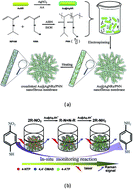
A novel type of metal nanoparticle-loaded smart nanofibrous membrane with fast thermo-responsiveness was fabricated by electrospinning an aqueous solution containing poly(N-isopropylacrylamide-co-N-hydroxymethylacrylamide) and monodispersed Au@Ag bimetallic nanorods with a core–shell structure, followed by heat treatment. The results obtained by electron microscopy show that the anisotropic nanoparticles are oriented along the axes of its constituent nanofibers. The membrane produced has high stability in aqueous media and remarkable thermo-responsiveness. It takes less than 10 seconds to reach its deswelling or swelling equilibrium state with the temperature alternating between 25 °C and 50 °C. The smart nanofibrous membrane with macroscopic mechanical strength can be used as a free-standing surface-enhanced Raman scattering (SERS) substrate with high Raman signal reproducibility for quantitative analysis, and its SERS efficiency can be readily elevated by raising the detection temperature across its phase transition temperature due to its fast thermo-response rate. Particularly, since the composite nanofibrous membrane possesses catalytic properties toward the reduction of 4-nitrothiophenol to 4-aminothiophenol by NaBH4, it has the ability to act as an in situ SERS monitor for the reaction, and it was deduced from the detected intermediate that the reaction proceeds via a condensation reaction route.
A stimulus-responsive nanofibrous membrane is a smart polymer material, which consists of orderly or randomly aligned nanofibers with diameters of tens to hundreds of nanometers.16 Compared to smart bulky hydrogels, these have much faster stimulus-responses owing to their constituent ultra thin fibers and porous structure, which is helpful for the transfer of the stimulus to the whole material. Their reported applications have demonstrated that such smart materials allow for convenient use in a device because of their macroscopic mechanical strength and flexible morphology.17,18 If used as a carrier for nanocatalysts, they can be readily recovered from a reaction system.19,20 A nanofibrous membrane is usually produced by an electrospinning technique, which features simplicity, flexible morphology tuning and a wide variety of usable polymers.19,21,22 Therefore, it could be expected that stimulus-responsive electrospun nanofibrous membranes can act as substitutes for smart bulky hydrogels and microgels for loading metal nanoparticles.
Within the field of metal nanoparticle-based nanoscience and nanotechnology, bimetallic nanoparticles with a core–shell structure have attracted increasing interest due to their superior physicochemical, optical and catalytic properties compared to their respective monometallic analogues.23,24 Among these, a bimetallic nanorod based on a gold nanorod (AuNR) core and a silver shell (Au@AgNR) is particularly attractive because of its potential applications in surface-enhanced Raman scattering (SERS) substrates and catalysts.23,25,26 Recently, in our group, monodispersed Au@AgNR prepared through a seed-mediated growth process by reduction of AgNO3, using AuNRs with a narrow size and shape distribution as seeds, was used as a SERS substrate with high Raman signal reproducibility for quantitative analysis.26 Its SERS efficiency is remarkably increased with its Ag shell thickness due to the enhancing surface electric field and the chemical enhancement associated with the electronic ligand effect. Furthermore, it has been reported that, compared to monometallic Ag nanoparticles, the bimetallic Au@Ag nanoparticles with a core–shell structure have much higher chemical stability owing to free electron transfer from the Au core to the Ag shell.27 This can avoid weakening of the Au@AgNRs'; SERS efficiency during usage and storage, otherwise their oxidized Ag surfaces would hinder the charge transfer to the analyte molecule, according to the chemical enhancement mechanism.28 In addition to SERS substrates, the nanorods can be used as nanocatalysts with high catalytic reactivity and selectivity for some chemical reactions owing to the synergistic catalytic effect of their constituent gold and silver components.29,30 For example, the results obtained by Monga et al. demonstrated that Au@AgNRs catalyze the selective formation of 3-nitroaniline in the reduction of 1,3-dinitrobenzene, compared to 1,3-phenylenediamine in the case of Au and Ag nanorods.31
Herein, we report the fabrication of a monodispersed Au@AgNR-loaded thermo-responsive composite nanofibrous membrane by electrospinning an aqueous solution containing poly(N-isopropylacrylamide-co-N-hydroxymethylacrylamide) and the nanorods, followed by heat treatment. Observations made using electron microscopy confirm that the Au@AgNRs are oriented along the axes of its constituent nanofibers. The composite nanofibrous membrane produced with a fast thermo-response and stable macroscopic morphology in aqueous media can be used as a smart free-standing SERS substrate with temperature-tunable efficiency. Particularly, it was found that this has the ability to in situ SERS monitor a catalyzed reaction, which is beneficial to understanding the reaction mechanism.
:
40, v/v). Deionized water used in synthesis and characterization was made using a Millipore Direct-Q system.
:
1. A certain amount of purified Au@AgNRs dispersed in 1 mL of deionized water were then added to 3 mL of PNN aqueous solution of 30 wt% concentration, followed by vigorous stirring at room temperature for 24 h to produce an electrospinning solution of Au@AgNRs with a concentration of 25 nM, 50 nM or 100 nM. Subsequently, the electrospinning solution was loaded into a 10 mL plastic syringe and electrospun at a positive voltage of 13 kV, a working distance of 15 cm (distance between the needle tip and the grounded metal plate), and a flow rate of 0.9 mL h−1 for 4 h at room temperature. Finally, the nanofibrous membrane formed was heated at 110 °C for 5 h. Keeping the polymer concentration constant in the spinning solution, three kinds of Au@AgNRs/PNN composite nanofibrous membranes were fabricated by changing the concentration of the Au@AgNRs (from 25 nM to 50 nM to 100 nM), which are labeled as HNF-1, HNF-2 and HNF-3, respectively.
TEM images of Au@AgNRs/PNN composite nanofibrous membranes were taken with a JOEL 2100F transmission electron microscope at a voltage of 200 kV. The samples were collected on copper grids during electrospinning, followed by heating at 110 °C for 5 h. Their SEM images were obtained with a Hitachi S-4800 field emission scanning electron microscope at a voltage of 5 kV. The samples were sputtered with gold.
UV-vis adsorption spectra of diluted Au@AgNRs/PNN composite nanofiber aqueous dispersions at various temperatures were measured using a Perkin Elmer Lambda 35 UV-vis spectrometer with a temperature controller. A quartz cell with a 1 cm optical path length was used. Fourier transform infrared (FTIR) spectra of uncrosslinked PNN nanofibrous membranes and Au@AgNRs/PNN composite nanofibrous membranes were measured with a Thermo Fisher Nicolet 6700 FTIR spectrometer using an ATR attachment with a horizontal Ge crystal.
SERS measurements were performed using a Renishaw inVia-Reflex micro-Raman system equipped with a multi-channel charge-coupled device detector and a DM2500M Leica confocal microscope upon excitation by a 532, 633 or 785 nm laser source. All the SERS spectra were the results of 10 s accumulations. In order to assess the SERS efficiency of an Au@AgNRs/PNN composite nanofibrous membrane (size: 0.7 cm × 0.7 cm), it was put in the quartz sample cell, then 200 μL of 4-NTP aqueous solutions of different concentrations were injected into the cell, before recording SERS spectra using a Raman microscope with a 50× objective. Temperature-dependent SERS spectra were measured for a piece of membrane (HNF-3, 0.7 cm × 0.7 cm) that was immersed in 10−9 M 4-NTP aqueous solution in a Linkam THMS600 temperature-controllable cell, which was equilibrated thermally for at least 5 min before SERS measurement.
![]() | ||
| Fig. 1 The fabrication route of thermo-responsive Au@AgNRs/PNN composite nanofibrous membrane (a) and its use in an in situ SERS monitored catalytic reaction (b). | ||
Its NIPAM/NMA composition determined from its 1H NMR spectrum (see Fig. S1(a) in the (ESI)†) is 100
:
20.4 (molar ratio), very close to the feeding ratio of 100
:
20, and its number-average molecular weight estimated by gel permeation chromatography using tetrahydrofuran as eluent is 1.27 × 10⁵ g mol−1. Fig. S1(b) in the ESI† shows the temperature-dependent absorbance (at 500 nm) of the diluted PNN aqueous solution, which exhibits a thermo-responsive hydrophilic-to-hydrophobic phase transition in aqueous media. Its lower critical solution temperature (LCST) is 41.8 °C, which is considerably higher than that of poly(N-isopropylacrylamide) (32 °C), due to the hydrophilicity of its constituting NMA moiety.34 Concurrently, monodispersed Au@AgNRs were prepared and purified by our recently reported method.26 A TEM image of the Au@AgNRs is shown in Fig. S2(a) in the ESI,† and we can note that they have excellent monodispersity and a well-defined core–shell structure. Their average length and diameter obtained by randomly measuring 50 particles are 81 ± 4 nm and 25 ± 2 nm, with the calculated aspect ratio being 3.2 and the average silver shell thicknesses of their lateral facets and two tips all being 2.7 ± 0.2 nm. The ultraviolet visible (UV-vis) spectrum of the diluted Au@AgNR aqueous dispersion and its optical image are shown in Fig. S2(b) in the ESI.† According to a previous report,26 the two bands at 708 nm and 505 nm are attributed to the longitudinal and transverse dipolar plasmon modes of the Au@AgNRs, respectively, and the band at 385 nm is ascribed to their octupolar plasmon mode. It can also be found from Fig. S2(b) in the ESI† that the longitudinal plasmon band of the Au@AgNRs is relatively narrow, further proving their monodispersity. In the electrospinning stage, PNN was mixed with an Au@AgNR aqueous dispersion to generate a spinning solution after stirring for 24 h. Subsequently, the solution was electrospun at a positive voltage of 13 kV, a working distance of 15 cm (distance between the needle tip and the grounded metal plate), and a flow rate of 0.9 mL h−1 at room temperature for 4 h. Finally, the nanofibrous membrane produced was heated at 110 °C for 5 h to form a crosslinking structure between the polymer chains within its constituent fibers, as illustrated in Scheme S1 in the ESI.†
Optical images of the Au@AgNRs/PNN composite nanofibrous membranes and the PNN nanofibrous membrane as control sample are shown in Fig. S3 in the ESI,† and it can be seen that the fabricated products are nonwoven membranes with an area of over 240 cm2 and a thickness of ca. 60 μm, and their colors in the dry state change from white to light green as their Au@AgNR content increases. Fig. 2 exhibits their scanning electron microscopy (SEM) and transmission electron microscopy (TEM) images at low and high magnification. From their SEM images, we can note that these membranes have three-dimensional networked structures consisting of many randomly deposited fibers, and the fibers are homogeneous in their diameters, without a beaded morphology. Their average diameters, determined by randomly measuring 50 fibers within each membrane, are 806 ± 112 nm, 682 ± 104 nm, 635 ± 34 nm and 607 ± 84 nm, decreasing as their Au@AgNR content increases. This behavior is presumably related to the increase in solution conductivity concomitant with the incorporation of Au@AgNRs.35 When observing the amplified SEM images of the nanofibers, the bright spots on their surfaces are the Au@AgNRs orienting along their axes, whose amounts are increased with the Au@AgNR concentration in their spinning solution. From their TEM images, we can clearly see that the Au@AgNRs are embedded within the fibers and are directionally aligned along their axis directions. As the Au@AgNR content increases within the nanofibers, the average end-to-end distance between the adjacent nanorods decreases from 146 nm to 89 nm to 41 nm. The low-magnification TEM images show that the oriented alignments are extremely uniform over long distances throughout the whole sample. In contrast to the spinning solution, where the Au@AgNRs are randomly dispersed, we can conclude that their oriented alignments inside the nanofibers result from the electrospinning process.36 There are two external fields that trigger such an alignment of the nanorods during the electrospinning process. One is the electric field supplied by the needle, and the other is the flow field formed during the formation of the fibers. When a high voltage was applied to the spinning solution, the whole solution was polarized. Furthermore, if the repulsive force within the charged solution was larger than its surface tension, a jet would erupt from the tip of the spinneret, producing the flow field. Shortly, the jet entered a bending instability stage, and the nanofibers were formed by the stretch and acceleration of the fluid filament in the region of instability as the solvent evaporated. During the formation of nanofibers, a sink-flow effect and strong shear forces were exerted on the Au@AgNRs, leading to their oriented alignment along the flow direction, which is the axial direction of the fibers. Even when a high concentration of the Au@AgNRs (100 nM) was present in the spinning solution, the nanorods were all directionally aligned, which demonstrates that electrospinning is a powerful technique for forming an oriented alignment of anisotropic nanorods on a large scale.
Subsequently, it is essential to investigate the effect of temperature change on the plasmonic properties of the Au@AgNRs embedded within the composite nanofibrous membrane. To this end, an aqueous dispersion of nanofibers was prepared by adding a small amount of the broken membrane (HNF-2) to water, followed by sonicating the solution for 5 h, and UV-vis spectra in the range 300–1100 nm at different temperatures were obtained, as shown in Fig. 3(d). Firstly, the longitudinal plasmon band of the Au@AgNRs embedded within the membrane becomes broader compared to that before electrospinning (Fig. S1(b) in the ESI†), which may arise from their plasmonic coupling due to their approaching inside its constituent nanofibers. Secondly, as the temperature rises from 39 °C to 47 °C, close to the temperature range of the area change of the membrane, both their transverse and longitudinal plasmon bands are abruptly enhanced, due to the refractive index increase of the dispersed nanofibers after the temperature-induced phase transition. Thirdly, their maximum wavelengths are both red-shifted within the same temperature range, which could be due to two reasons. One is that the collapsed polymer network around the Au@AgNRs could increase their surrounding refractive index.39 However, as investigated by Schmidt et al.,40 this contribution to the red-shift of the wavelengths should be limited owing to a slight variation in the refractive index before and after the phase transition. Another is that the temperature-triggered phase transition of the composite nanofibers could reduce the distances between the loaded Au@AgNRs. As reported by Sánchez-Iglesias, in the gold-nanorod-pair system, side-to-side interactions lead to a red-shift of the transverse plasmon band and a blue-shift of the longitudinal plasmon band, while tip-to-tip ones would red-shift both bands.41 In our system, both the tip-to-tip and side-to-side distances between the Au@AgNRs should be shortened after the temperature-driven phase transition of their lodged nanofibers due to their directional alignment along the fibers' axes, as evidenced above. However, it can be deduced from the red-shift of their longitudinal plasmon band that their tip-to-tip interactions are dominant over their side-to-side ones, due to the possibility that the longitudinal percent shrinkage of the nanofibers is larger than the transverse one.
O symmetric stretching and the phenyl ring stretching modes, respectively.42 From Fig. S6(a) in the ESI,† we can see that the membrane as a substrate exhibits the strongest SERS efficiency with an excitation wavelength of 785 nm, since this is the closest to the longitudinal plasmon resonance wavelength of its entrapped Au@AgNRs, resulting in the strongest surface electric field enhancement effect. Au@AgNRs/PNN composite nanofibrous membranes with three different Au@AgNR contents were used as substrates to measure the SERS spectra of a 10−7 M 4-NTP aqueous solution at 25 °C, and the results are given in Fig. S6(b) in the ESI.† A plot of their 1334 cm−1 peak intensities against the Au@AgNRs concentrations in their spinning solutions is depicted in Fig. S6(c) in the ESI.† It can be seen that their SERS efficiency is nearly proportionally increased with the Au@AgNR concentration, revealing that their SERS efficiency comes from the bimetallic nanorods. The composite nanofibrous membranes were used as substrates to measure the SERS spectra of different concentrations of 4-NTP solution from 10−13 M to 10−7 M at 25 °C; the result for HNF-2 is given in Fig. 4(a) and the others in Fig. S7 in the ESI.† As the 4-NTP concentration increases, its SERS intensity is progressively raised. Even on the HNF-1 substrate, a SERS signal of 10−12 M 4-NTP can be clearly observed (see Fig. S7(b) in the ESI†). When the membrane, HNF-3, was used as a SERS substrate, 10−13 M 4-NTP in aqueous solution could be detected (Fig. S7(e) in the ESI†). A plot of the 1334 cm−1 peak intensities in Fig. 4(a) versus the logarithmic concentrations (log(c)) of 4-NTP is shown in Fig. 4(b) and can be fitted into a line with a correlation coefficient of 0.993 (n = 9). This result shows that there is a good linear relationship between the SERS intensity and the logarithmic concentration of the analyte, indicating that the SERS signal based on the Au@AgNRs/PNN composite nanofibrous membrane as substrate is highly reproducible. A high SERS signal reproducibility should arise from the monodispersed and homogeneous distribution of the Au@AgNRs within its constituent nanofibers. In addition, fifteen measurements of 10−6 M 4-NTP aqueous solution were carried out on the same membrane (HNF-2), and the obtained SERS spectra are exhibited in Fig. 4(c). The estimated relative standard deviation (RSD) of the intensities of their 1334 cm−1 peaks is 3.87%, further confirming high reproducibility of the SERS signals from the membrane. These results mean that the Au@AgNRs/PNN composite nanofibrous membrane could be used as a SERS substrate for quantitative analysis.
The effect of temperature on the SERS efficiency of the Au@AgNRs/PNN composite nanofibrous membrane was investigated using 10−9 M 4-NTP aqueous solution as the analyte, as illustrated in Fig. 5(a). One can observe that all the peaks in its SERS spectrum are remarkably enhanced as the temperature is increased from 20 °C to 50 °C. A plot of the 1334 cm−1 peak intensity as a function of temperature is exhibited in Fig. 5(b). A marked change occurs within the temperature range from 35 °C to 45 °C, in agreement with that for the area change of the composite nanofibrous membrane with temperature. It can be inferred from this that the temperature-driven SERS signal enhancement possibly results from plasmonic coupling between approaching Au@AgNRs after temperature-triggered adhesion between their constituent nanofibers, as previously discussed.13 This result implies that the detection sensitivity based on the Au@AgNRs/PNN composite nanofibrous membrane as a SERS substrate can be improved by elevating the detection temperature. This estimation has been confirmed through SERS detecting of 10−13 M 4-NTP in aqueous solution at 50 °C using HNF-2 as substrate, as shown in Fig. 5(c) and (d). By comparing the results obtained at 25 °C and 50 °C, the low detection limit of 4-NTP at the higher detection temperature is lowered by one order of magnitude. Because of its fast thermo-response rate, the swelling or deswelling equilibrium of the membrane as a SERS substrate at each detection temperature can be quickly reached, which is beneficial with respect to shortening the measurement time.
N stretching, and C–H in-plane bending modes of 4,4′-DMAB, respectively.42 Their intensities initially increase with reaction time (from black to green spectra) and then gradually decrease to near-zero (from green to yellow ones), corroborating its character as an intermediate. In addition to the peaks of 4,4′-DMAB, a new peak at 1590 cm−1 shows up almost simultaneously and gradually dominates the spectrum, which is attributed to the amino vibrational mode of the product, 4-ATP. The results indicate that the reduction of 4-NTP into 4-ATP by NaBH4, catalyzed by the Au@AgNRs/PNN composite nanofibrous membrane, proceeds via a condensation route. The mechanism can be explained by the speculation that the transformation of 4-NTP to 4,4′-DMAB may be caused by plasmon-induced charge transfer under the laser irradiation.45 Concretely, the reaction could be catalyzed via electron transfer between the laser-irradiation-induced plasmon resonance surface of Au@AgNR and the nitro group of 4-NTP through the molecule, because of its chemisorption on the Ag surface via its thiol group.
Footnote |
| † Electronic supplementary information (ESI) available. See DOI: 10.1039/c6ra04247c |
| This journal is © The Royal Society of Chemistry 2016 |