Issue 57, 2019

Nuclear localization of dirhodium(ii) complexes in breast cancer cells by X-ray fluorescence microscopy

Abstract

The cellular distribution of three dirhodium(II) complexes with a paddlewheel structure was investigated using synchrotron-based X-ray fluorescence microscopy and cell viability studies. Complexes with vacant axial sites displayed cytotoxic activity and nuclear accumulation whereas complexes in which the axial positions were blocked showed little to no toxicity nor uptake.
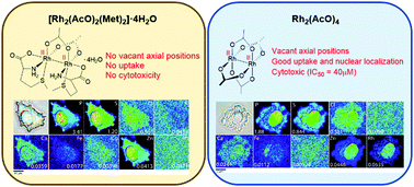

Graphical abstract: Nuclear localization of dirhodium(ii) complexes in breast cancer cells by X-ray fluorescence microscopy

Supplementary files

Article information

Article type
Communication
Submitted
21 Jan 2019
Accepted
05 Jun 2019
First published
05 Jun 2019

Chem. Commun., 2019,55, 8223-8226

Nuclear localization of dirhodium(II) complexes in breast cancer cells by X-ray fluorescence microscopy

A. Enriquez Garcia, B. Lai, S. G. Gopinathan, H. H. Harris, C. S. Shemanko and F. Jalilehvand, Chem. Commun., 2019, 55, 8223 DOI: 10.1039/C9CC00521H

To request permission to reproduce material from this article, please go to the Copyright Clearance Center request page.

If you are an author contributing to an RSC publication, you do not need to request permission provided correct acknowledgement is given.

If you are the author of this article, you do not need to request permission to reproduce figures and diagrams provided correct acknowledgement is given. If you want to reproduce the whole article in a third-party publication (excluding your thesis/dissertation for which permission is not required) please go to the Copyright Clearance Center request page.

Read more about how to correctly acknowledge RSC content.

Social activity

Spotlight

Advertisements