Abstract
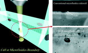
Microfluidic frameworks known as micro-total-analysis-systems or lab-on-a-chip have become versatile tools in cell biology research, since functional biochips are able to streamline dynamic observations of various cells. Glass or polymers are generally used as the substrate due to their high transparency, chemical stability and cost-effectiveness. However, these materials are not well suited for the microscopic observation of cell migration at the fluid boundary due to the refractive index mismatch between the medium and the biochip material. For this reason, we have developed a new method of fabricating three-dimensional (3D) microfluidic chips made of the low refractive index fluoric polymer CYTOP. This novel fabrication procedure involves the use of a femtosecond laser for direct writing, followed by wet etching with a dilute fluorinated solvent and annealing, to create high-quality 3D microfluidic chips inside a polymer substrate. A microfluidic chip made in this manner enabled us to more clearly observe the flagellum motion of a Dinoflagellate moving in circles near the fluid surface compared to the observations possible using conventional microfluidic chips. We believe that CYTOP microfluidic chips made using this new method may allow more detailed analysis of various cell migrations near solid boundaries.


 Please wait while we load your content...