Photoacids as a new fluorescence tool for tracking structural transitions of proteins: following the concentration-induced transition of bovine serum albumin†
Abstract
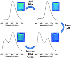
Spectroscopy-based techniques for assessing structural transitions of proteins follow either an intramolecular chromophore, as in absorption-based circular dichroism (CD) or fluorescence-based tryptophan emission, or an intermolecular chromophore such as fluorescent probes. Here a new fluorescent probe method to probe the structural transition of proteins by photoacids is presented, which has a fundamentally different photo-physical origin to that of common fluorescent probes. Photoacids are molecules that release a proton upon photo-excitation. By following the steady-state and time-resolved emission of the protonated and de-protonated species of the photoacid we probe the environment of its binding site in bovine serum albumin (BSA) in a wide range of weight concentrations (0.001–8%). We found a unique concentration-induced structural transition of BSA at pH2 and at concentrations of >0.75%, which involves the exposure of its hydrophobic core to the solution. We confirm our results with the common tryptophan emission method, and show that the use of photoacids can result in a much more sensitive tool. We also show that common fluorescent probes and the CD methodologies have fundamental restrictions that limit their use in a concentration-dependent study. The use of photoacids is facile and requires only a fluorospectrometer (and preferably, but not mandatorily, a time-resolution emission system). The photoacid can be either non-covalently (as in this study) or covalently attached to the molecule, and can be readily employed to follow the local environment of numerous (bio-)systems.


 Please wait while we load your content...