Attomolar protein detection using a magnetic bead surface coverage assay†
Abstract
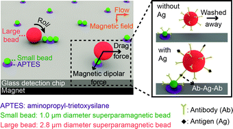
We demonstrate a microfluidic method for ultra-sensitive

* Corresponding authors
a
Laboratory of Microsystems, Ecole Polytechnique Fédérale de Lausanne (EPFL), CH-1015, Lausanne, Switzerland
E-mail:
martin.gijs@epfl.ch
Fax: +41 (0) 21 693 59 50
Tel: +41 (0) 21 693 77 31
We demonstrate a microfluidic method for ultra-sensitive

 Please wait while we load your content...
Something went wrong. Try again?
H. C. Tekin, M. Cornaglia and M. A. M. Gijs, Lab Chip, 2013, 13, 1053 DOI: 10.1039/C3LC41285G
To request permission to reproduce material from this article, please go to the Copyright Clearance Center request page.
If you are an author contributing to an RSC publication, you do not need to request permission provided correct acknowledgement is given.
If you are the author of this article, you do not need to request permission to reproduce figures and diagrams provided correct acknowledgement is given. If you want to reproduce the whole article in a third-party publication (excluding your thesis/dissertation for which permission is not required) please go to the Copyright Clearance Center request page.
Read more about how to correctly acknowledge RSC content.
 Fetching data from CrossRef.
This may take some time to load.
Loading related content
