
 Open Access Article
 This Open Access Article is licensed under a Creative Commons Attribution-Non Commercial 3.0 Unported Licence
Sensitized near-infrared lanthanide emission in chalcogenide perovskites†
Jinan H.
Al Shuhaib
 a,
Isabel J.
Ferrer
ab,
José R.
Ares
a,
Salvatore
Cianci
c,
Federico
Tuzi
c,
Elena
Blundo
c,
Antonio
Polimeni
c,
Antonio
Benayas
abd,
Riccardo
Marin
*abde and
Fabrice
Leardini
*ab
a,
Isabel J.
Ferrer
ab,
José R.
Ares
a,
Salvatore
Cianci
c,
Federico
Tuzi
c,
Elena
Blundo
c,
Antonio
Polimeni
c,
Antonio
Benayas
abd,
Riccardo
Marin
*abde and
Fabrice
Leardini
*ab
aDepartamento de Física de Materiales, Universidad Autónoma de Madrid, E-28049 Madrid, Spain. E-mail: riccardo.marin@uam.es; fabrice.leardini@uam.es
bInstituto Nicolás Cabrera, Universidad Autónoma de Madrid, E-28049 Madrid, Spain
cDipartimento di Fisica, Sapienza Università di Roma, I-00185, Rome, Italy
dNanomaterials for Bioimaging Group (nanoBIG), Universidad Autónoma de Madrid, E-28049 Madrid, Spain
eInstitute for Advanced Research in Chemistry (IAdChem), Universidad Autónoma de Madrird, E-28049 Madrid, Spain
First published on 28th November 2024
Abstract
Semiconductor materials capable of hosting luminescent lanthanide ions (Ln3+) and sensitize their emission are scarce. Halide perovskites are prime systems for this purpose, yet they often feature toxic elements (e.g., lead) in their composition and have reduced stability. The discovery of alternative semiconductors that feature host-to-Ln3+ energy transfer mechanisms – while being more stable and environmentally benign – would thus broaden the applicability of this class of luminescent materials. Herein, we report near-infrared (NIR) emitting phosphors made of BaZrS3 chalcogenide perovskite doped with Ln3+ ions (Ln = Yb, Er, Nd). We chose BaZrS3 because it features (i) crystallographic sites that can accommodate Ln3+ ions, (ii) high light absorption coefficient in the visible, and (iii) stability. The phosphors were prepared via sulfurization of Ln3+-doped BaZrO3 microparticles obtained by a microwave-assisted procedure. The so-obtained Ln3+-doped BaZrS3 display low-temperature NIR emission characteristic of each Ln3+ ion when exciting the matrix. Following photoluminescence studies on doped and undoped BaZrS3 as a function of temperature, we propose an energy level scheme that explains the rich NIR photoluminescence displayed by these phosphors. The obtained results pave the way for the optimization of Ln3+-doped BaZrS3 for optical applications and are expected to spur the study of other ternary chalcogenides sensitization of Ln3+ luminescence.
1. Introduction
Lanthanide (Ln3+) ions feature rich 4f energy level schemes that support linear and non-linear photoluminescence with narrow emission lines and long luminescence lifetimes. These properties made Ln3+ ions the focus of interest for different photonic applications including bioimaging, luminescence sensing, energy conversion, and anticounterfeiting.1–3 Yet, Ln3+ ions present an important drawback in the context of photoluminescence: an inefficient photon absorption due to the quantum-mechanically forbidden nature of 4f–4f electronic transitions.4 One approach that has been proposed to circumvent this limitation is the incorporation of Ln3+ ions into a semiconductor host matrix that can effectively absorb excitation light and transfer it to the Ln3+ emitters (i.e., sensitized emission). However, classical semiconductors (e.g., CdSe, ZnSe, GaN) feature crystal sites with low coordination numbers (CNs = 4). This makes Ln3+ incorporation challenging, since these ions prefer CNs ≥ 6.4,5Over the past years, halide perovskites (ABX3, with X = Cl, Br, I) have emerged as state-of-the-art host semiconductor materials to support Ln3+ sensitized emission.5–7 Their success stems from the availability of octahedral B sites (CN = 6) where Ln3+ ions can be doped alongside the presence of shallow, localized energy states that have been suggested to support efficient matrix-to-Ln3+ energy transfer.8 However, ABX3 generally suffer from poor stability, and they often feature Pb2+ in their composition (as B element). All in all, there is therefore a limited availability of stable semiconductor materials that can be efficiently doped with Ln3+ ions and support their sensitized emission.
More recently, the sensitization of Ln2+ photoluminescence from chalcogenide perovskites has been reported. Specifically, visible light emission from Eu2+-doped SrHfS3 has been demonstrated both in powder9 and in thin films.10 These demonstrations open alluring prospects, since chalcogenide perovskites present some advantages over halide perovskites, such as non-toxicity and robust stability.
In this work, we expand the scope of the research on Ln3+-doped semiconductors introducing a new family of near-infrared (NIR)-emitting Ln3+-doped sulfide perovskites based on BaZrS3. We start from BaZrO3: a member of the ABO3 perovskite family, where A is a group II cation (i.e., Ca2+, Sr2+, or Ba2+) in a cuboctahedral site (CN = 12) and B is a group IV transition metal (i.e., Ti4+, Zr4+, or Hf4+) in an octahedral site (CN = 6). Both sites are suitable for hosting Ln3+ ions. Yet, in BaZrO3, Ln3+ ions preferentially reside at the B site (occupied by Zr4+) with introduction of oxygen vacancies for charge compensation.1,3,11–13 However, BaZrO3 is an insulator with a direct band gap >3.5 eV, making it a poor visible light absorber.3,14,15 To push its absorption in the visible range, a CS2-assisted sulfurization process can be carried out, to obtain the semiconductor material BaZrS3. Here we show that Ln3+-doped BaZrO3 microparticles can be sulfurized via this approach, obtaining the first example of NIR luminescent, Ln3+-doped BaZrS3 microparticles. After a thorough spectroscopical, compositional, morphological, and structural characterization, the photoluminescence of the Ln3+-doped BaZrS3 microparticles was investigated as a function of temperature (from 10 K up to room temperature). The semiconductor-sensitized emission of Yb3+, Er3+, and Nd3+ was observed and a tentative interpretation of the mechanisms underpinning the optical properties of the microparticles is provided.
2. Experimental details
2.1. Chemicals
Commercial BaZrO3 powders were acquired from Sigma Aldrich. Barium nitrate (BaNO3, 99.95%), zirconyl chloride hydrate (ZrOCl2·xH2O, 99.9%), and lanthanide oxides (Nd2O3, 99.9%; Er2O3, 99.9%; Yb2O3, 99.9%), hydrochloric acid (HCl, 37%) were purchased from Alfa Aesar. Sodium hydroxide (NaOH, >97%) was purchased from Thermo Scientific Chemicals. CS2 (≥99.5%) used for sulfurization was from Emsure. Ethanol (96%) was purchased from Labbox España.2.2. Synthesis methods
BaZrO3 (BZO) powders were prepared via a microwave-assisted hydrothermal method. The synthesis was performed using a microwave (MW) reactor (CEM Discover 2.0) by adopting a procedure previously reported in the literature.12 Briefly, 0.5 mmol of Ba(NO3)2 (130.7 mg) and ZrOCl2·8H2O (162.1 mg) were added to a 35-mL Pyrex vessel lined with Teflon, along with 10 mL of a 1 M NaOH solution (1![[thin space (1/6-em)]](https://www.rsc.org/images/entities/char_2009.gif) :1 H2O:ethanol) and a stirring bar. The pH was adjusted to 10 with additional NaOH solution. After stirring for 30 min at room temperature (sonication was occasionally used to promote dissolution of the salts), the vessel was capped and placed in the MW reactor. The mixture was heated to 200 °C over the course of 10 min (18 °C min−1 heating rate approximately), and kept at that temperature for 30 min. After cooling to room temperature, a white precipitate was collected via centrifugation (5 min, 3000 rcf) and washed at least 4 times with ethanol to remove unreacted precursors and excess NaOH. The powder was placed in an oven at 60 °C overnight to dry and later crushed in an agate mortar. For Ln3+-doped BZO samples (Ln = Nd, Er, Yb), 2% of the total amount of Zr4+ was substituted with the respective Ln3+, using chloride salts as precursors. The following quantities were used: 0.49 mmol ZrOCl2·8H2O (158.9 mg) and 0.01 mmol of either NdCl3·6H2O (3.6 mg), ErCl3·6H2O (3.8 mg), or YbCl3·6H2O (3.9 mg). Lanthanide chlorides were prepared from the respective oxides, dissolving them in diluted HCl and drying them overnight at 60 °C in an open atmosphere. Undoped and doped BZO powders were calcined at 1000 °C for 3 h, and labelled as BZO, BZO-Nd, BZO-Er, and BZO-Yb (BZO-Ln taken altogether).
:1 H2O:ethanol) and a stirring bar. The pH was adjusted to 10 with additional NaOH solution. After stirring for 30 min at room temperature (sonication was occasionally used to promote dissolution of the salts), the vessel was capped and placed in the MW reactor. The mixture was heated to 200 °C over the course of 10 min (18 °C min−1 heating rate approximately), and kept at that temperature for 30 min. After cooling to room temperature, a white precipitate was collected via centrifugation (5 min, 3000 rcf) and washed at least 4 times with ethanol to remove unreacted precursors and excess NaOH. The powder was placed in an oven at 60 °C overnight to dry and later crushed in an agate mortar. For Ln3+-doped BZO samples (Ln = Nd, Er, Yb), 2% of the total amount of Zr4+ was substituted with the respective Ln3+, using chloride salts as precursors. The following quantities were used: 0.49 mmol ZrOCl2·8H2O (158.9 mg) and 0.01 mmol of either NdCl3·6H2O (3.6 mg), ErCl3·6H2O (3.8 mg), or YbCl3·6H2O (3.9 mg). Lanthanide chlorides were prepared from the respective oxides, dissolving them in diluted HCl and drying them overnight at 60 °C in an open atmosphere. Undoped and doped BZO powders were calcined at 1000 °C for 3 h, and labelled as BZO, BZO-Nd, BZO-Er, and BZO-Yb (BZO-Ln taken altogether).
Undoped and Ln3+-doped BaZrS3 powders were prepared by sulfurization of the corresponding oxides by using CS2 as sulfur source according to the approach described in our previous work.16 Briefly, sulfurization was carried out in sealed silica ampoules (10 mm in diameter, 170 mm in length, and 2.5 mm in wall thickness) at temperatures ≥900 °C. Before sulfurization, the ampoules were cleaned by oxygen plasma in a Zepto plasma surface treatment machine (Diener electronic GmbH & Co. KG). The oxide powders were placed at the bottom of the ampoule and CS2 was added using a Pasteur pipette. All this procedure was conducted inside a glove box (JACOMEX, Model Campus) and an Ar-filled glove bag to prevent introducing oxygen or moisture in the ampoules. The amount of CS2 was limited to reach a maximum pressure of 10 bar inside the ampoule when heating at high temperatures. The mass of the oxide precursors was kept at approximately 130 mg to ensure an excess of CS2 after the complete sulfurization of BZO. The ampoules were later evacuated using a diffusion pump to a residual pressure in the range of 10−6 mbar and sealed with a blowtorch. The bottom part of the ampoules was immersed in a liquid nitrogen bath to prevent the evaporation of CS2 during the sealing process. Subsequently, the ampoules were heated in a tube furnace (Carbolite) at different temperatures ranging between 900 and 1100 °C for 6 days and then allowed to cool down naturally. A temperature gradient of about 50 °C was present between both ends of the ampoules. During the cooling process, the CS2 excess condensed at the cold side of the ampoules. The ampoules were opened in a ventilated hood and the obtained powders were ground in an agate mortar. All samples were stored and handled in air. Sulfurized undoped samples were labelled according to the sulfurization temperature used (in °C units), namely BZS-900 to BZS-1100. Doped sulfides were prepared by sulfurization of BZO-Ln at 1000 °C and were labelled with the name of the dopant element (BZS-Nd, BZS-Er, BZS-Yb; BZS-Ln taken altogether). Note that the undoped BZS series was obtained sulfurizing commercial BZO powders, while the doped BZS powders were obtained from sulfurization of the doped BZO microparticles prepared via MW-assisted synthesis.
2.3. Characterization
The crystal structure of the samples was characterized by X-ray Powder Diffraction (XRPD) using a Bruker D8 diffractometer with power settings of 45 kV and 40 mA. Diffraction patterns were recorded with Cu Kα1 radiation (λ = 1.5406 Å) using a θ–2θ Bragg–Brentano configuration with a step of 0.02° and integration time of 2 s. Rietveld refinements were performed using Fullprof Suite software on scans from 10° to 80°.Chemical composition and morphological characterizations were carried out by energy dispersive X-ray analysis (EDX) with a Quantax system coupled to a scanning electron microscope (SEM) Hitachi S3000 model. EDX spectra and SEM images were acquired with 15 kV accelerating voltage and a working distance of 15 mm at different magnifications ranging from 100× to 6000×.
Raman spectra were recorded at room temperature in a Witec ALPHA 300AR instrument using a confocal microscope with a 100× objective lens (N. A. = 0.9). A green laser with an excitation wavelength of 532.3 nm and a power of 0.2 mW was used.
Thermogravimetric analyses (TGA) were carried out on a Q600-TA instrument. Samples were heated in alumina crucibles from 25 to 1000 °C at a constant heating rate of 10 °C min−1 under Ar flow.
A portion of the powders was pressed into pellets with a diameter and thickness of 5 and 1 mm, respectively, by applying a uniaxial stress of 0.75 GPa using a hydraulic cold press. These pellets were used to measure the diffuse reflectance and photoluminescence (PL).
Diffuse reflectance spectra were collected using a PerkinElmer Lambda 1050 UV-vis spectrometer with an integrating sphere.
PL measurements were carried out at variable temperatures between 10 and 290 K in a He closed-cycle cryostat by Oxford Instruments (CCC1104 model). The samples were excited by a laser with wavelength equal to 405 nm (provided by a single frequency single longitudinal mode laser by Integrated Optics) or 532 nm (provided by a single frequency Nd:YVO4 laser, DPSS series by Lasos), as specified in the text. The laser was focussed by a standard lens resulting in a laser power density equal to 0.8 W cm−2. The luminescence was dispersed by a 75-cm focal length Acton monochromator by Princeton Instruments equipped with a diffraction grating having 300 grooves per mm and blazed at 1 μm. The intensity of the dispersed light was measured by an InGaAs linear array detector (OMA-V, Roper Scientific) cooled at −100 °C.
3. Results and discussion
3.1. Optimization of the sulfurization conditions of BaZrO3
The optimization of the sulfurization process was carried out on commercial BaZrO3 powders (cubic crystal structure, space group Pm![[3 with combining macron]](https://www.rsc.org/images/entities/char_0033_0304.gif) m, Fig. 1a and b). This first step is critical since the sulfurization of BaZrO3 yields different phases depending on the chalcogen precursors (elemental S, H2S or CS2) and employed conditions (e.g., open flow reactor or closed ampoules, sulfurization temperature and time).17,18 Based on our previous experience, we chose CS2 as sulfurization agent and explored the effect of sulfurization temperature – from 900 to 1100 °C – in closed ampoules (Fig. 1c). The chemical composition analysis carried out by EDX on the sulfurized powders reveals that the S/Ba and S/Zr ratios vary as a function of the sulfurization temperature (Fig. 1d and Fig. S1, ESI†). This trend agrees with the results obtained from XRPD analysis.
m, Fig. 1a and b). This first step is critical since the sulfurization of BaZrO3 yields different phases depending on the chalcogen precursors (elemental S, H2S or CS2) and employed conditions (e.g., open flow reactor or closed ampoules, sulfurization temperature and time).17,18 Based on our previous experience, we chose CS2 as sulfurization agent and explored the effect of sulfurization temperature – from 900 to 1100 °C – in closed ampoules (Fig. 1c). The chemical composition analysis carried out by EDX on the sulfurized powders reveals that the S/Ba and S/Zr ratios vary as a function of the sulfurization temperature (Fig. 1d and Fig. S1, ESI†). This trend agrees with the results obtained from XRPD analysis.
 | ||
| Fig. 1 (a) XRPD pattern of commercial BZO (red circles), its Rietveld refinement (black line), alongside the residuals (blue line) and the position of the Bragg reflexes for cubic BaZrO3 (green vertical lines, JCPDS 96-153-2744). (b) Schematic crystal structure of cubic BaZrO3. (c) Schematic representation of the sulfurization process of BZO with CS2. (d) S/Ba and S/Zr ratios obtained by EDX analyses as a function of the sulfurization temperature. (e) XRPD pattern of BZS obtained from the sulfurization of BZO at 1000 °C (yellow circles), its Rietveld refinement (black line), alongside the residuals (blue line) and the position of the Bragg reflections for cubic BaZrS3 (green vertical lines, JCPDS 01-073-0847). (f) Schematic crystal structure of BaZrS3. | ||
Indeed, the high oxygen and low sulfur contents (S/Ba and S/Zr ratios between 1.5 and 1.8) in the samples prepared at 900 and 950 °C follow from the presence of a mixture of crystalline phases: ZrO2 (both in monoclinic and tetragonal phases) and other unidentified phases (Fig. S2b and c, ESI†). These latter phases contain S according to EDX analyses (Fig. 1d and Fig. S1b, c, ESI†). However, the crystal structures of BZS-900 and BZS-950 are not the same, as evidenced by XRPD measurements and the different optical bandgaps observed in both samples (see Fig. S5, ESI†). The presence of unidentified phases when sulfurizing BaZrO3 with CS2 at 900 °C was also reported by Clearfield.19
The diffraction patterns of BZS-1000 and BZS-1050 can be indexed to a single BaZrS3 phase with an orthorhombic distorted perovskite structure with space group Pnma (JCPDS 01-073-0847; Fig. 1e, f and Fig. S2d, e, ESI†). This phase features Zr4+ sites with distorted octahedra (B site) and Ba2+ in the higher-coordination A site, as it occurs for the parent BaZrO3 compound (Fig. 1b and f). The S/Ba and S/Zr ratios in BZS-1000 and BZS-1050 were close to 2.5: A value indicative of a sub-stoichiometric sulfur content, and hence suggesting the presence of sulfur vacancies in the crystal structure. This off-stoichiometry has been previously observed in BaZrS3 perovskites,20,21 and mirrors the oxygen vacancies generally observed in BaZrO3.22
Upon further increasing the sulfurization temperature to 1100 °C, the S/Ba and S/Zr ratios decrease to 1.8 and 2.1, respectively (Fig. 1d and Fig. S1f, ESI†) and the XRPD patterns reveal the presence of two crystalline phases (Fig. S2f, ESI†): a Ba3Zr2S7 Ruddlesden–Popper phase (Cccm, JCPDS 01-080-1999) and monoclinic ZrO2 (P21/c, JCPDS 01-078-1807). The appearance of Ba3Zr2S7 phase at this sulfurization temperature has been also reported by Saeki et al.23
According to the above results, a temperature between 1000 and 1050 °C is thus optimal for the preparation of pure-phase BaZrS3via CS2-assisted sulfurization of BaZrO3. Therefore, these conditions were used to sulfurize Ln3+-doped BaZrO3 powders.
3.2. Suitability of BaZrS3 as matrix for Ln3+-doped phosphors
We subsequently investigated the vibrational modes of the parent BaZrO3 and the sulfurized BaZrS3 phases via Raman spectroscopy (Fig. 2a), observing a good match with the data reported in the literature.21,24–29 Importantly, BaZrS3 shows a higher density of phonon (hω) modes at lower energies compared to BaZrO3. The more intense (i.e. statistically abundant) phonon modes in BaZrS3 are found at energy values <400 cm−1. In this regard, BaZrS3 is similar to – if not better than – state-of-the-art low-phonon energy fluoride matrices used to prepare efficient Ln3+-doped phosphors, like NaYF4 and LiYF4.30–32 This consideration is pivotal in the context of Ln3+-based phosphor preparation, since multiphonon relaxation (MPR) is a major contributor of photoluminescence quenching. The lower the hω value featured by the matrix, the more phonons are required to non-radiatively depopulate Ln3+ excited states, thus maximizing the probability of radiative relaxation instead.33 | ||
| Fig. 2 (a) Raman spectra of commercial BaZrO3 (BZO) and BaZrS3 obtained after sulfurization of BaZrO3 at 1000 °C (BZS-1000). Dotted lines are guidelines to follow the position of the different modes, and symbols indicate the previously reported Raman peak positions for BZO and BZS (red triangles,24 red circles,25 yellow triangles,26 yellow circles27). (b) Diffuse reflectance spectra of BZO and BZS-1000 samples. (c) Tauc plots (direct allowed transitions) obtained from the corresponding Kubelka–Munk functions of BZO and BZS-1000 samples. The lines are the linear fits used to calculate the optical energy bandgaps. | ||
The most noticeable change introduced by sulfurization is the change of the material color from white to dark brown (Fig. S4, ESI†). This is a result of the extension of the absorption range of the material (Fig. 2b), passing from a bandgap of (3.7 ± 0.1) to (1.77 ± 0.05) eV for BaZrO3 and BaZrS3, respectively (Fig. 2c). These values were extracted from diffuse reflectance spectra using the Kubelka–Munk methodology34 considering direct allowed transitions, and they agree with reported data for BaZrO33 and BaZrS3.35,36 This extension of the absorption range passing from oxide to sulfide sets the conditions for matrix-assisted visible sensitization of NIR emission of luminescent Ln3+ ions doped in BaZrS3.
Lastly, the stability of BaZrS3 was studied. For that, TGA measurements were conducted under Ar atmosphere (Fig. S6a, ESI†), observing negligible weight loss (<0.1%) below 400 °C, and an overall excellent thermal stability up to 1000 °C, with a total weight loss below 1.4%. The XRPD pattern of the sample recorded after this analysis (Fig. S6b, ESI†) showed that the dominant phase was still BaZrS3, although traces of other phases appeared, which might have formed by reaction with O2 or CO2 traces in the TGA setup and partial decomposition of the material. Previous results by Niu et al.37 also showed the high stability of BaZrS3 even when heated up to 500 °C in air. In addition, BaZrS3 also presents excellent stability when stored in air for long periods, as evidenced from the comparison of XRPD patterns obtained from a fresh sample and after one year of storage in air (Fig. S7, ESI†). Despite a tendency to degrade when immersed in water (Fig. S8, ESI†), BaZrS3 shows an overall good stability and is hence an ideal candidate for its use in lighting and, more generally, high-temperature applications.
3.3. Incorporation of Ln3+ ions into BaZrS3
For the preparation of Ln3+-doped BaZrS3, we firstly synthesized Ln3+-doped BaZrO3 microparticles (Ln = Nd, Er, Yb) via a MW-assisted hydrothermal approach. This synthetic procedure yielded particles with a size of few micrometers and a decaoctahedral morphology (Fig. 3a, c, e, and Fig. S9, ESI†) that is typical of BaZrO3 powders.38 EDX analysis – conducted recording spectra from several particles of each sample – yielded the chemical compositions given in Table 1. Ba:(Zr + Ln) ratios close to 1 were observed for all the samples, while O:Ba ratios lower than 3 were encountered. These values underscore the presence of oxygen vacancies, which are usually observed in BaZrO3.22,39 The concentrations of Ln3+ were close to the nominal value of 2% (considering substitutional doping at the Zr4+ site), hence confirming the incorporation of the Ln3+ ions within the host BaZrO3 matrix. Moreover, EDX mapping of the BZO-Ln samples reveal a homogeneous distribution of the different elements (including the Ln3+ ions) in the obtained powders (Fig. S10, ESI†). The diffraction patterns of all the samples can be indexed to a single perovskite BaZrO3 phase with the expected cubic structure (Pmm; Fig. S12 and Table S1, ESI†). The crystal structure of the host material is thus preserved after incorporation of the Ln3+ ions, which are expected to mainly substitute Zr4+ ions at the octahedral site (B-site) coordinated by 6 O2−.3,12,13,40 However, some substitutional doping of Ln3+ ions in cuboctahedral Ba2+ sites (i.e., A sites) can also occur.11,39 The incorporation of Ln3+ ions (BZO-Ln) is accompanied by a slight shift of the diffraction peaks towards lower angles compared to undoped BaZrO3 (BZO). Rietveld refinements of the diffraction patterns (Fig. S12, ESI†) show an expansion of the lattice parameter of the Ln3+-doped samples compared to the undoped one (Table 1). This effect can be ascribed to the larger ionic radius of Ln3+ (98, 89 and 87 pm for Nd3+, Er3+, and Yb3+, respectively) compared to Zr4+ (72 pm) in octahedral coordination.41 Similar results have been previously reported for Ln3+-doped BaZrO3 samples.12,13
 | ||
| Fig. 3 SEM images of BZO-Yb (a), BZS-Yb (b), BZO-Nd (c), BZS-Nd (d), BZO-Er (e) and BZS-Er (f). Scale bars are 10 μm. XRD patterns of BZO-Ln series (g) and of BZS-Ln series (h). Vertical grey lines in (g) and (h), respectively indicate the position of the Bragg reflections for BaZrO3 and BaZrS3. | ||
| Sample name | V (Å3) | Chemical composition | Sample name | V (Å3) | Chemical composition |
|---|---|---|---|---|---|
| a The uncertainty on the atomic concentrations measured by EDX is approximately 1 at%. | |||||
| BZO | 73.655 | Ba1.02Zr1.00O2.67 | BZS-1000 | 495.08 | Ba0.99Zr1.00S2.50 |
| BZO-Nd | 73.732 | Ba1.11Zr0.97Nd0.03O2.58 | BZS-Nd | 495.58 | Ba0.99Zr0.98Nd0.02S2.43 |
| BZO-Er | 73.777 | Ba1.06Zr0.98Er0.02O2.67 | BZS-Er | 497.04 | Ba0.94Zr0.98Er0.02S2.60 |
| BZO-Yb | 73.790 | Ba1.06Zr0.98Yb0.02O2.56 | BZS-Yb | 496.76 | Ba0.95Zr0.99Yb0.01S2.50 |
After confirming the successful incorporation of the Ln3+ ions in the oxide matrix, we sulfurized the materials – using the optimal conditions described in Section 3.1 – to obtain the Ln3+-doped BaZrS3 phosphors. SEM images show that the decaoctahedral morphology exhibited by parent oxide particles was lost after sulfurization, with the sulfide particles being bigger and presenting an irregular shape (Fig. 3). This observation suggests that gaseous/liquid intermediates might form during the sulfurization process, which are subsequently deposited/solidified, thus producing irregular particles with a larger size.
The average chemical formulas determined from EDX analyses (Table 1) indicate a stoichiometry close to that of the expected BaZrS3 phase yet sulfur-deficient (S/Ba ≈ 2.5), which is indicative of the presence of sulfur vacancies in the perovskite structure. Most crucially, the Ln3+ ions were retained in the lattice of the host matrix after sulfurization with a content comparable to that found in parent BZO-Ln powders. As in the case of the BZO-Ln samples, EDX maps obtained from the BZS-Ln particles show a homogeneous distribution of the composing elements in the samples (Fig. S11, ESI†). The diffraction patterns of all sulfurized samples presented a single crystalline phase characteristic of BaZrS3. It is expected that Ln3+ remains at the B site (this time surrounded by 6 S rather than 6 O) after the sulfurization process. The retention of Ln3+ ions during the sulfurization is also supported by the results of the Rietveld analysis (Fig. S12 and Table S2, ESI†), which shows an increase in the cell volume upon incorporation of the Ln3+ ions in the crystalline structure (Table 1). This effect mirrors the one observed for the BZO-Ln series.
The occurrence of a BaZrS3 perovskite structure in the BZS-Ln samples was further confirmed by Raman measurements (Fig. S13 and Table S3, ESI†). Moreover, the Raman spectra recorded on different particles were repeatable, thus highlighting a good homogeneity of the powders.
To summarize, we have shown that Ln3+-doped BaZrS3 perovskite microparticles can be prepared by sulfurization of the corresponding doped oxides.
3.4. Optical properties of BaZrS3 doped with Ln3+ ions
We then moved to investigate the optical properties of BZS-Ln powders and their capability to support matrix-sensitized emission of the Ln3+ ions.First, values of bandgap between 1.75–1.85 eV were extracted from the diffuse reflectance spectra of the BZS-Ln series (Fig. S14, ESI†), in agreement with the value obtained for the undoped sample (BZS, 1.77 eV). This broad, visible absorption of the matrix – characterized by an absorption coefficient >104 cm−1 above 2.0 eV36 – is an important prerequisite for matrix-mediated sensitization of the NIR emission of the doped Ln3+ ions.
Subsequently, we investigated the emission of BZS and BZS-Ln powders under either 405 (3.06 eV) or 532 nm (2.33 eV) excitation (Fig. 4) focusing on the NIR wavelength range. The undoped material (BZS) displayed a broadband emission covering the 0.8–1.6 eV range when excited at 405 nm. Temperature-dependent photoluminescence measurements show a distinct quenching of the high- and low-energy side of the band, suggesting the presence of two radiative relaxation pathways. The high-energy deexcitation channel (centered at approximately 1.25 eV) is dominant at 10 K, while only the low-energy route (centered at approximately 1.10 eV) remains observable approaching room temperature. The presence of two emission components in the NIR range is consistent with the results reported by Márquez et al.42 Note that both bands are located at energies well below the one expected for excitonic emission, considering a bandgap close to 1.8 eV. This observation hints at the involvement of localized, intra-bandgap energy states in the electronic transitions that underpin this luminescence signal. The nature of these states is not clear, yet, based on the results reported in the sections above, we hypothesize that they might be related to sulfur vacancies. The integrated intensity of the band vs. temperature was fitted according to the Mott–Seitz model43,44 with two non-radiative components:
 | (1) |
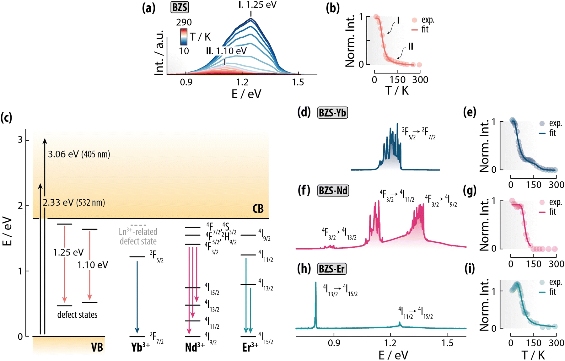 | ||
| Fig. 4 (a) Photoluminescence of BSZ (undoped BaZrS3) as a function of temperature and (b) intensity vs. temperature plot obtained integrating over the whole range shown in (a). (c) Proposed energy level scheme for the doped and undoped samples. For Ln3+ ions, only the energy levels that are in the range <1.8 eV are shown. The emission spectra recorded at 10 K for BZS-Yb, BZS-Nd, and BZS-Er are shown in (d), (f), and (h), alongside the intensity vs. temperature plots obtained for each sample – shown respectively in (e), (g), and (i). Details about the data treatment for obtaining the integrated intensity for (e), (g), and (i) can be found in the ESI.† All the fittings were carried out using eqn (1). All spectra were collected under 405-nm excitation, save for the BZS-Yb sample, which was excited at 532 nm. | ||
Upon doping the material with Ln3+ ions, the NIR photoluminescence changes drastically (Fig. 4d, f and h). To that end, BZS-Yb displays the expected Yb3+:2F5/2 → 2F7/2 emission band, while the intrinsic broad luminescence of the matrix completely disappears. Note that Yb3+ cannot be directly excited at 532 nm, moreover the emission of this ion in BZS-Yb is observed also under 405-nm excitation (Fig. S15, ESI†). These observations confirm the sensitization of Yb3+ emission upon excitation of the semiconductor matrix. Intriguingly, close inspection of the Yb3+ emission band reveals more signals than the expected four Stark components even at 10 K, alongside a thermal quenching trend entailing at least two steps (Fig. 4e). This behaviour indicates that at the very least two populations of Yb3+ ions that are optically non-equivalent are present in the BaZrS3 crystal structure (additional discussion on this topic can be found in the ESI†).
For BZS-Nd, we moved from 532 to 405-nm excitation. This is because Nd3+ can be directly excited with the former wavelength (green) but shows negligible absorption at the latter (violet). As such, the Nd3+ emission lines observed in BZS-Nd under 405-nm excitation are the result of energy transfer from the semiconductor matrix to the dopant ions. The NIR luminescence spectrum displayed by BZS-Nd is more complex than the one of BZS-Yb, featuring both the emission of the Ln3+ ion and broader bands that closely resemble the emission of the undoped material. Each contribution has a distinctive thermal quenching behaviour (Fig. 4g and Fig. S17, ESI†), possibly underscoring the presence of temperature-dependent (back) energy transfer mechanisms between the energy levels of Nd3+ and the ones of the matrix (Fig. 4c).
Lastly, BZS-Er was excited at 405 nm. Note that Er3+ is directly excited at this wavelength, leading to population of the 2H9/2 excited state. Yet, the minimal contribution of the matrix luminescence (Fig. S18, ESI†) to the overall emission of BZS-Er suggest that the part of optical energy that is absorbed by the matrix is effectively funnelled to the dopant Er3+ ions (Fig. 4h). As in the previous Ln3+-doped samples, a two-step thermal quenching of the PL signal is observed (Fig. 4i). This sample is also the only one showing anti-thermal quenching behaviour between 10 and 50 K. This trend is compatible with thermally-induced detrapping of charge carriers from (shallow) trap/defect states followed by capture by deeper emitting states – here belonging to Er3+.45
Therefore, the typical NIR emission bands of each Ln3+ ion were observed under excitation of the host matrix. It is expected that substitutional doping of Zr4+ with Ln3+ ions is accompanied by the formation of additional sulfur vacancies due to charge compensation. For energetic reasons, these vacancies should reside in the proximity of the introduced Ln3+ ions, thus possibly supporting localized energy transfer processes. A similar phenomenon has been observed in Ln3+-doped CsPbCl3 nanocrystals, where 2 Ln3+ replace 3 Pb2+, leaving an unoccupied site (Pb2+ vacancy, VPb); this localized, energetically favorable Ln–VPb–Ln defect complex appears to be fundamental in supporting, for example, efficient quantum cutting in Yb3+-doped CsPbCl3.8 Modelling and additional experimental studies (e.g., lifetime measurements) are required to pinpoint the exact nature of the semiconductor-to-Ln3+ transfer process in BaZrS3.
4. Conclusions
We have herein reported the first example of Ln3+ ion doping into BaZrS3 and matrix-to-Ln3+ near infrared (NIR) sensitized emission. We prepared these phosphors via sulfurization of Ln3+-doped BaZrO3 powders, which were in turn obtained through a microwave-assisted strategy.We initially investigated the optimal sulfurization conditions of BaZrO3 to obtain pure-phase BaZrS3. We observed that different phases can be obtained when varying the sulfurization temperature and determined an optimal range of sulfurization temperatures between 1000 and 1050 °C. The so-obtained BaZrS3 presents good stability both at high temperature and at ambient conditions over time. Moreover, it features a band gap energy of approximately 1.8 eV, which ensures efficient visible light absorption. Not less importantly, BaZrS3 presents low-energy phonon modes. All the above features set the conditions for the preparation of visible-excited, NIR-emitting Ln3+-doped BaZrS3 phosphors.
We therefore prepared BaZrS3 phosphors doped with Nd3+, Er3+, and Yb3+ and investigated their NIR photoluminescence under violet and green light excitation. Characteristic emission lines of Nd3+, Er3+ and Yb3+ ions were observed. The analysis of the photoluminescence spectra acquired as a function of the temperature (10 K to room temperature) on both doped and undoped samples shows a rich spectroscopy of these materials. Specifically, two broad, defect state-related, emission bands were observed alongside the sensitized narrow emission of each doped Ln3+ ion. These results pave the way towards future applications of perovskite sulfides as visible light absorbing phosphors and sensitizers of Ln3+ NIR emission, moving from the cryogenic range towards room temperature.
Data availability
Data for this article, including the original datasets of the X-ray powder diffraction, Raman, reflectance, Tauc plots, and photoluminescence (all in .txt format) are available at Zenodo at https://doi.org/10.5281/zenodo.13937075.Conflicts of interest
There are no conflicts to declare.Acknowledgements
The authors acknowledge the technical assistance of Mr F. Moreno, and the use of SIdI and Segainvex facilities at Universidad Autónoma de Madrid. This research has been funded by Spanish Ministerio de Ciencia, Innovación y Universidades (MICIU) under PID2021-126098OB-I00/AEI/FEDER10.13039/501100011033 grant. R. M. is also grateful to the Spanish MICIU for support to research through a Ramón y Cajal Fellowship (RYC2021-032913-I) and Project PID2022-14210NA-I00 (NAMSTEPS) funded by MICIU/AEI/10.13039/501100011033 and by FEDER, EU. A. B. acknowledges funding from Comunidad de Madrid (Spain) through TALENTO grants ref. 2019-T1/IND-14014 and ref. 2023-5A/IND-28937. A. P. and E. B. acknowledge support by Sapienza University of Rome (progetti medi 2022).References
- R. Marin, G. Brunet and M. Murugesu, Shining new light on multifunctional lanthanide single-molecule magnets, Angew. Chem., Int. Ed., 2021, 60, 1728–1746 CrossRef CAS PubMed.
- A. Satapathy and E. Sinha, Optical band gap and photoluminescence studies of samarium-doped barium zirconate perovskite prepared by solid state reaction route, J. Appl. Spectrosc., 2018, 84, 948–953 CrossRef CAS.
- R. Borja-Urby, L. Diaz-Torres, P. Salas, M. Vega-Gonzalez and C. Angeles-Chavez, Blue and red emission in wide band gap BaZrO3: Yb3+, Tm3+, Mater. Sci. Eng. B, 2010, 174, 169–173 CrossRef CAS.
- R. Marin and D. Jaque, Doping lanthanide ions in colloidal semiconductor nanocrystals for brighter photoluminescence, Chem. Rev., 2021, 121, 1425–1462 CrossRef CAS PubMed.
- W. J. Mir, T. Sheikh, H. Arfin, Z. Xia and A. Nag, Lanthanide doping in metal halide perovskite nanocrystals: spectral shifting, quantum cutting and optoelectronic applications, NPG Asia Mater., 2020, 12, 9 CrossRef CAS.
- A. G. Bispo-Jr, A. J. de Morais, C. M. S. Calado, I. O. Mazali and F. A. Sigoli, Lanthanide-doped luminescent perovskites: A review of synthesis, properties, and applications, J. Lumin., 2022, 252, 119406 CrossRef CAS.
- H. Shao, L. Li, X. Wu, L. Xu, B. Dong, D. Zhou, X. Bai and H. Song, Multicolor emission from lanthanide ions doped lead-free Cs3Sb2Cl9 perovskite nanocrystal, J. Rare Earths, 2024, 42, 940–946 CrossRef CAS.
- T. J. Milstein, D. M. Kroupa and D. R. Gamelin, Picosecond Quantum Cutting Generates Photoluminescence Quantum Yields Over 100% in Ytterbium-Doped CsPbCl3 Nanocrystals, Nano Lett., 2018, 18, 3792–3799 CrossRef CAS PubMed.
- Y. Liang, J. Li, Z. Chen, G. Li, M. Li, M. Jia, X. Chen, X. Li, Y. Han and Z. Shi, Tapping the Light Emitting Potential of Chalcogenide Perovskite SrHfS3 via Eu2+ Doping, Adv. Optical Mater., 2024, 12, 2301977 CrossRef CAS.
- Y. Han, J. Fang, Y. Liang, H. Gao, J. Yang, X. Chen, Y. Yuan and Z. Shi, Preparation of chalcogenide perovskite SrHfS3 and luminescent SrHfS3:Eu2+ thin films, Appl. Phys. Lett., 2024, 124, 131902 CrossRef CAS.
- D. Han, K. Shinoda and T. Uda, Dopant Site Occupancy and Chemical Expansion in Rare Earth-Doped Barium Zirconate, J. Am. Ceram. Soc., 2014, 97, 643–650 CrossRef CAS.
- R. Borja-Urby, L. Diaz-Torres, P. Salas, C. Angeles-Chavez and O. Meza, Strong broad green UV-excited photoluminescence in rare earth (RE = Ce, Eu, Dy, Er, Yb) doped barium zirconate, Mater. Sci. Eng. B, 2011, 30, 1388–1392 CrossRef.
- X. Liu and X. Wang, Preparation and luminescence properties of BaZrO3: Eu phosphor powders, Opt. Mater., 2007, 30, 626–629 CrossRef CAS.
- R. Borja-Urby, L. A. Diaz-Torres, I. Garcia-Martinez, D. Bahena-Uribe, G. Casillas, A. Ponce and M. Jose-Yacaman, Crystalline and narrow band gap semiconductor BaZrO3: Bi-Si synthesized by microwave-hydrothermal synthesis, Catal. Today, 2015, 250, 95–101 CrossRef CAS.
- I. Leonidov, V. Tsidilkovski, E. Tropin, M. Vlasov and L. Putilov, Acceptor doping, hydration and band-gap engineering of BaZrO3, Mater. Lett., 2018, 212, 336–338 CrossRef CAS.
- J. H. Al Shuhaib, J. F. Fernandez, J. Bodega, J. R. Ares, I. J. Ferrer and F. Leardini, Synthesis, optical band gap and thermoelectric properties of Sr1+xTiS3−y chalcogenide perovskites, Mater. Res. Bull., 2023, 167, 112405 CrossRef.
- C. Comparotto, A. Davydova, T. Ericson, L. Riekehr, M. V. Moro, T. Kubart and J. Scragg, Chalcogenide Perovskite BaZrS3: Thin Film Growth by Sputtering and Rapid Thermal Processing, ACS Appl. Energy Mater., 2020, 3, 2762–2770 CrossRef CAS.
- J. Xu, Y. Fan, W. Tian, L. Ye, Y. Zhang, Y. Tian, Y. Han and Z. Shi, Enhancing the optical absorption of chalcogenide perovskite BaZrS3 by optimizing the synthesis and post-processing conditions, J. Solid State Chem., 2022, 307, 122872 CrossRef CAS.
- A. Clearfield, The Synthesis and Crystal Structures of some Alkaline Earth Titanium and Zirconium Sulfides, Acta Cryst., 1961, 16, 135–142 CrossRef.
- S. Niu, H. Zhao, Y. Zhou, H. Huyan, B. Zhao, J. Wu, S. B. Cronin, H. Wang and J. Ravichandran, Mid-wave and Long-Wave Infrared Linear Dichroism in a Hexagonal Perovskite Chalcogenide, Chem. Mater., 2018, 30, 4897–4901 CrossRef CAS.
- X. Wei, et al., Realization of BaZrS3 chalcogenide perovskite thin films for optoelectronics, Nano Energy, 2020, 68, 104317 CrossRef CAS.
- T. S. Bjørheim, M. Arrigoni, S. W. Saeed, E. Kotomin and J. Maier, Surface Segregation Entropy of Protons and Oxygen Vacancies in BaZrO3, Chem. Mater., 2016, 28, 1363–1368 CrossRef.
- M. Saeki, Y. Yajima and M. Onoda, Preparation and Crystal Structures of New Barium Sulfides, J. Solid State Chem., 1991, 92, 286–294 CrossRef CAS.
- C. Toulouse, et al., Lattice dynamics and Raman spectrum of BaZrO3 single crystals, Phys. Rev. B, 2019, 100, 134102 CrossRef CAS.
- D.-H. Gim, Y. Sur, Y. H. Lee, J. H. Lee, S. Moon, Y. S. Oh and K. H. Kim, Pressure-Dependent Structure of BaZrO3 Crystals as Determined by Raman Spectroscopy, Materials, 2022, 15, 4286 CrossRef CAS.
- S. Perera, et al., Chalcogenide perovskites – an emerging class of ionic semiconductors, Nano Energy, 2016, 22, 129–135 CrossRef CAS.
- N. Gross, Y.-Y. Sun, S. Perera, H. Hui, X. Wei, S. Zhang, H. Zeng and B. A. Weinstein, Stability and Band-Gap Tuning of the Chalcogenide Perovskite BaZrS3 in Raman and Optical Investigations at High Pressures, Phys. Rev. Appl., 2017, 8, 044014 CrossRef.
- J. Pandey, D. Ghoshal, D. Dey, T. Gupta, A. Taraphder, N. Koratkar and A. Soni, Local ferroelectric polarization in antiferroelectric chalcogenide perovskite BaZrS3 thin films, Phys. Rev. B, 2020, 102, 205308 CrossRef CAS.
- S. P. Ramanandan, et al., Understanding the growth mechanism of BaZrS3 chalcogenide perovskite thin films from sulfurized oxide precursors, J. Phys. Energy, 2023, 5, 014013 CrossRef.
- G. Chen, C. Yang and P. N. Prasad, Nanophotonics and Nanochemistry: Controlling the Excitation Dynamics for Frequency Up- and Down-Conversion in Lanthanide-Doped Nanoparticles, Acc. Chem. Res., 2013, 46, 1474–1486 CrossRef CAS PubMed.
- P. Loiko, et al., Emission properties of Tm3+-doped CaF2, KY3F10, LiYF4, LiLuF4 and BaY2F8 crystals at 1.5 μm and 2.3 μm, J. Lumin., 2020, 225, 117279 CrossRef CAS.
- R. Shi and A.-V. Mudring, Phonon-Mediated Nonradiative Relaxation in Ln3 + -Doped Luminescent Nanocrystals, ACS Mater. Lett., 2022, 4, 1882–1903 CrossRef CAS.
- M. Szalkowski, M. Dudek, Z. Korczak, C. Lee, L. Marciniak, E. M. Chan, P. J. Schuck and A. Bednarkiewicz, Predicting the impact of temperature dependent multi-phonon relaxation processes on the photon avalanche behavior in Tm3 +: NaYF4 nanoparticles, Optical Mater.: X, 2021, 12, 100102 CAS.
- G. Kortum, W. Braun, D. Chem and G. Herzog, Principles and Techniques of Diffuse-Reflectance Spectroscopy, Angew. Chem., Int. Ed. Engl., 1963, 2, 333–341 CrossRef.
- S. Niu, et al., Bandgap Control via Structural and Chemical Tuning of Transition Metal Perovskite Chalcogenides, Adv. Mater., 2017, 29, 1604733 CrossRef PubMed.
- Y. Nishigaki, et al., Extraordinary Strong Band-Edge Absorption in Distorted Chalcogenide Perovskites, Sol. RRL, 2020, 4, 1900555 CrossRef CAS.
- S. Niu, J. Milam-Guerrero, Y. Zhou, K. Ye, B. Zhao, B. C. Melot and J. Ravichandran, Thermal stability study of transition metal perovskite sulfides, J. Mater. Res., 2018, 33, 4135–4143 CrossRef CAS.
- M. L. Moreira, J. Andrés, J. A. Varela and E. Longo, Synthesis of Fine Micro-sized BaZrO3 Powders Based on a Decaoctahedron Shape by the Microwave-Assisted Hydrothermal Method, Cryst. Growth Des., 2009, 9, 833–839 CrossRef CAS.
- P. G. Sundell, M. E. Björketun and G. Wahnström, Thermodynamics of doping and vacancy formation in BaZrO3 perovskite oxide from density functional calculations, Phys. Rev. B, 2006, 73, 104112 CrossRef.
- I. Ahmed, S.-G. Eriksson, E. Ahlberg, C. S. Knee, H. Götlind, L.-G. Johansson, M. Karlsson, A. Matic and L. Börjesson, Structural study and proton conductivity in Yb-doped BaZrO3, Solid State Ion., 2007, 178, 515–520 CrossRef CAS.
- R. D. Shannon, Revised Effective Ionic Radii and Systematic Studies of Interatomic Distances in Halides and Chalcogenide, Acta Cryst., 1976, A32, 751–767 CrossRef CAS.
- J. A. Márquez, et al., BaZrS3 Chalcogenide Perovskite Thin Films by H2S Sulfurization of Oxide Precursors, J. Phys. Chem. Lett., 2021, 12(8), 2148–2153 CrossRef PubMed.
- R. W. Gurney and N. F. Mott, Luminescence in solids, Trans. Faraday Soc., 1939, 35, 69–73 RSC.
- F. Seitz, An interpretation of crystal luminescence, Trans. Faraday Soc., 1939, 35, 74–85 RSC.
- P. Dang, W. Wang, H. Lian, G. Li and J. Lin, How to Obtain Anti-Thermal-Quenching Inorganic Luminescent Materials for Light-Emitting Diode Applications, Adv. Opt. Mater., 2022, 10, 2102287 CrossRef CAS.
Footnote |
| † Electronic supplementary information (ESI) available. See DOI: https://doi.org/10.1039/d4tc04446k |
| This journal is © The Royal Society of Chemistry 2025 |