
 Open Access Article
 This Open Access Article is licensed under a Creative Commons Attribution-Non Commercial 3.0 Unported Licence
Lab on chip for medical and clinical applications
Bhagyashree
Gupte
a,
Umesh
Jadhav
a,
Suresh
Gosavi
*b and
Shweta
Jagtap
 *c
*c
aDepartment of Microbiology, Savitribai Phule Pune University, Pune 411007, India
bDepartment of Physics, Savitribai Phule Pune University, Pune 411007, India. E-mail: swg@physics.unipune.ac.in
cDepartment of Electronic and Instrumentation Science, Savitribai Phule Pune University, Pune 411007, India. E-mail: shweta.jagtap@gmail.com
First published on 5th September 2025
Abstract
Lab-on-a-chip (LoC) devices represent systems where microfluidics converges with state-of-the-art technologies, playing an immense role in reshaping clinical and biomedical sciences. This review deeply explores the design principles and diverse applications of LoC devices, ranging from point-of-care diagnostics to entire human-on-a-chip devices. Notably, LoC devices showcase remarkable adaptability and versatility. While LoC devices offer many advantages over conventional laboratory assessment methodologies including small sample size, reduced assay time and cost-effectiveness, the field faces many challenges in terms of designing, standardizing and large-scale production of the devices. In the end, while shedding light on how LoC devices stand at the forefront of the innovative technologies in the field of clinical and biomedical sciences, the review also emphasizes on their applications and integration with state-of-the-art technologies like AI and machine learning, along with their limitations and the further necessary developments for their widespread acceptance.
1. Introduction
1.1 Overview of lab-on-a-chip technology
Lab-on-a-chip (LoC) technology represents a pioneering amalgamation of fluidics, electronics, optics, and biosensors that performs various laboratory functions at a miniaturizing scale on a single chip that ranges from millimeters to a few square centimeters processing small volumes of fluids typically ranging from 100 nL to 10 μL.1–3 By consolidating multiple laboratory processes such as sampling, sample pretreatment, chemical reactions, product separation and isolation, detection, and data analysis onto a single chip, LoC systems minimize reliance on bulky instrumentation and extensive manual intervention, thereby enhancing automation and operational efficiency.2,4 Although laboratory techniques are very precise, due to the time-consuming analyses, trained technician demand, and large sample and reagent volume requirements leading to cost escalation, the compact LoC has gained importance in recent years.5,6 Their miniaturized design reduces reagent and sample volumes, lowering costs while enabling high-throughput analysis. The primary advantage of these systems stems from their compactness, which offers portability, minimal consumption of samples, reagents, and power, and significantly shorter assay times.2,4 As mentioned in Fig. 1 the concept of LoC technology developed in a sequential manner. The development of LoC technology dates back to the 1970s, when Terry et al. introduced a miniaturized gas chromatography analyzer on a silicon wafer.7,8 The concept gained prominent recognition since the conceptual work on miniaturized total chemical analysis systems (μTAS) by Manz et al. in 1990 while a groundbreaking advancement was accomplished by Harrison and Manz in 1993 with their pioneering discovery of on-chip capillary electrophoresis; since then the field of LoC technology has experienced intensive development in many biotechnological areas spanning from basic theoretical models and academic proof-of-concept studies to commercial applications over the subsequent decades.2,9–11 In 1998, Whitesides and colleagues introduced soft lithography for microfluidics using PDMS, which significantly advanced the field by enabling high-fidelity replication of microscale features, optical transparency down to 280 nm, and rapid fabrication with low curing temperature and time.12 In the early 2000s, droplet microfluidics emerged as a promising technique for studying droplets ranging in size from a few microns to several hundred microns.13 In 2004, Shuler and co-workers applied micromachining techniques to create multi-compartment cell culture systems, where fluid flow between tissue compartments was regulated by microchannels; this “cell-on-a-chip” approach laid the foundation for organ-on-a-chip.14 The organ-on-a-chip concept began to take form in 2005, when Huh and his team developed a foundational lung-on-a-chip design using PDMS channels above and below a porous membrane to mimic the human lung environment.15 In 2007, the Whitesides group introduced paper-based microfluidics (μPADs), which use capillary action to move liquids, offering low-cost and portable diagnostic devices. Since then, μPADs have become a transformative tool for point-of-care diagnostics, food safety, environmental monitoring, and toxicology due to their affordability, portability, and ease of use.16 In 2012, Kaigala and colleagues presented open-space microfluidics, a new approach that eliminates sealed channels and enables localized chemistry and analysis directly on biological surfaces.17 In 2015, droplet-based microfluidic technologies such as Drop-seq and inDrop were introduced, facilitating high-throughput single-cell RNA sequencing by encapsulating single cells with barcoded beads in nanoliter droplets.18 Finally, in December 2022, the FDA Modernization Act 2.0 approved the use of alternative non-animal testing methods, including organ-on-a-chip, for generating drug safety and efficacy data.19 | ||
| Fig. 1 Schematic illustration of key milestones in LoC technology. | ||
Although the early focus of the development of LoC was to develop devices with applications such as DNA analysis, protein separation, and cell sorting, demonstrating the potential of miniaturized laboratory processes, in recent years their application has broadened to include personalized medicine, real-time monitoring of biological processes, and high-throughput screening for drug discovery.4 Furthermore, in recent years LoC has advanced through integration of biosensing technologies which has enabled the convergence of chemical and biological components into a single platform. It has expanded capabilities to include portability, disposability, real-time detection, unprecedented accuracies, and simultaneous analysis of different analytes in a single device.20 The integration of artificial intelligence (AI) and machine learning (ML) with LoC systems has enhanced diagnostic accuracy and reliability enabling predictive analytics for disease outbreaks, treatment responses, and complications, while automating workflows from sample handling to data interpretation to reduce human intervention and error.4
This tutorial review provides a comprehensive overview of LoC technology—from its developmental origins and engineering principles to its current applications in biomedical research and diagnostics—while highlighting the transformative role of AI integration in expanding performance, accessibility, and clinical relevance. By tracing the trajectory of LoC innovation and discussing future directions, we aim to offer both newcomers and experienced practitioners a consolidated understanding of the field's technological, analytical, and translational landscape.
2. Fundamentals of lab-on-a-technology
2.1 Basics of microfluidics
As briefly described above, LoC systems operate based on microfluidics technology, which is based on the science and engineering of manipulating and processing small volumes of fluids in microchannels measuring between 1 and 1000 micrometers.2,3,21,22 Fluid flow behaves significantly differently at micro and macro scale levels. Micro scale flows are typically smooth and are commonly referred to as laminar flow. Parameters that are often ignored in macro flows, such as surface force, shear force, diffusion, air dampening, and viscosity, become crucial in micro scale flows.6,21,23In microfluidics, gravitational forces take a backseat whereas surface tension and capillary forces play a dominant role. Microfluidic devices are governed by laminar flow, with surface areas becoming more prominent than volume. Transporting fluid into microchannels involves various methods, with pressure-driven flow and electro-osmotic flow standing out as two crucial approaches.6 To translate these principles into practical devices, microfluidics relies on a set of integrated components, each fulfilling a distinct role.
3. Microfluidic platforms and components
Fluidic platforms and components of LoC devices are the basic building blocks of these devices.24 Within a well-defined and cost-effective fabrication technology, the microfluidic platform offers a range of fluidic unit operations that are designed for easy combination. Such unit operations include transport, metering, valving, mixing, and separation as well as amplification of particles. Additionally, these operations involve reagent storage and incubation. These platforms enable the implementation of various application-specific systems including miniaturization, integration, automation, and parallelization of biochemical assays in a convenient and adaptable manner, based on the same fabrication technology.25,263.1 Microfluidic platforms
The LoC devices are intended to perform very specific functions, hence while selecting materials for the microfluidic platform special attention should be paid to device application, required function, and the degree of integration. The material selection not only influences the intrinsic properties of the devices and the methods of fabrication, but also influences the choice of components.2,6,24 While selecting these materials in addition to cost, other factors like flexibility, air permeability, electric conductivity, solvent compatibility, optical transparency, and biocompatibility of the material should be carefully considered.2 Common materials for microfluidic platforms include the following (Table 1).| Material | Pros | Cons | Diagnostic application |
|---|---|---|---|
| Silicon | Well-characterized for surface modifications via silanol (–Si) groups. Chemically inert and offers high design flexibility31 | High production cost and optical opacity in the visible range, limiting imaging and fluorescence-based detection in biological assays27,32 | Utilized in nucleic acid detection through integrated PCR and hybridization microarrays34 |
| High elasticity complicates fabrication of reliable valves and pumping components32 | Applied in organ-on-chip platforms for drug toxicity assessment31 | ||
| Being electrically conductive, it may interfere with electro-osmotic pumping33 | |||
| Glass | Exhibits low nonspecific adsorption and background fluorescence. Highly compatible with biological samples. Cost-effective, user-friendly, thermally stable (>1400 °C), chemically resistant, and optically transparent27,32 | Requires high bonding temperatures and voltages, which present challenges during device manufacturing33 | Applied in point-of-care diagnostics, cell-based assays, nucleic acid analysis, drug delivery studies, and immunoassay/molecular biology platforms32 |
| Polymer (PDMS) | Non-toxic, biocompatible, optically transparent, gas-permeable, flexible, and lightweight. Easy room-temperature bonding with low fabrication cost. Enables cell culture on microchannel surfaces and allows fabrication of intricate structures at both micro- and macro-scales27,35,36 | Hydrophobic nature, absorption of hydrophobic analytes, scalability issues in mass production, and attenuation of acoustic waves27 | Widely used in organ-on-chip models for studying drug interactions, disease mechanisms, and real-time cellular responses. Applied in development of blood flow models36 |
| Not ideal for high-pressure or chemically intensive, long-duration experiments37 | |||
| Epoxy resin | A thermosetting polymer with excellent biocompatibility, mechanical strength, chemical resistance, thermal stability, and toughness. Supports fabrication of microfluidic devices quickly and economically without cleanroom facilities.28 Highly scalable, compatible with soft and photolithography, and reliable for extended experiments37 | Direct 3D printing is challenging due to long curing times38 | Applied in DNA amplification and point-of-care diagnostic chips28 |
| Paper | Intrinsic porosity and microstructure enable capillary-driven flow, making it highly suitable for microfluidics. Mechanical and conductive properties can be tuned via solvent treatments. Natural white color aids in colorimetric assays. Environmentally friendly—easy to recycle and dispose32 | Lacks optical transparency, unsuitable for absorbance spectroscopy, and incompatible with open-channel applications. Sample recovery is difficult due to adsorption onto fibers. Natural variability introduces measurement errors. Enzyme and reagent stability is often poor on paper substrates39 | Employed for detecting infectious and non-infectious diseases, monitoring biomarker levels for health assessment, and identifying antibiotic resistance39 |
Out of the abovementioned polymers, PDMS, a silicon-based elastomer has gained special attention due to its simple fabrication requirements and compatible characteristics such as chemical, physical, and biological resistance. In addition to being cost effective, PDMS possesses some noteworthy properties such as biocompatibility, transparency, high elasticity, low surface tension, gas and water permeability, rapid fabrication, and ease of implementation. Despite the numerous advantages, PDMS does have certain drawbacks like low pressure resistance and a high absorption capacity, necessitating the need for surface modification.2,6,24,27
In addition to the most used materials, various metals, metal-thin films, and hydrogels are trying to pave their path in the realm of microfluidics.6
3.1.5.1 PDMS–COC hybrid platform. Hybrid platforms combining cyclic olefin copolymer (COC) and PDMS leverage the complementary properties of both materials. COC offers optical transparency and biocompatibility essential for imaging, while PDMS enables better gas permeability to perfusion culture. Such COC–PDMS devices have been applied in diverse microfluidic applications including lipidic mesophase screening, cell analysis, microfluidic mixing, and microphysiological systems such as aorta-on-a-chip and liver-on-a-chip models. In recent work, a hybrid COC–PDMS microfluidic platform (HCP) was used to culture Huh7 hepatoma cells in a perfusion environment, demonstrating its suitability for proof-of-concept experiments in liver-on-a-chip development, particularly when using immortalized cell lines for reproducibility.41
3.1.5.2 Paper–polymer hybrid platform. Paper/polymer hybrid microfluidic devices have emerged as low-cost platforms for biological and biomedical applications, combining the ubiquity, recyclability, and 3D porous structure of paper with the durability of polymers. Paper offers inherent capillary-driven flow without external pumps, compatibility with various biological samples, ease of functionalization for biomolecule binding, and visual contrast for colorimetric assays. Despite challenges such as nonspecific adsorption, evaporation, and limitations in optical detection, integrating paper into polymeric regions enhances performance and stability (Table 2).40
| Application | Platform type | Key details | Performance metrics |
|---|---|---|---|
| DNA extraction & PCR integration42 | Paper/photopolymer resin | One-step DNA extraction from diverse biological samples | LOD: 104 copies per mL (HBV nucleic acids), <2 min extraction |
| Filter paper/PDMS/PMMA43 | DNA extraction + PCR amplification from human whole blood | DNA yield: 5.6–21.8 ng (0.25–1 μL blood), <7 min | |
| Chitosan-modified filter paper44 | Enhanced DNA capture efficiency | 98% (K562 human DNA), 95% (bacteriophage DNA), >30× enrichment of phage DNA | |
| Nucleic acid extraction & LAMP integration | Polysulfone membrane/PMMA45 | Plasma separation for nucleic acid amplification | 275 ± 33.5 μL plasma from 1.8 mL blood in 7 min |
| Paper/PDMS40 | Instrument-free diagnosis of bacterial meningitis and whooping cough-causing bacterium, B. pertussis | LOD: 5 copies per LAMP zone (*B. pertussis*), 45 min | |
| Paper/PDMS/glass46 | Multiplexed foodborne pathogen detection46 | LOD: 21.5 (S. aureus) & 20.9 (V. parahaemolyticus) copies per μL; 100-fold PCR sensitivity47 | |
| DNA hybridization40 | Paper/PMMA SpinChip | ssDNA probe-functionalized GO nanosensors for multiplex LAMP | LOD: 6 (N. meningitidis), 12 (S. pneumoniae) DNA copies per assay |
| Protein analysis – LFAs40 | Paper/polymer with electro-wetting valves48 | Colorimetric detection of T7 bacteriophage | LOD: 108 PFU mL−1, 40 min |
| Paper/plastic | Sandwich LFA for dengue NS1 antigen | LOD: 84.66 ng mL−1 | |
| Nitrocellulose/PDMS | Immunoassays without pumps, cavity-induced microstreaming | 18 min assay, smartphone app | |
| Paper/plastic | Smartphone-based urine analyte detection | Glucose, protein, pH, RBC | |
| Protein analysis – ELISA40 | Paper/PMMA (56 microwell) | Rapid biomarker detection via office scanner | Comparable to commercial ELISA, <1 h |
| Paper/plastic | Multiplex detection of protein biomarkers, viruses, nucleic acids | — | |
| PMMA/paper PnP device | High-sensitivity immunoassays, analyte enrichment | 10× sensitivity increase | |
| Whole-cell detection40 | Paper/PDMS/glass | Aptamer-functionalized GO biosensor for foodborne pathogens | LOD: 61 CFU mL−1 & 800 CFU mL−1, 10 min |
| Paper/PDMS | AST of uropathogens, species-specific enzymes, chromogenic medium | 83.3–100% agreement with conventional | |
| 3D cell culture40 | Paper/glass | 3D culture with impedimetric quantification of cancer cells | Up to 3 day non-invasive measurement |
| Paper/PMMA | Wax-printed paper microreactors with diffusion channels | Studied cell proliferation, EGFR phosphorylation | |
| Paper/PDMS/PMMA | 3D culture under chemical gradients | HeLa cell viability & protein expression profiling |
3.1.5.3 Emerging unconventional hybrid platforms in LoC devices. In addition to the commonly used and well-established hybrid platforms, recent innovations have explored unconventional material combinations such as paper/tape, paper/glass microcapillary, and thread-based systems, offering new avenues for low-cost, integrated diagnostics. Paper/tape devices enable simple, reconfigurable 3D structures for rapid nucleic acid extraction, amplification, and detection, as demonstrated by an HPV DNA assay completed in under an hour. Paper/glass microcapillary systems integrate nucleic acid capture, purification, and LAMP detection with minimal user intervention, while thread-based hybrids leverage the wicking properties of cotton or polyester threads to enhance sample delivery and enable sensitive or semi-quantitative assays for biomarkers such as CEA, ferritin, BSA, and nitrite. These emerging platforms, though less conventional, highlight the growing diversity of material strategies in LoC development and their potential to expand accessibility in resource-limited settings.40
In the above section we learnt about the types of microfluidic platforms based on the materials; now let's dive deep into the classification of platforms according to the main liquid propulsion principle. Based on main liquid propulsion principles, the microfluidic platforms are divided into five types namely, capillary flow platform, pressure driven platform, centrifugal platform, electrokinetic platform, and acoustic platform.24–26
| Platform | Characteristics | Applications |
|---|---|---|
| Linear actuated devices | • Liquid flow is controlled by mechanical displacement of liquid by means of a plunger | It is used in POCT to determine blood parameters. Additionally, it's used in a lab-in-a-tube analyzer for amplification-based nucleic acid tests |
| • Liquid control is confined to a linear, one-dimensional liquid flow | ||
| • Usually, liquid calibrants and reaction buffers are prestored in pouches | ||
| Pressure driven laminar flow | • Liquid transport relies on pressure gradient, resulting in hydrodynamically stable laminar flow | Phase transfer magnetophoresis technology operates through pressure driven laminar flow and is used for continuous DNA extraction. Additionally, it finds its application in the integrated detection of bacteria, DNA purification, PCR, and fluorescence readouts |
| • Pressure is applied through diverse internal or external sources like syringes, pumps, gas expansion, pneumatic displacement of membranes, etc. | ||
| • Samples and reagents are introduced onto chip inlets either batch-wise or in a continuous mode | ||
| Microfluidic large-scale integration | • This microfluidic channel circuitry incorporates chip-integrated microvalves, utilizing flexible membranes positioned between a liquid-guiding layer and a pneumatic control-channel layer | These platforms find application in LoC designed for nucleic acid extraction from minimal cell volume. Furthermore, these platforms are utilized in tasks like protein crystallization, immunoassays, automated cell culturing, DNA synthesis |
| • The status of microvalves, whether closed or open, is determined by the pneumatic pressure applied to the control-channels | ||
| • By combining multiple microvalves, more complex units like micropumps, mixers, multiplexers, etc. can be assembled with hundreds of units on one single chip | ||
| Segmented flow microfluidics | • This platform operates based on multiphase fluid flow through microchannels | These platforms find diverse applications including single cell analysis, single organism analysis, DNA analysis, drug screening, protein crystallization, and chemical synthesis |
| • Dispersed liquid phase droplets are immersed in the second continuous phase (usually gas or liquid) forming micro-confinements ranging from a few picolitres to microliters | ||
| • These droplets can be transported through pressure gradient or can be merged, split, sorted, or processed without any dispersion in the microfluidic channels |
The platform comprises three essential components: a plastic disposable rotor, a cartridge, and an analyzer instrument. The rotor is responsible for specimen processing, the cartridge contains preloaded dried reagents, and the analyzer assists actuation and readout.25 The channel arrangement allows modulation of microfluidic flow by adjusting the rotation speed of the axis, which allows controlled movement of the fluids without the need for actively controllable components such as pumps and valves. As the rotating disks are difficult to bring into contact with each other mechanically and active components are difficult to set up, the centrifugal platforms are operated with the help of siphon valves, capillary valves, or hydrophobic barrier valves. These valves are triggered by modulating the rotation speed.24
Centrifugal microfluidic platforms are applicable in diverse fields including integrated plasma separation, DNA extraction, protein-based and nucleic acid-based assays, clinical chemistry assays, chromatography, and protein crystallization. The modular set up and the disposable and easily exchangeable cartridges are the two major advantages of the centrifugal platforms. However, challenges arise from the need for additional actuation or sensing function during rotation, limited flexibility, and the absence of completely portable systems. These drawbacks restrict the widespread use of this platform.26
3.1.9.1 Electrokinetic platform. Use of electrokinetic platforms stands as one of the earliest LoC technologies. They work on the fundamental principle that individual components within a sample will move in an electric field as per their charge-to-mass ratio, thereby defining their respective electrokinetic mobility.24 Fluid propulsion on the electrokinetic platform relies on the movement of the liquid layer right at the interface to the solid phase.25 In addition to the movement of charged particles through externally applied electric fields, small capillaries such as microfluidic channels also exert a secondary effect on the bulk liquid causing lateral movement known as electroosmotic flow (EOF) and it can be used to separate different types of molecules or particles within the bulk liquid.24 Depending on buffers and/or sample involved, several electrokinetic effects such as electroosmosis, electrophoresis, dielectrophoresis, and polarization superimpose each other.26 Silicon or glass is commonly preferred as the material for these platforms. These platforms are typically employed in the analysis of DNA and proteins.25 Among these electrokinetic effects, dielectrophoresis (DEP) has gained particular prominence due to its versatility and unique ability to manipulate particles regardless of their net charge.
Dielectrophoresis (DEP) is a microfluidic technique that separates dielectric particles by applying a non-uniform electric field (NUEF), inducing polarization forces. Unlike electrophoresis, it does not require particles to be charged. Its key advantages include being label-free, fast, accurate, efficient, and easily integrated into microfluidic systems, making it valuable for bio-molecular diagnostics and cell separation. However, challenges remain in achieving higher levels of system integration, optimizing efficiency (especially regarding Joule heating in DC-iDEP), and simplifying the fabrication of complex 3D electrode structures. Despite these drawbacks, DEP is widely applied for tasks such as cell separation, plasma separation, and bacteria isolation.49
These platforms present two primary benefits: pulse-free pumping without any moving part thanks to the electroosmotic actuation of liquid flows, and prevention of dispersion in EOF flow, thus averting the broadening of the sample plugs during chromatographic separation. However, the drawbacks include the necessity for high-performance detection technologies due to reduced volumes and technical challenges arising from the development of pH-gradients over time or during operation.25
3.1.9.2 Electrowetting. The electrowetting effect was first described by Lippmann in 1875. Here, the droplets are immersed in a second immiscible continuous phase, either gas or liquid, as stable microconfinements. These droplets rest on a hydrophobic surface featuring a one- or two-dimensional array of individually addressable electrodes. Application of voltage between the electrode and the liquid droplet induces the increased wettability of a solid surface through polarization and electric fields. This phenomenon, known as “electrowetting-on-dielectric” (EWOD), serves as a tool to control the contact angle of liquids on surfaces. By adjusting voltages between adjacent electrodes, droplets can be generated, transported, split, merged, and processed. These unit operations are freely programmable for each individual droplet allowing real-time control of assay. The electrowetting technique finds application in enzymatic assays crucial for diagnostic purposes and glucose concentration measurement. Additionally, these systems also play a role in automated sample preparation of peptides and proteins intended for matrix-assisted laser desorption/ionization mass spectroscopy.25,26
The foremost advantage of the EWOD technique lies in its significant potential to manipulate numerous single droplets in parallel. Notably, this platform excels in handling nanoliter liquid volumes with high precision and offers flexibility in programming the droplet movement. Despite its low sample and reagent requirements, the development of portable systems has been hindered by the bulky electronic instrumentation requirements to operate the platform. Moreover, variation in the wetting abilities of different patient materials influences droplet transport behavior, resulting in differences in volume or movement speed.26
Recently in a study conducted by Javanifar et al. in 2025, they introduced a green microfiltration approach within a LoC format designed to filter Escherichia coli (E. coli) by integrating positively charged electrospun polyacrylonitrile (PAN) fibers into a microfluidic chip. In this context, the microfiltration technique involves using the charged PAN nanofibers as a functional filter medium to selectively capture or retain E. coli cells as fluid flows through the microstructured channels of the LoC system.51
4. Fabrication
The fabrication of LoC devices typically refers to manufacturing and device assembly prior to its use. There is a plethora of fabrication methods available, and the selection of the fabrication method is influenced by factors like functional needs and platform materials as shown in Table 4.52| Material | Fabrication method |
|---|---|
| Glass | Photolithography, chemical etching, micromachining, powder blasting, water jet cutting |
| Rigid polymer | Micromachining, injection molding, hot embossing, 3D printing |
| Soft polymer (PDMS) | Photolithography, injection molding, hot embossing, 3D printing53 |
| Paper | Wax patterning, alkyl ketene dimer printing, flexographic printing, shaping/cutting |
4.1 Molding
Molding is widely used for producing disposable microfluidic devices. Molding-based fabrication methods are divided into three types, replica molding, injection molding, and hot embossing. Replica molding, which is a soft lithography technique that uses PDMS cast over patterned silicon molds bonded to polymeric or glass substrates, offers low-cost, rapid prototyping, but is limited to aqueous applications due to poor solvent compatibility. Injection molding enables high throughput, reproducible production of thermoplastic devices by injecting molten polymer into heated molds followed by cooling below the glass transition temperature before demolding. While it supports mass production with excellent reproducibility, it is restricted to thermoplastics due to involved high-cost.54Hot embossing, which imprints using heat and pressure, provides accurate replication across various thermoplastics but is time-consuming and demands expensive mold fabrication.54
4.2 3D printing
3D printing is a layer-by-layer manufacturing technology that uses a 3D digital model which can be interpreted and created by the printer. This approach offers rapid prototyping for microfluidics. Among its methods, fused deposition modeling (FDM) is a cost-effective, compact, fast, and easy to operate fabrication method that supports various biocompatible thermoplastics such as ABS, PLA, polycarbonate, polyamide, polystyrene, and nanocomposites. FDM is used to create PDMS casting molds and has been applied in fabricating capillary valves for centrifugal microfluidic discs, reusable reactionware for chemical synthesis, and microfluidic immunosensors. Stereolithography (SL) uses UV or structured light to polymerize resin layer by layer. It enables rapid fabrication of fine features, though biocompatible resins essential for microfluidics. SL has been applied to create micromixers, cell separation chips, gradient generators, microneedles, GelMa cell-encapsulation structures, and active components like pumps and valves. Multi Jet Modeling (MJM) ejects light-cured photosensitive resin droplets offering high accuracy and multi-material capability for drug transport and cell viability studies, multi-material microfluidic valves with elastic membranes, and non-planar PDMS channels, but at high cost. Two-photon polymerization (2PP) uses a near-infrared laser for nanometer-sized feature fabrication supporting applications such as micromixers, parallel cell counters, and microlens arrays inside a glass substrate though it is slow and costly.554.3 Nanofabrication
Nanofabrication enables nanoscale features in microfluidics through top-down and bottom-up approaches. Top-down techniques include extreme ultraviolet lithography (13 nm light) for nanopillars, electron beam lithography for sub-10 nm nanochannels, biosensors, and tissue engineering (high precision but slow and costly), and nanoimprint lithography for low-cost, high-throughput multiscale channels with roll-to-roll potential. Bottom-up methods like anodic aluminum oxidation create uniform porous hexagonal cells (4–200 nm) and ultrathin membranes for cell biology and diagnostics, offering rapid, low-cost production.555. Simulation
Fundamentally, LoC consists of a system of wells and channels etched onto platforms, coupled with nanostructured, biofunctionalized surfaces targeted towards analysis.56 Microfluidic channels are pivotal to LoC systems, and the development of LoC particularly depends on the presence and correct functionality of microfluidic channels and other liquid handling components. Given the high cost involved in the production of LoC systems, it's imperative to validate the design before fabrication to prevent unnecessary repeated fabrication.57 Additionally, understanding the analytical processes during chip operation, along with the chip layout and manufacturing process, is essential to evaluate design trade-offs and constraints. As shown in Fig. 2, Pfeiffer et al. developed a system-level LoC simulator to account the complexities. They combined Kirchoffian network analysis and topological sorting from electrical circuit simulation with the sequential-modular structure of process flowsheet simulation. It employed fast, accurate physiochemical models and allows simulation of complex designs in only seconds.58 By elucidating the functioning of microfluidic systems, simulation enables theoretical optimization of design, potentially saving time and cost. Simulation is particularly valuable when microfluidic channels are expected to serve additional functions beyond transport, offering insight into their anticipated performance before embarking on expensive and time-consuming production. In essence, simulation is an inexpensive means to ensure whether an LoC will exhibit the envisioned behavior.57 | ||
| Fig. 2 The LoC simulation process: a) partial library of LoC units, b) channel topology constructed from the library of unit, c) resistor network representation of the channel topology, and d) directed acyclic graph representation of the channel topology. Reproduced from ref. 58 with permission from Elsevier, copyright 2006. | ||
The myriad of available simulation software programs available in the market with diverse functionalities need a thoughtful approach before embarking on the simulation process. Setting up a simulation involves a thorough assessment of the problem, identifying the need for simulation, understanding the reason behind its application, and defining the expected results. It helps in making decisions about the choice of simulation software to employ. The currently available simulation software options on the market include finite element analysis (FEA) software, COMSOL Multiphysics, Coventor, ANSYS (fluent), CFD-ACE+, and Flow 3D; each software is crafted to fulfill distinct roles.57
6. Components of lab on chip devices
As discussed earlier, the selection of platforms for LoC systems depends on the intended use of the device. The choice of platform influences the selection of functional building blocks of the LoC system.Lab-on-a-chip systems are designed to execute a multitude of standard laboratory functions, which include crude sample handling, sample and reagent mixing and reacting, separation, and subsequently detecting analytes. To implement these functions, different standard functional components can be incorporated onto a single chip. As illustrated in Fig. 3, LoC components are classified into eight major groups as follows namely injector, preparator, transporter, mixer, reactor, separator, detector, controller, and power supply. It also summarizes the function of each component of LoC systems.59
 | ||
| Fig. 3 Overview of LoC components and their functions.59 | ||
As highlighted by Lim et al. (2010), following the integration of the core functional components in a LoC system, particular attention must be given to the detector, since it ultimately defines the device's analytical performance. The selection of an appropriate detection mechanism influences not only sensitivity and specificity but also the overall design, integration approach, and applicability of the system, warranting a detailed comparison of the main available methods.59
Working principles of detection mechanisms
LoC detectors operate by employing transducers that acquire a physical signal from the analyte and convert it into an electrical signal for analysis. Optical detection, which is the most prevalent detection method in LoC, uses light–matter interactions, such as fluorescence emission from dye-labeled targets, absorption changes in a label-free environment, or refractive index shifts in micro-ring resonators.60 On the other hand, electrochemical biosensors transduce biological element-target detection events into detectable electrochemical signals. In biosensing measurements, the inherent electrochemical properties of the biological system are used to find access to valuable information.61 Mass spectrometry identifies compounds by determining the mass-to-charge ratio of ionized analytes and is widely used in proteomics analysis.60Table 5 explains the advantages and limitations of two primarily used detection mechanisms i.e., optical detection and electrochemical detection, in LoC systems.| Detection mechanism | Optical detection60 | Electrochemical detection61 |
|---|---|---|
| Advantages | High sensitivity and specificity, often capable of single-cell analysis | High sensitivity even in small sample volumes due to high surface-to-volume ratio of miniaturized electrodes |
| Wide range of label-free and labeled methods (e.g., fluorescence, SPR, interferometry) allowing flexible assay design | Portable, low-cost instrumentation possible (e.g., screen-printed electrodes) | |
| Enables real-time detection. Low cost and small form factors are the other advantages | Suitable for on-site applications | |
| Limitations | Demand for complex laser and optical systems makes miniaturization and integration challenging | Performance strongly dependent on electrode material, type of modification, and geometry |
| Optical interference (e.g., scattering, absorption) from complex samples can reduce performance | ||
| Applications | Biomolecular detection, medical diagnostics, food and environmental monitoring, early detection of cancer | Glucose monitoring (enzymatic and non-enzymatic sensors) |
| Detection of pathogens and nucleic acids via electrochemical immunosensors and aptasensors |
7. Advantages and disadvantages of lab on chip technology
While LoC offers a spectrum of advantages over the conventional laboratory techniques, it has faced a myriad of challenges over the developmental years. The challenges include issues such as surface roughness, the influence of capillary forces, chemical interaction between materials, and the necessity for temperature monitoring and control, which subsequently result in experimental complications.6,62 Furthermore, additional challenges associated with LoC include the application of devices on a large scale, working with substantial volumes, fabrication processes at reduced cost, enhancing the user-friendliness of the devices and their integration with analytical techniques.637.1 Challenges beyond technological innovation
Bringing the microfluidic LoC devices into a hospital setting or to commercialization presents a complex set of challenges that extend beyond technological innovations. Before the devices enter the market, they face a multitude of hurdles including regulatory issues, commercialization and clinical adoption. Moreover, seamless integration into the existing clinical laboratory infrastructure demands consideration of pre- and post-analytical workflows—key elements of the total testing process (TTP) that ensure accuracy and reliability.648. Applications of lab on chip technology
The advent of LoC technology represents a paradigm shift in medical diagnostics, enabling the examination of physiological samples at point-of-care facilities or in remote and resource-poor locations instead of the conventional method of sending them to a laboratory resulting in significant reduction in diagnosis time. Consequently, it benefits the patients in early detection and receiving timely treatment, thereby improving the overall healthcare outcomes.56 Moreover, LoC technology finds extensive applications, primarily focusing on areas such as genomics, biochemical analysis, proteomics and cell research, biosensors, biomedical sciences, and drug development.6,73 The following section covers a few applications of LoC in detail. Fig. 4 indicates the design of lab on chip systems for multifaceted applications. The following section covers a few applications of LoC in detail. | ||
| Fig. 4 Designing lab on chip systems for multifaceted applications. | ||
8.1 Point of care testing
Point of care testing (POCT) refers to a clinical laboratory testing carried out near the patient's location without the need for a permanent, dedicated facility and can be performed outside the clinical laboratories. In contrast to the multistep lab testing, POCT has only 3 primary steps: pre-clinical, analytical, and post-analytical (Fig. 5). resulting in notable reduction in test TAT.6 The reduction in TAT not only circumvents the delay in receiving treatment, but also plays a crucial role in reducing mortality, morbidity, and the quality of life. Additionally, the other advantages of PCOT include – 1) simplified operation, 2) elimination of the need for trained staff, 3) cost-effectiveness, and 4) easy bulk fabrication.74–78 In recent years, POCT has gained increasing attention especially in limited resource areas which has led to the development of various methods, devices, and biosensors that can fulfill the need for POC diagnostic tools.79 Additionally, COVID-19 has fueled the increase in the use of POCT to help control the pandemic.76 | ||
| Fig. 5 Steps involved in point-of-care testing.76 | ||
POCT encompasses a diverse array of testing modalities customized to specific applications. POCT diagnostics are predominantly based on two prevalent technologies, lateral flow assay (LFA) technology and nucleic acid amplification. Lateral flow assay technology can be further divided into nucleic acid-based assays and immune-based assays. Immune-based assays employ antibodies to detect antigens, proteins, and hormones, while nucleic acid-based assays utilize DNA/RNA oligonucleotides or aptamers as biorecognition elements.78
LFA usually consists of four components namely, a sample pad, conjugate pad, detection zone and absorption pad (Fig. 6).78 The diagram below illustrates the typical LFA strip.
 | ||
| Fig. 6 Schematic representation of the steps involved in LFAT. Reproduced from ref. 78 with permission from Frontiers in Bioengineering and Biotechnology, copyright 2021. | ||
LFAs find application in diagnosis and prognosis of diseases like cancer, rapid detection tests as well as in the diagnosis of certain bloodborne diseases. While LFA is a widely used technique to detect a variety of pathogens and proteins, due to its poor detection techniques, nucleic acid amplification tests (NAATs) have gained popularity in the POCT area. NAATs involve three steps: sample preparation, amplification, and detection (Fig. 7). NAATs are applied in the detection of various infectious diseases like Mycoplasmas pneumonia, Bordetella pertussis, and Legionella Pneumonia, various strains of Influenza virus, SARS-Cov-2, etc. The technique allows rapid detection of C. difficile, Strep B, and Streptococcus A in approximately 2 minutes. Moreover, it is also used to determine the viral load in HIV positive patients, and it aids in the diagnosis of STDs like chlamydia and gonorrhea within 30 minutes.78
 | ||
| Fig. 7 Schematic representation of the steps involved in NAAT. Reproduced from ref. 80 with permission from Journal of Biomechanics, copyright 2021. | ||
8.2 Organ on a chip
Another innovation of microfluidics is organ-on-a-chip (OoC). OoCs are systems containing engineered or natural miniature tissues grown inside microfluidic chips. OoCs are designed to provide a suitably in vivo-like environment to guide a collection of cells to assemble into a 3D tissue capable of replicating one or more organ-level functions or to culture organotypic tissue to retain function.81 Organ on chip platforms hold significant promise as alternatives to animal models or traditional cell cultures, both of which poorly recapitulate human pathophysiology and human level responses.82Organ-on-a-chip (OoC) technology offers distinct advantages over conventional 2D/3D cell culture and animal models by more accurately replicating human-relevant physiology and disease states.83 It offers high physiological relevance by closely mimicking human organ structure, mechanics, and biochemistry, while providing a dynamic microenvironment with controlled fluid flow to simulate processes like blood circulation. It supports integration of multiple cell types for complex studies, enables real-time, high-resolution monitoring, and improves the accuracy of drug efficacy and safety predictions. By reducing reliance on animal models, it eliminates interspecies variability and ethical concerns. Its customizable, scalable design suits disease-specific and high-throughput applications, and it delivers human-relevant pharmacokinetic and pharmacodynamic data in hours to days instead of weeks to months.81 OoC devices are mainly divided into four categories namely single-organ chips, multi-organ chips, solid organ chips and barrier organ chips. Table 6 explains the examples of organ on chips falling under each category and their applications.
| Type of OoC | Examples | Applications |
|---|---|---|
| Single-organ chips | Gut-on-a-chip, liver-on-a-chip, lung-on-a-chip, skin-on-a-chip, brain/BBB-on-a-chip, heart-on-a-chip | Drug absorption studies, disease modeling, metabolism and toxicity testing, barrier function studies, neurovascular interaction, contractility and electrophysiology analysis |
| Multi-organ chips (body-on-achip) | Liver–kidney chip, liver–gut–skin–lung chip, cancer multi-OoC systems | Studying systemic drug metabolism, multi-organ toxicity, pharmacokinetics, tumour efficacy/toxicity evaluation |
| Solid organ chips | Liver chip, tumour chip, pancreas chip, bone chip, cartilage chip | Modeling parenchymal/mesenchymal tissues, drug metabolism, cancer biology, bone and cartilage physiology |
| Barrier tissue chips | Vascular endothelium chip, gut epithelium chip, corneal epithelium chip, skin epithelium chip | Studying selective transport, barrier integrity, infection models, permeability assays |
In a recent study conducted by Larson et al., the liver-chip model offered several advantages over conventional methods for assessing the hepatotoxicity of cannabinoids, such as cannabidiol (CBD), cannabinol (CBN), cannabichromene (CBC), and cannabigerol (CBG). This model offers a human-relevant alternative to animal models, accurately reproducing known toxicities such as acetaminophen-induced hepatotoxicity and differentiating the toxicity profiles of various cannabinoids. It enables detailed mechanistic insights by revealing distinct modes of action—such as reduced mitochondrial function, oxidative stress, inflammation, and cellular toxicity—and highlighting varying effects on reactive oxygen species and mitochondrial function in hepatocytes and non-parenchymal cells. Serving as a comprehensive evaluation tool, it supports live toxicity screening and allows assessment of cellular morphology alongside effluent-based biomarkers like albumin, LDH, ALT, AST, and cytokines, facilitating a deeper understanding of cannabinoid-induced liver injury.84
8.3 Advancement of lab-on-a-chip technology in cancer research and diagnostics
Cancer ranks among the primary contributors to global mortality, and the high mortality rates are likely linked to delayed diagnosis, ultimately resulting in delayed treatment. While methods such as tissue biopsy and various imaging techniques exist to assess tumor heterogeneity, their inherent limitations constrain their long-term use, particularly in patients who develop resistance to the treatment.85The customizable nature of LoC has been demonstrated as a groundbreaking technology, paving the way for various opportunities in cancer research. It has significantly contributed to the development of preclinical cancer models, identification of cancer biomarkers, screening of anti-cancer drugs, investigating tumor heterogeneity and producing nano-drugs.86
 | ||
| Fig. 8 Schematic representation of tumor-on-a-chip platforms developed based on photolithotrophy and 3D bioprinting. Reproduced from ref. 88 with permission from Microsystems & Nanoengineering, copyright 2021. | ||
The section below details a few single-organ and multiorgan TOCs.
Ayuso et al. pioneered the development of single organ in vitro models based on LoC technology, effectively replicating ductal carcinoma in situ (DCIS) (Fig. 9a). The design facilitated the generation of a microenvironment characterized by hypoxia and nutrient deprivation, enabling the selective targeting of hypoxic DCIS cells.87,91 Another example is where Hassell et al. established in vitro human orthotopic models of non-small-cell lung cancer (NSCLC) (Fig. 9b). These models simulate organ microenvironment-specific cancer growth, tumor dormancy, and responses to tyrosine kinase inhibitor (TKI) therapy. By leveraging the mechanical actuation features of this technology, the researchers discovered an unknown sensitivity of lung cancer cell growth, invasion, and TKI therapeutic responses to physical indicators associated with breathing motions. These effects seemed to be mediated by changes in signaling through epidermal growth factor receptor (EGFR) and MET protein kinase. These findings may shed light on observed resistance to therapy in cancer patients with minimal residual disease in aerated and mobile lung regions. Additionally, this model serves as an experimental platform for investigating cancer persister cells and mechanisms of tumor dormancy in vitro.90 Shen et al. introduced HCC-on-a-chip (Fig. 9c), a model designed to replicate cancer. The hepatic stellate cells are one of the key components in the HCC microenvironments and play an important role in tumor progression and drug resistance. Blood vessels are essential for nutrition supply and metabolite elimination. HCC-on-a-chip is a three-channel microfluidic device developed to culture three distinct cell types in a spatially organized manner to mimic HCC microenvironments. This approach allows us to study the roles of diverse cells in remodeling the TME by altering the secretome, proteome and metabolome. Critical events including tumor cell proliferation, endothelial cell invasion, tumor cell drug resistance, NK cell infiltration and exhaustion were assessed in the HCC-on-a-chip.92
 | ||
| Fig. 9 Schemic representation of single-organ and multiorgan tumor-on-chip. a) Scheme of the microfluidic model for DCIS. Reproduced from ref. 91 with permission from EBioMedicine, copyright 2018. b) Scheme for tumor lung chip for NSCLC. Reproduced from ref. 90 with permission from Cell Reports, copyright 2017. c) Scheme for tumor liver chip for HCC. Reproduced from ref. 92 with permission from Acta Pharmaceutica Sinica B, copyright 2023. d) Scheme for multiorgan tumor on chip for lung cell metastases. Reproduced from ref. 88 with permission from Microsystems & Nanoengineering, copyright 2021. | ||
In the above section we have discussed about tumor-on-a-chip developed to mimic primary tumors. Moreover, this section discusses multi-organ tumor-on-a-chip models developed to study metastatic cancer. On these chips, different organoids are separated by specific biomaterials like PDMS, and they relate to each other by channels and controllable fluids. Xu et al. designed and constructed a multi-organ microfluidic chip to mimic lung cancer metastasis to the brain, bone, and liver. The system consists of four organs: one upstream lung and three downstream parallel brain, bone, and liver organs (Fig. 9d).86,88
8.4 Cancer diagnostics
Cancer can be detected by LoC devices using various biomarkers such as circulating tumor cells (CTCs), circulating tumor DNA (ctDNA), exosomes, non-coding RNA (ncRNA) and various cellular metabolites or proteins.86Deng et al. designed and developed an integrated microfluidic system to simplify the separation, purification, and single-cell secretory omics analysis of whole blood CTCs (Fig. 10a). This system can process 1 ml of whole blood samples in less than 2 hours with a separation efficiency of more than 70%. The platform can also classify CTCs into specific phenotypes based on surface markers and conduct single-cell secretory omics analysis on these subsets.93 Pahattuge et al. created a smart chip that combines modules for CTC sorting, cell counting, and immunofluorescence imaging. This mart chip offers fully automatic operation facilitating seamless separation and detection of CTCs in blood while eliminating the need for human interference (Fig. 10b).94 In chip developed by Cho H. et al. gold nanoparticles were modified with antibodies and Raman signal molecules to label CTCs (Fig. 10c). The CTCs captured on the chip can be characterized and detected in situ by surface-enhanced Raman technology. This method has high sensitivity and can distinguish common CTCs and circulating tumor stem cells (CTSCs) according to the difference in Raman signal peaks.95
 | ||
| Fig. 10 Schematic representation of on-chip CTC detection. a) Chip designed to simplify the separation, purification, and single-cell secretory omics analysis of whole blood CTCs. b) Chip designed to combine modules for CTC sorting, cell counting, and immunofluorescence imaging. c) A chip with modified gold nanoparticles with antibodies and Raman signal molecules to label CTCs. Reproduced from ref. 94 with permission from ACS Sensors, copyright 2021. | ||
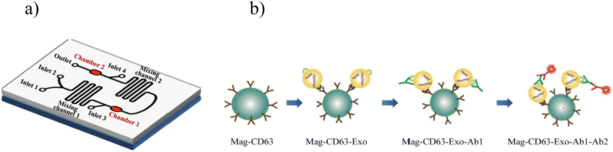 | ||
| Fig. 11 On-chip exosome detection. a) Schematic representation of the microfluidic chip. b) Workflow for the immunogenetic capture and detection of exosomes. Reproduced from ref. 96 with permission from PLoS One, copyright 2017. | ||
To better illustrate the similarities and distinctions between these two LoC-based approaches, a comparative summary of CTC and exosome detection is presented in Table 7.
| Parameter | Circulating tumor cells (CTCs) | Exosomes |
|---|---|---|
| Target | Whole intact tumor cells97 | Extracellular nanovesicles carrying a wide range of biomolecules including DNA, RNA, proteins, and lipids97,98 |
| Size | ∼15–25 μm (ref. 99) | ∼50–100 nm in diameter98 |
| Abundance | Rare97 | High abundance98 |
| Separation methods | Size and deformity based, affinity-based (EpCAM)97 | Viscoelastic flow sorting, acoustic nanofiltration, membrane-based filtration, immunoaffinity, trapping on nanowires, and deterministic lateral displacement (DLD)98 |
| Detection technique | Fluorescence-based, flow cytometry based, optical biosensing techniques, photoacoustic cytometry, imaging cytometry60 | Immunomagnetic beads, electrochemical, 3D self-assembled herringbone nanopatterns, fluorescence of photonic crystals using a filtration system, fluorescence, alternating current electrokinetic (ACE) & fluorescence, microfluidics integrated with surface-enhanced Raman spectroscopy (SERS) and electrohydrodynamic force with SERS using nanomixing fluid flow98 |
| Platform materials | PDMS, nanomaterials such as gold nanoparticles and graphene oxide nanosheets100 | PDMS, paper-based platform98 |
| Clinical applications | Cancer prognosis, diagnosis, metastasis monitoring, personalized drug testing97 | Early and rapid cancer diagnosis, advances understanding of intercellular communication and disease mechanisms, monitors treatment response as well as disease progression along with disease recurrence98 |
| Advantages | Enables small-volume handling with integrated sorting and analysis, preserves CTC viability, and processes large blood volumes to improve capture of rare CTCs97 | Greatly improves the accuracy and sensitivity of exosome analysis at the point of care for early disease detection, exploring personalized medicine, and thereby, improving patient outcomes98 |
| Key challenges | Lack of standardization, unvalidated diagnostics and lack of complete understanding of clinical significance, challenges in terms of sensitivity and specificity e.g., rarity, marker heterogeneity, false negatives in EpCAM-only capture97 | Need for expensive instruments for exosome separation, heterogenicity, sample impurity, reproducibility issues, need for standardization and validation98 |
Immunoaffinity. Tajudin et al. developed an integrated acoustic immunoaffinity-capture (IAI) platform which is a novel microfluidic system designed for rapid, direct detection of protein biomarkers, such as prostate specific antigen (PSA), from whole blood samples. It seamlessly integrates acoustophoresis-based plasma separation from undiluted whole blood with a miniaturized immunoaffinity-capture system utilizing a porous silicon antibody microarray. This integrated approach significantly reduces assay time, enabling detection of clinically relevant PSA levels within 15 minutes, and improves overall efficiency by minimizing conventional sample preparation steps and automating the process. The platform demonstrates a linear response and high sensitivity, showcasing its potential for advanced point-of-care diagnostics.101
8.5 LoC in drug development and discovery
The pharmaceutical industry struggles with a significant hurdle in the form of the limited efficacy of current drug development and discovery approaches. Drug development is a time consuming process with a success rate as low as 10.4% from phase I. Typically, it takes almost 12–15 years for drug development, from discovery to marketing approval. Drug screening is a time consuming process requiring an extensive involvement of animals and humans raising several ethical concerns.102In contrast, LoC devices employ minuscule amounts of reagents, facilitating efficient heat and mass transfer due to the high surface to volume ratio. These devices capitalize on diverse flow regimens with molecular diffusion serving as the predominant mechanism as well as the concept of parallelization. Control in LoC devices is achieved through active and passive mixers, while throughput enhances throughput capacity. Furthermore, these devices offer distinct advantages compared to conventional methods, such as rapid drug development, portability, and user-friendly systems with a high degree of functional integration for drug development.102 They also enable more precise modelling of physiological conditions, benefiting both fundamental research and drug development, and enable systematic high-volume testing across various aspects of drug discovery. Advancement in tissue-on-a-chip and organ-on-a-chip holds promise in accelerating early stages of drug discovery and mitigating the extensive need for animal testing. The following section outlines notable examples of LoC approaches that play a crucial role in the field of drug discovery.103
Li et al. developed the heart-on-a-chip using the elastic material PDMS, and microchannel structure that simulated a blood vessel when integrated onto the chip. The cells were cultured from human umbilical vein endothelial cells (HUVECs) inside the channel, subjected to varying pressures and shear stresses. The generated vessel mimics serve as a valuable platform for assessing the safety and effects of antihypertensive drugs.102,105
 | ||
| Fig. 12 Drug screening with the human-on-a-chip approach. Reproduced from ref. 106 with permission from Trends in Cell Biology, copyright 2011. | ||
9. Integrating artificial intelligence and machine learning with LoC
The integration of artificial intelligence with LoC devices has received significant interest in the last few years due to constantly emerging machine learning and deep learning methods. AI has paved its way in various fields of healthcare including research, clinical diagnostics, precision medicine, and drug discovery and development including clinical trials.107 By combining the miniaturization and precision of microfluidics with the computational power of AI, these platforms can automate complex processes, enhance analytical accuracy, optimize device performance, and provide real-time decision support. This synergy not only improves assay sensitivity, specificity, and throughput but also facilitates scalability, remote accessibility, and seamless integration into clinical and research environments. The following are key roles AI can play in advancing LoC and microfluidic systems.9.1 AI roles in LoC/microfluidic systems
1. Facilitates design automation & optimizes device performance.108,1092. Enables automated control of microsystem parameters such as flow, thermal, particle movement, and droplet manipulation.109
3. Identifies patterns in complex biological datasets and supports advanced cell analysis and personalized medicine approaches.109
4. Performs rapid data processing and high-precision image analysis.110,111
5. Enables rapid error detection.111
6. Supports cloud-based data analytics and evidence-based tele-reporting.110
7. Analyzes high-throughput datasets.72
8. Automates the calibration process during device fabrication.109
9. Enhances sensitivity, specificity and multiplexing in POC diagnostics.111
10. Aids in development of personalized drug formulations.109
Rizzuto et al. designed a microfluidic device integrated with machine learning technology to assess red blood cell (RBC) deformability by immobilizing them in a planar orientation (Fig. 13). The set up allows the visual inspection of RBCs' ability to regain their original shape after passing through the micro constrictions. The objective in creating this device is to adapt the physiological spleen filtration process for in vitro study and monitoring of blood diseases through RBC shape analysis. Subsequently, a microfluidic device replicating the slits of the spleen red pulp area and video data analysis are combined for the characterization of RBCs in rare hereditary hemolytic anemia and it consists of a main channel branched until forming eight parallel microchannels. Each microchannel contains a row of filtering funnel-shaped micro constrictions to mimic the inter endothelial slit (IES) section of the spleen. A healthy RBC deforms its shape and recovers it soon after passing the slits. In contrast, in an RHHA patient the capacity of the RBC to return to the original shape is compromised. Then, two cooperative learning approaches are used for the analysis: the majority voting scheme, in which the most voted label for all the cell images is the class assigned to the entire video; and the maximum sum of scores to decide the maximally scored class to assign. The proposed platform shows the capability to discriminate healthy controls and patients with an average efficiency of 91%, but also to distinguish between RHHA subtypes, with an efficiency of 82%.112
 | ||
| Fig. 13 Schematic representation of RBC shape analysis. A) Sample collection, B) sample preparation, C) RBC perfusion, and D) video analysis. Reproduced from ref. 112 with permission from Scientific Reports, copyright 2021. | ||
Timely and accurate diagnosis is a key to getting treatment on time. AI plays this dual role in diagnosis of minimal residual disease (MRD) which can otherwise lead to relapse or repeat of the cancer. In 2020, Uslu et al. designed a biochip that incorporates micron size immunomagnetic beads together with micropad arrays, which thus requires automated detection and quantification of not only cells but also the micropads and the immunomagnetic beads.113 The main purpose of the biochip is to capture target cells having different antigens simultaneously. In this proposed study, a digital image processing-based method to quantify the leukemia cells, immunomagnetic beads and micropads was developed as a readout method for the biochip. Color, size-based object detection and object segmentation methods were implemented to detect structures in the images acquired from the biochip by a bright field optical microscope. It has been shown that manual counting and flow cytometry results are in good agreement with the developed automated counting. The average precision is 85% and the average error rate is 13% for all images of patient samples, while the average precision is 99% and the average error rate is 1% for cell culture images. With the optimized micropad size, the proposed method can reach up to 95% precision rate for patient samples with an execution time of 90 s per image.113–116
Furthermore, Deng et al. proposed a model that integrates organ-on-a-chip technology with artificial intelligence in drug evaluation. This innovative approach encompasses a wide range of applications including identification and validation of drug targets, designing of novel drugs, quantification of the structure–activity relationship, drug repurposing, enhancement of research and development (R&D) efficiency, as well as evaluation of absorption, distribution, metabolism, excretion, and toxicity. Furthermore, the proposed model extends its utilization in aggregating and analyzing biomedicine information and refining the decision-making process to recruit patients for clinical trials. As per their proposed model (Fig. 14), AI plays a crucial role in both data extraction and analysis, as well as playing a vital role in experimental design and control.107
 | ||
| Fig. 14 Schematic representation of organ-on-a-chip integrated with artificial intelligence in drug evaluation. Reproduced from ref. 112 with permission from Scientific Reports, copyright 2021. | ||
OoCs have been extensively employed to replicate nearly all organs in humans for purposes like drug testing, disease modeling, personalized medicine, and others. To enhance the physiological relevance of OoCs, various factors are considered and incorporated such as cell types, stimulations, and materials. Ultimately, the OoCs-AI combination will offer benefits in terms of experimental design and control, as well as data extraction and analysis. This integration holds considerable promise for drug evaluation using OoCs. Abbreviations: iPSCs: induced pluripotent stem cells; ECM: extracellular matrix.107
Despite substantial progress of OoCs in the academic realm, where some OoC platforms have been transitioned successfully into commercial products, a host of challenges prevent their widespread integration into industrial settings. OoCs continue to face marginalization within the pharmaceutical industry. To overcome the gap and promote the incorporation of OoCs in the drug development process, it is imperative to foster ongoing engagement, and discussions with OoC developers, end users, and regulatory bodies.107
Beyond mimicking human physiology in organ-on-a-chip systems, AI also drives innovations in droplet microfluidics, enabling real-time sorting and high-throughput analysis. Droplet sorting is a pivotal component of droplet microfluidics, enabling the isolation of target droplets for downstream analysis with high precision and throughput.117 Artificial intelligence (AI) significantly enhances droplet microfluidics by enabling precise control, prediction, and optimization of droplet formation. Machine learning models, such as ANNs, CNNs, Bayesian inference, and XGBoost, can accurately predict droplet size, stability, and formation performance, incorporating factors such as flow rate and surfactant concentration. Tools like DAFD leverage community-fed datasets and federated learning to create adaptable, universal predictive models. AI-driven image analysis, including YOLO-based detectors and deep learning techniques, enables high-speed (up to 100 FPS) droplet detection, classification, and tracking under various microscopy conditions. Neural networks can reverse-predict experimental parameters (e.g., flow rate, concentration) from droplet images with high accuracy, assess mixing efficiency, and detect physical changes such as freezing or nucleation temperatures. Moreover, AI facilitates real-time decision-making and automated quality control in digital microfluidic platforms, supports intelligent sorting and routing of droplets via reinforcement learning or evolutionary algorithms, and improves target isolation and purification when integrated with active sorting components.109
Demonstrating this potential, Anagnostidis et al. (2022) developed a deep learning-guided, image-based real-time droplet sorting system using convolutional neural networks (CNNs) to classify droplets into empty, single-object, or multiple-object categories, enabling precise, on-demand selection of micro-objects such as single mammalian cells, beads, and 3D cell cultures. Bright-field imaging combined with FPGA-triggered acquisition enabled high-speed image capture, and CNN outputs controlled dielectrophoretic deflection for sorting at rates of up to 40 droplets per second. Training required only a few hundred manually labeled images per class, augmented to thousands via rotations and flips, achieving over 90% accuracy for single-cell identification in under 10 minutes on a single GPU. The approach supported complex tasks such as identifying single cells in mixed populations, alternating between specific droplet types, and enriching proliferating spheroids by four-fold. Unlike traditional fluorescence-based sorting, this AI method enabled robust morphological screening, tolerated droplet polydispersity and varied focal planes, and could be adapted for multiple object types without changing network architecture.118 Together, these advances highlight AI's transformative role in enabling high-throughput, morphology-driven, and multiplexed droplet sorting for applications in single-cell analysis, organoid screening, and precision microfluidics.109,118
In addition to optimizing droplet sorting, AI also plays a pivotal role in enabling advanced image recognition within LoC systems, expanding their analytical and diagnostic potential. The integration of artificial intelligence (AI) into LoC platforms, particularly in smartphone-based mobile health (mHealth) systems, has significantly advanced image recognition capabilities for point-of-care diagnostics. These platforms combine microfluidic chips for automated sample handling, compact optical imaging modules (bright field, fluorescence, lens-free, or lensed systems), and AI algorithms for real-time image analysis. Traditional machine learning models such as support vector machines (SVM), k-nearest neighbor (KNN), random forest, and bootstrap aggregation have been employed for image denoising, region-of-interest detection, and quantitative analyses (e.g., colorimetric assays, fluorescence counting, pH classification), offering robust performance under varying environmental and device conditions. More recently, deep learning approaches, particularly convolutional neural networks (CNNs) and their advanced architectures (MobileNet, U-Net, Inception v3, ResNet, GANs), have enabled automated feature extraction for image enhancement, segmentation, classification, and regression, eliminating the need for manual feature engineering. These methods have been successfully applied to enhance smartphone microscope images to near bench-top quality, identify diseased cells, classify viral detection patterns, and quantify biomarkers. Furthermore, data augmentation using generative adversarial networks (GANs) and unsupervised learning strategies addresses the challenge of limited annotated datasets, improving model generalizability across diverse LoC applications. Collectively, AI integration transforms LoC systems into portable, cost-effective, and highly accurate diagnostic tools, capable of autonomous biomedical image processing and rapid, on-site decision-making across a wide range of clinical and environmental settings.119
Point-of-care testing (POCT) has become fundamental to modern healthcare, offering rapid diagnostic results directly at or near the patient and thereby facilitating timely clinical decision-making. The integration of artificial intelligence (AI) into POCT markedly enhances its impact by enabling rapid and accurate diagnostics through real-time synthesis of diverse patient data, which can improve clinical outcomes and increase survival rates, especially during time-sensitive situations such as infectious disease outbreaks. AI-driven predictive analytics optimize device uptime and resource allocation, leading to cost efficiencies and expanded testing capacity, particularly in resource-limited environments. Furthermore, AI-powered portable and mobile health platforms extend access to high-quality diagnostics into underserved regions, empowering frontline healthcare workers to deliver timely, precise care and thus bridging significant gaps in healthcare delivery.120 Notably, deep learning models such as convolutional neural networks (CNNs) exhibit superior performance over traditional algorithms by effectively modeling complex, nonlinear relationships within high-dimensional datasets typical of multiplexed biomarker assays and image-based tests.121 These advanced models not only provide increased diagnostic accuracy, robustness to imaging variability, and enhanced feature extraction from high-resolution or noisy data, but also contribute to greater sensitivity in critical applications—including reduced false negatives in myocardial infarction detection and improved classification in complex assessments like COVID-19 immunity profiling.121 As assay complexity grows, AI-driven approaches ensure the scalability, adaptability, and reliability essential for effective POCT across diverse settings.120,121
Despite the transformative advantages that AI brings to point-of-care testing, integration of AI into POCT faces significant bottlenecks spanning regulatory, technical, ethical, and practical domains. The evolving nature of AI/ML models challenges current regulatory frameworks (e.g., FDA SaMD, EU AI Act) that struggle with approvals and post-market oversight of continuously learning algorithms, especially in low- and middle-income countries (LMICs) lacking established pathways and infrastructure.120 Ensuring high-quality, diverse, and clinically representative datasets is difficult and costly, with issues in ground-truth labeling, sample reliability, and potential bias that undermine model validity and generalizability.121 Many AI models remain “black-boxes”, impeding clinician trust due to limited interpretability, and existing explainable AI (XAI) methods may trade accuracy for transparency or provide inadequate insights, particularly with small datasets. Strict data privacy laws (GDPR, HIPAA) require robust encryption and secure data management; cyber-security concerns and high implementation costs further complicate deployment, especially in resource-limited settings.120 Effective integration also depends on comprehensive, context-sensitive provider training and seamless interoperability with legacy systems, but high staff turnover and inconsistent connectivity can impede both, particularly in rural environments. Regulatory fragmentation and inconsistent ethical oversight lead to further challenges in bias mitigation and equitable access, and advanced regions enforce strict validation, while frameworks are often lacking in LMICs.120 Substantial initial investments, ongoing maintenance costs, energy constraints, and limited technical support restrict scalability and sustainability, even when long-term benefits are projected.121
In summary, realization of AI's promise in POCT is constrained by regulatory and data-related hurdles, transparency and explainability limitations, training and infrastructural deficits, and unresolved ethical and equity concerns. Overcoming these challenges will require interdisciplinary collaboration, adaptive policy evolution, inclusive dataset curation, and focus on human-centered innovation.120,121
10. Future perspectives and conclusion
LoC technology integrates fluidics, electronics, optics, and biosensors into a miniaturized system, enabling laboratory functions with minimal sample volume, reduced reagent use, faster analysis, improved process control, and cost-effectiveness.5,6Over time, LoC has advanced significantly, with innovations such as OoC models and, more recently, the integration of AI and machine learning. Importantly, these developments are not merely technical milestones but are shaping the clinical and medical utility of LoC platforms. The integration of LoC and AI is set to accelerate drug development and discovery, enable rapid and accurate detection of infectious diseases, and advance transfusion medicine by providing objective assessments of stored red blood cells. Thus, the future of LoC lies in its role as an AI-enabled clinical tool, with the potential to revolutionize diagnostics, therapeutic monitoring, and personalized medicine, offering a clear path from technological innovation to direct healthcare impact.
Author contributions
Bhagyashree Gupte: resources, data curation, investigation, methodology, visualization and writing – original draft. Umesh Jadhav: resources, data curation, investigation, methodology, visualization, conceptualization, supervision, validation, project administration and writing – review. Suresh Gosavi: supervision, validation, review editing. Shweta Jagtap: resources, data curation, investigation, methodology, visualization, conceptualization, supervision, validation, project administration and writing – review and editing.Conflicts of interest
The authors declare no conflict of interest.Data availability
The data sets generated during and/or analyzed during the current study are available from the corresponding author on reasonable request.References
- S. Gupta, K. Ramesh, S. Ahmed and V. Kakkar, Lab-on-chip technology: A review on design trends and future scope in biomedical applications, Int. J. Bio-Sci. Bio-Technol., 2016, 8(5), 311–322 Search PubMed.
- A. Francesko, V. F. Cardoso and S. Lanceros-Méndez, Lab-on-a-chip technology and microfluidics, in Microfluidics for Pharmaceutical Applications, William Andrew Publishing, 2019, pp. 3–36 Search PubMed.
- Y. Zhu, L. Sun, X. Wu, Z. Gu and Y. Zhao, Engineered human organoids for biomedical applications, Adv. Funct. Mater., 2024, 34(11), 2310961 CrossRef CAS.
- C. M. Ardila, Advancing healthcare through laboratory on a chip technology: Transforming microorganism identification and diagnostics, World J. Clin. Cases, 2025, 13(3), 97737 Search PubMed.
- S. Mukherji and D. Mondal, Lab-on-chip (LOC) devices for point of care (POC) applications, in Medical biosensors for point of care (POC) applications, Woodhead Publishing, 2017, pp. 99–131 Search PubMed.
- P. Sengupta, K. Khanra, A. R. Chowdhury and P. Datta, Lab-on-a-chip sensing devices for biomedical applications, in Bioelectronics and medical devices, Woodhead Publishing, 2019, pp. 47–95 Search PubMed.
- S. C. Terry, A gas chromatography system fabricated on a silicon wafer, PhD thesis, Department of Electrical Engineering, Stanford University, 1975 Search PubMed.
- S. C. Terry, J. H. Jerman and J. B. Angell, A gas chromatographic air analyzer fabricated on a silicon wafer, IEEE Trans. Electron Devices, 1979, 26(12), 1880–1886 Search PubMed.
- A. Manz, N. Graber and H. M. Widmer, Miniaturized total chemical analysis systems: a novel concept for chemical sensing, Sens. Actuators, B, 1990, 1(1–6), 244–248 CrossRef CAS.
- D. J. Harrison, P. G. Glavina and A. Manz, Towards miniaturized electrophoresis and chemical analysis systems on silicon: an alternative to chemical sensors, Sens. Actuators, B, 1993, 10(2), 107–116 CrossRef CAS.
- D. Wartmann, M. Rothbauer, O. Kuten, C. Barresi, C. Visus, T. Felzmann and P. Ertl, Automated, miniaturized, and integrated quality control-on-chip (QC-on-a-chip) for cell-based cancer therapy applications, Front. Mater., 2015, 2, 60 Search PubMed.
- S. O. Catarino, R. O. Rodrigues, D. Pinho, J. M. Miranda, G. Minas and R. Lima, Blood cells separation and sorting techniques of passive microfluidic devices: From fabrication to applications, Micromachines, 2019, 10(9), 593 CrossRef PubMed.
- Z. Jiang, H. Shi, X. Tang and J. Qin, Recent advances in droplet microfluidics for single-cell analysis, TrAC, Trends Anal. Chem., 2023, 159, 116932 CrossRef CAS.
- A. Sin, K. C. Chin, M. F. Jamil, Y. Kostov, G. Rao and M. L. Shuler, The design and fabrication of three-chamber microscale cell culture analog devices with integrated dissolved oxygen sensors, Biotechnol. Prog., 2004, 20(1), 338–345 CrossRef CAS PubMed.
- D. Huh, H. Fujioka, Y. C. Tung, N. Futai, R. Paine III, J. B. Grotberg and S. Takayama, Acoustically detectable cellular-level lung injury induced by fluid mechanical stresses in microfluidic airway systems, Proc. Natl. Acad. Sci. U. S. A., 2007, 104(48), 18886–18891 CrossRef CAS PubMed.
- L. M. Fu, Microfluidic Paper-Based Devices, Micromachines, 2025, 16(3), 307 CrossRef PubMed.
- G. V. Kaigala, R. D. Lovchik and E. Delamarche, Microfluidics in the “open space” for performing localized chemistry on biological interfaces, Angew. Chem., Int. Ed., 2012, 51(45), 11224–11240 CrossRef CAS PubMed.
- A. M. Klein and E. Macosko, InDrops and Drop-seq technologies for single-cell sequencing, Lab Chip, 2017, 17(15), 2540–2541 RSC.
- US Food and Drug Administration, Roadmap to reducing animal testing in preclinical safety studies [Internet], FDA, Silver Spring (MD), 2024, [cited 2025 Sep 4], Available from: https://www.fda.gov/science-research/about-science-research-fda/roadmap-reducing-animal-testing Search PubMed.
- G. Luka, A. Ahmadi, H. Najjaran, E. Alocilja, M. DeRosa, K. Wolthers, A. Malki, H. Aziz, A. Althani and M. Hoorfar, Microfluidics integrated biosensors: A leading technology towards lab-on-a-chip and sensing applications, Sensors, 2015, 15(12), 30011–30031 CrossRef CAS PubMed.
- M. Mour, D. Das and A. N. Mullick, Characteristics of fluid flow through microchannels, in AIP Conference Proceedings, American Institute of Physics, 2010, vol. 1298, 1, pp. 71–79 Search PubMed.
- G. M. Whitesides, The origins and the future of microfluidics, Nature, 2006, 442(7101), 368–373 CrossRef CAS PubMed.
- W. E. Svendsen, Basic microfluidics theory, in Lab-on-a-Chip Devices and Micro-Total Analysis Systems: A Practical Guide, Springer International Publishing, Cham, 2014, pp. 17–26 Search PubMed.
- C. Neumann and B. E. Rapp, Fluidic Platforms and Components of Lab-on-a-Chip devices, in Lab-on-a-Chip Devices and Micro-Total Analysis Systems: A Practical Guide, Springer International Publishing, Cham, 2014, pp. 83–139 Search PubMed.
- S. Haeberle and R. Zengerle, Microfluidic platforms for lab-on-a-chip applications, Lab Chip, 2007, 7(9), 1094–1110 RSC.
- D. Mark, S. Haeberle, G. Roth, F. Von Stetten and R. Zengerle, Microfluidic lab-on-a-chip platforms: requirements, characteristics and applications, Microfluidics based microsystems: fundamentals and applications, 2010, pp. 305–376 Search PubMed.
- C. E. Staicu, F. Jipa, E. Axente, M. Radu, B. M. Radu and F. Sima, Lab-on-a-chip platforms as tools for drug screening in neuropathologies associated with blood–brain barrier alterations, Biomolecules, 2021, 11(6), 916 CrossRef CAS PubMed.
- H. Mansour, E. A. Soliman, A. M. El-Bab and A. L. Abdel-Mawgood, Development of epoxy resin-based microfluidic devices using CO2 laser ablation for DNA amplification point-of-care (POC) applications, Int. J. Adv. Manuf. Technol., 2022, 120(7), 4355–4372 CrossRef.
- A. W. Martinez, S. T. Phillips, M. J. Butte and G. M. Whitesides, Patterned paper as a platform for inexpensive, low-volume, portable bioassays, Angew. Chem., 2007, 119(8), 1340–1342 CrossRef.
- Anushka, A. Bandopadhyay and P. K. Das, Paper based microfluidic devices: a review of fabrication techniques and applications, Eur. Phys. J.: Spec. Top., 2023, 232(6), 781–815 CAS.
- J. B. Nielsen, R. L. Hanson, H. M. Almughamsi, C. Pang, T. R. Fish and A. T. Woolley, Microfluidics: innovations in materials and their fabrication and functionalization, Anal. Chem., 2019, 92(1), 150–168 CrossRef PubMed.
- S. Aralekallu, R. Boddula and V. Singh, Development of glass-based microfluidic devices: A review on its fabrication and biologic applications, Mater. Des., 2023, 225, 111517 CrossRef CAS.
- S. M. Scott and Z. Ali, Fabrication methods for microfluidic devices: An overview, Micromachines, 2021, 12(3), 319 CrossRef PubMed.
- G. Ventimiglia, M. Pesaturo, A. Malcolm and S. Petralia, A miniaturized silicon lab-on-chip for integrated PCR and hybridization microarray for high multiplexing nucleic acids analysis, Biosensors, 2022, 12(8), 563 CrossRef CAS PubMed.
- L. Lin and C. K. Chung, PDMS microfabrication and design for microfluidics and sustainable energy application, Micromachines, 2021, 12(11), 1350 CrossRef PubMed.
- R. A. Lima, The Impact of Polydimethylsiloxane (PDMS) in Engineering: Recent Advances and Applications, Fluids, 2025, 10(2), 41 CrossRef CAS.
- J. Lee and M. Kim, Polymeric microfluidic devices fabricated using epoxy resin for chemically demanding and day-long experiments, Biosensors, 2022, 12(10), 838 CrossRef CAS PubMed.
- Z. Cheng, Y. Gu, S. Li, Y. Wang, H. Chen, J. Cheng and P. Liu, Enclosed casting of epoxy resin for rapid fabrication of rigid microfluidic chips, Sens. Actuators, B, 2017, 252, 785–793 CrossRef CAS.
- S. Nishat, A. T. Jafry, A. W. Martinez and F. R. Awan, Paper-based microfluidics: Simplified fabrication and assay methods, Sens. Actuators, B, 2021, 336, 129681 CrossRef CAS.
- W. Zhou, M. Dou, S. S. Timilsina, F. Xu and X. Li, Recent innovations in cost-effective polymer and paper hybrid microfluidic devices, Lab Chip, 2021, 21(14), 2658–2683 RSC.
- G. Kulsharova, A. Kurmangaliyeva, E. Darbayeva, L. Rojas-Solórzano and G. Toxeitova, Development of a hybrid polymer-based microfluidic platform for culturing hepatocytes towards liver-on-a-chip applications, Polymers, 2021, 13(19), 3215 CrossRef CAS PubMed.
- R. Tang, H. Yang, J. R. Choi, Y. Gong, J. Hu, T. Wen, X. Li, B. Xu, Q. Mei and F. Xu, Paper-based device with on-chip reagent storage for rapid extraction of DNA from biological samples, Microchim. Acta, 2017, 184(7), 2141–2150 CrossRef CAS.
- W. Gan, B. Zhuang, P. Zhang, J. Han, C. X. Li and P. Liu, A filter paper-based microdevice for low-cost, rapid, and automated DNA extraction and amplification from diverse sample types, Lab Chip, 2014, 14(19), 3719–3728 RSC.
- W. Gan, Y. Gu, J. Han, C. X. Li, J. Sun and P. Liu, Chitosan-modified filter paper for nucleic acid extraction and “in situ PCR” on a thermoplastic microchip, Anal. Chem., 2017, 89(6), 3568–3575 CrossRef CAS PubMed.
- C. Liu, M. Mauk, R. Gross, F. D. Bushman, P. H. Edelstein, R. G. Collman and H. H. Bau, Membrane-based, sedimentation-assisted plasma separator for point-of-care applications, Anal. Chem., 2013, 85(21), 10463–10470 CrossRef CAS PubMed.
- B. Pang, K. Fu, Y. Liu, X. Ding, J. Hu, W. Wu, K. Xu, X. Song, J. Wang, Y. Mu and C. Zhao, Development of a self-priming PDMS/paper hybrid microfluidic chip using mixed-dye-loaded loop-mediated isothermal amplification assay for multiplex foodborne pathogens detection, Anal. Chim. Acta, 2018, 1040, 81–89 CrossRef CAS PubMed.
- K. M. Chandrashekhar, S. Isloor, B. H. Veeresh, R. Hegde, D. Rathnamma, S. Murag, B. M. Veeregowda, H. A. Upendra and N. R. Hegde, Limit of detection of genomic DNA by conventional PCR for estimating the load of Staphylococcus aureus and Escherichia coli associated with bovine mastitis, Folia Microbiol., 2015, 60(6), 465–472 CrossRef CAS PubMed.
- F. He, J. Grimes, S. D. Alcaine and S. R. Nugen, A hybrid paper and microfluidic chip with electrowetting valves and colorimetric detection, Analyst, 2014, 139(12), 3002–3008 RSC.
- H. Zhang, H. Chang and P. Neuzil, DEP-on-a-chip: Dielectrophoresis applied to microfluidic platforms, Micromachines, 2019, 10(6), 423 CrossRef PubMed.
- K. Aran, A. Fok, L. A. Sasso, N. Kamdar, Y. Guan, Q. Sun, A. Ündar and J. D. Zahn, Microfiltration platform for continuous blood plasma protein extraction from whole blood during cardiac surgery, Lab Chip, 2011, 11(17), 2858–2868 RSC.
- R. Javanifar, S. Dabagh, M. Kaya, S. B. Sengel, A. Ebrahimi, H. Ghorbanpoor and H. Avci, A novel green Microfiltration approach by developing a Lab-on-a-Chip System: A case study for Escherichia coli, Sep. Purif. Technol., 2025, 353, 128411 CrossRef CAS.
- G. D. Kipling, S. J. Haswell and N. J. Brown, A considered approach to lab-on-a-chip fabrication, in Lab-on-a-Chip Devices and Micro-Total Analysis Systems: A Practical Guide, Springer International Publishing, Cham, 2014, pp. 53–82 Search PubMed.
- F. Alghannam, M. Alayed, S. Alfihed, M. A. Sakr, D. Almutairi, N. Alshamrani and F. N. Al, Recent progress in PDMS-based microfluidics toward integrated organ-on-a-chip biosensors and personalized medicine, Biosensors, 2025, 15(2), 76 CrossRef CAS PubMed.
- G. Ballacchino, E. Weaver, E. Mathew, R. Dorati, I. Genta, B. Conti and D. A. Lamprou, Manufacturing of 3D-printed microfluidic devices for the synthesis of drug-loaded liposomal formulations, Int. J. Mol. Sci., 2021, 22(15), 8064 CrossRef CAS PubMed.
- B. K. Gale, A. R. Jafek, C. J. Lambert, B. L. Goenner, H. Moghimifam, U. C. Nze and S. K. Kamarapu, A review of current methods in microfluidic device fabrication and future commercialization prospects, Inventions, 2018, 3(3), 60 CrossRef.
- F. Gorjikhah, S. Davaran, R. Salehi, M. Bakhtiari, A. Hasanzadeh, Y. Panahi, M. Emamverdy and A. Akbarzadeh, Improving “lab-on-a-chip” techniques using biomedical nanotechnology: a review, Artif. Cells, Nanomed., Biotechnol., 2016, 44(7), 1609–1614 CrossRef CAS PubMed.
- M. Dimaki and F. Okkels, Design and simulation of lab-on-a-chip devices, in Lab-on-a-Chip Devices and Micro-Total Analysis Systems: A Practical Guide, Springer International Publishing, Cham, 2014, pp. 27–51 Search PubMed.
- A. J. Pfeiffer, X. He, T. Mukherjee and S. Hauan, A lab-on-a-chip simulation framework, in Computer Aided Chemical Engineering, Elsevier, 2006, vol. 21, pp. 1631–1636 Search PubMed.
- Y. C. Lim, A. Z. Kouzani and W. Duan, Lab-on-a-chip: a component view, Microsyst. Technol., 2010, 16(12), 1995–2015 CrossRef.
- L. A. García-Hernández, E. Martínez-Martínez, D. Pazos-Solís, J. Aguado-Preciado, A. Dutt, A. U. Chávez-Ramírez, B. Korgel, A. Sharma and G. Oza, Optical detection of cancer cells using lab-on-a-chip, Biosensors, 2023, 13(4), 439 CrossRef PubMed.
- B. Rezaei and N. Irannejad, Electrochemical detection techniques in biosensor applications, in Electrochemical biosensors, Elsevier, 2019, pp. 11–43 Search PubMed.
- A. A. Dos-Reis-Delgado, A. Carmona-Dominguez, G. Sosa-Avalos, I. H. Jimenez-Saaib, K. E. Villegas-Cantu, R. C. Gallo-Villanueva and V. H. Perez-Gonzalez, Recent advances and challenges in temperature monitoring and control in microfluidic devices, Electrophoresis, 2023, 44(1–2), 268–297 CrossRef CAS PubMed.
- J. Castillo-León, Microfluidics and lab-on-a-chip devices: history and challenges, in Lab-on-a-Chip Devices and Micro-Total Analysis Systems: A Practical Guide, Springer International Publishing, Cham, 2014, pp. 1–15 Search PubMed.
- K. Fibben, E. K. Williams, J. D. Roback, W. A. Lam and D. N. Alter, From Lab-on-a-Chip to Lab-on-a-Chip-in-the-Lab: a perspective of clinical laboratory medicine for the microtechnologist, Lab Chip, 2025, 2566–2577 RSC.
- Y. Zhang, S. D. Ramasundara, R. E. Preketes-Tardiani, V. Cheng, H. Lu and L. A. Ju, Emerging microfluidic approaches for platelet mechanobiology and interplay with circulatory systems, Front. Cardiovasc. Med., 2021, 8, 766513 CrossRef CAS PubMed.
- M. Piergiovanni, S. B. Leite, R. Corvi and M. Whelan, Standardisation needs for organ on chip devices, Lab Chip, 2021, 21(15), 2857–2868 RSC.
- M. Mendes, A. S. Morais, A. Carlos, J. J. Sousa, A. C. Pais, S. M. Mihăilă and C. Vitorino, Organ-on-a-chip: Quo vademus? Applications and regulatory status, Colloids Surf., B, 2025, 114507 CrossRef CAS PubMed.
- J. M. Cooper, Challenges in lab-on-a-chip technology, Front. Lab Chip Technol., 2022, 1, 979398 CrossRef.
- M. Hügle, G. Dame, O. Behrmann, R. Rietzel, D. Karthe, F. T. Hufert and G. A. Urban, A lab-on-a-chip for preconcentration of bacteria and nucleic acid extraction, RSC Adv., 2018, 8(36), 20124–20130 RSC.
- B. Johannsen, L. Müller, D. Baumgartner, L. Karkossa, S. M. Früh, N. Bostanci, M. Karpíšek, R. Zengerle, N. Paust and K. Mitsakakis, Automated pre-analytic processing of whole saliva using magnet-beating for point-of-care protein biomarker analysis, Micromachines, 2019, 10(12), 833 CrossRef PubMed.
- H. Cong and N. Zhang, Perspectives in translating microfluidic devices from laboratory prototyping into scale-up production, Biomicrofluidics, 2022, 16(2), 021301 CrossRef CAS.
- J. Meneses, F. Conceição, A. D. Van Der Meer, S. de Wit and L. Moreira Teixeira, Guiding organs-on-chips towards applications: a balancing act between integration of advanced technologies and standardization, Front. Lab Chip Technol., 2024, 3, 1376964 CrossRef.
- B. Yılmaz and F. Yılmaz, Lab-on-a-Chip technology and its applications, in Omics Technologies and Bio-Engineering, Academic Press, 2018, pp. 145–153 Search PubMed.
- S. Arshavsky-Graham and E. Segal, Lab-on-a-chip devices for point-of-care medical diagnostics, Adv. Biochem. Eng./Biotechnol., 2020, 247–265 CrossRef PubMed.
- B. Crocker, E. L. Lewandrowski, N. Lewandrowski, K. Gregory and K. Lewandrowski, Patient satisfaction with point-of-care laboratory testing: report of a quality improvement program in an ambulatory practice of an academic medical center, Clin. Chim. Acta, 2013, 424, 8–11 CrossRef CAS PubMed.
- M. C. Larkins and A. Thombare, Point-of-care testing, StatPearls Publishing, 2023 Search PubMed.
- E. Lee-Lewandrowski, D. Corboy, K. Lewandrowski, J. Sinclair, S. McDermot and T. I. Benzer, Implementation of a point-of-care satellite laboratory in the emergency department of an academic medical center: impact on test turnaround time and patient emergency department length of stay, Arch. Pathol. Lab. Med., 2003, 127(4), 456–460 CrossRef PubMed.
- S. Sachdeva, R. W. Davis and A. K. Saha, Microfluidic point-of-care testing: commercial landscape and future directions, Front. Bioeng. Biotechnol., 2021, 8, 602659 CrossRef PubMed.
- L. N. Thwala, S. C. Ndlovu, K. T. Mpofu, M. Y. Lugongolo and P. Mthunzi-Kufa, Nanotechnology-based diagnostics for diseases prevalent in developing countries: current advances in point-of-care tests, Nanomaterials, 2023, 13(7), 1247 CrossRef CAS PubMed.
- K. Jiang, D. S. Jokhun and C. T. Lim, Microfluidic detection of human diseases: From liquid biopsy to COVID-19 diagnosis, J. Biomech., 2021, 117, 110235 CrossRef PubMed.
- C. M. Leung, P. De Haan, K. Ronaldson-Bouchard, G. A. Kim, J. Ko, H. S. Rho, Z. Chen, P. Habibovic, N. L. Jeon, S. Takayama and M. L. Shuler, A guide to the organ-on-a-chip, Nat. Rev. Methods Primers, 2022, 2(1), 33 CrossRef CAS.
- C. G. Alver, E. Drabbe, M. Ishahak and A. Agarwal, Roadblocks confronting widespread dissemination and deployment of Organs on Chips, Nat. Commun., 2024, 15(1), 5118 CrossRef CAS PubMed.
- S. K. Srivastava, G. W. Foo, N. Aggarwal and M. W. Chang, Organ-on-chip technology: Opportunities and challenges, Biotechnol. Notes, 2024, 5, 8–12 CrossRef CAS PubMed.
- J. D. Larson, S. Jadalannagari, J. Chaff, J. Velez, J. Sliz, L. Ewart, D. Webster and J. Hu, Hepatotoxicity evaluation of cannabidiol, cannabinol, cannabichromene and cannabigerol using a human quad culture liver chip, Sci. Rep., 2025, 15(1), 28132 CrossRef CAS PubMed.
- S. Singh, P. S. Podder, M. Russo, C. Henry and S. Cinti, Tailored point-of-care biosensors for liquid biopsy in the field of oncology, Lab Chip, 2023, 23(1), 44–61 RSC.
- Q. R. Guo, L. L. Zhang, J. F. Liu, Z. Li, J. J. Li, W. M. Zhou, H. Wang, J. Q. Li, D. Y. Liu, X. Y. Yu and J. Y. Zhang, Multifunctional microfluidic chip for cancer diagnosis and treatment, Nanotheranostics., 2021, 5(1), 73 CrossRef PubMed.
- B. Firatligil-Yildirir and O. Yalcin-Ozuysal, Recent advances in lab-on-a-chip systems for breast cancer metastasis research, Nanoscale Adv., 2023, 5(9), 2375–2393 RSC.
- X. Liu, J. Fang, S. Huang, X. Wu, X. Xie, J. Wang, F. Liu, M. Zhang, Z. Peng and N. Hu, Tumor-on-a-chip: from bioinspired design to biomedical application, Microsyst. Nanoeng., 2021, 7(1), 50 CrossRef CAS PubMed.
- N. Del Piccolo, V. S. Shirure, Y. Bi, S. P. Goedegebuure, S. Gholami, C. C. Hughes, R. C. Fields and S. C. George, Tumor-on-chip modeling of organ-specific cancer and metastasis, Adv. Drug Delivery Rev., 2021, 175, 113798 CrossRef CAS PubMed.
- B. A. Hassell, G. Goyal, E. Lee, A. Sontheimer-Phelps, O. Levy, C. S. Chen and D. E. Ingber, Human organ chip models recapitulate orthotopic lung cancer growth, therapeutic responses, and tumor dormancy in vitro, Cell Rep., 2017, 21(2), 508–516 CrossRef CAS PubMed.
- J. M. Ayuso, A. Gillette, K. Lugo-Cintron, S. Acevedo-Acevedo, I. Gomez, M. Morgan, T. Heaster, K. B. Wisinski, S. P. Palecek, M. C. Skala and D. J. Beebe, Organotypic microfluidic breast cancer model reveals starvation-induced spatial-temporal metabolic adaptations, EBioMedicine, 2018, 37, 144–157 CrossRef PubMed.
- P. Shen, Y. Jia, W. Zhou, W. Zheng, Y. Wu, S. Qu, S. Du, S. Wang, H. Shi, J. Sun and X. Han, A biomimetic liver cancer on-a-chip reveals a critical role of LIPOCALIN-2 in promoting hepatocellular carcinoma progression, Acta Pharm. Sin. B, 2023, 13(11), 4621–4637 CrossRef CAS PubMed.
- Y. Deng, Y. Zhang, S. Sun, Z. Wang, M. Wang, B. Yu, D. M. Czajkowsky, B. Liu, Y. Li, W. Wei and Q. Shi, An integrated microfluidic chip system for single-cell secretion profiling of rare circulating tumor cells, Sci. Rep., 2014, 4(1), 7499 CrossRef CAS PubMed.
- T. N. Pahattuge, I. M. Freed, M. L. Hupert, S. Vaidyanathan, K. Childers, M. A. Witek, K. Weerakoon-Ratnayake, D. Park, A. Kasi, M. F. Al-Kasspooles and M. C. Murphy, System modularity chip for analysis of rare targets (SMART-chip): Liquid biopsy samples, ACS Sens., 2021, 6(5), 1831–1839 CrossRef CAS PubMed.
- H. Y. Cho, M. K. Hossain, J. H. Lee, J. Han, H. J. Lee, K. J. Kim, J. H. Kim, K. B. Lee and J. W. Choi, Selective isolation and noninvasive analysis of circulating cancer stem cells through Raman imaging, Biosens. Bioelectron., 2018, 102, 372–382 CrossRef CAS PubMed.
- S. Fang, H. Tian, X. Li, D. Jin, X. Li, J. Kong, C. Yang, X. Yang, Y. Lu, Y. Luo and B. Lin, Clinical application of a microfluidic chip for immunocapture and quantification of circulating exosomes to assist breast cancer diagnosis and molecular classification, PLoS One, 2017, 12(4), e0175050 CrossRef PubMed.
- T. A. Allen, The role of circulating tumor cells as a liquid biopsy for cancer: advances, biology, technical challenges, and clinical relevance, Cancers, 2024, 16(7), 1377 CrossRef CAS PubMed.
- K. R. Khondakar, M. A. Kachouei, F. E. Erukainure and M. A. Ali, Prospects in cancer diagnosis: exosome-chip for liquid biopsy, ECS Sens. Plus, 2023, 2(4), 043403 CrossRef CAS.
- R. A. Harouaka, M. Nisic and S. Y. Zheng, Circulating tumor cell enrichment based on physical properties, J. Lab. Autom., 2013, 18(6), 455–468 CrossRef CAS PubMed.
- I. Najafipour, P. Sadeh, A. M. Amani, H. Kamyab, S. Chelliapan, S. Rajendran, A. B. Peñaherrera-Pazmiño and S. Jamalpour, Dielectrophoresis-based microfluidics for detection and separation of circulating tumor cells, Sens. Actuators Rep., 2025, 100304 CrossRef.
- A. A. Tajudin, K. Petersson, A. Lenshof, A. M. Swärd-Nilsson, L. Åberg, G. Marko-Varga, J. Malm, H. Lilja and T. Laurell, Integrated acoustic immunoaffinity-capture (IAI) platform for detection of PSA from whole blood samples, Lab Chip, 2013, 13(9), 1790–1796 RSC.
- N. Khalid, I. Kobayashi and M. Nakajima, Recent lab-on-chip developments for novel drug discovery, Wiley Interdiscip. Rev.: Syst. Biol. Med., 2017, 9(4), e1381 Search PubMed.
- P. Neužil, S. Giselbrecht, K. Länge, T. J. Huang and A. Manz, Revisiting lab-on-a-chip technology for drug discovery, Nat. Rev. Drug Discovery, 2012, 11(8), 620–632 CrossRef PubMed.
- F. Gielen, L. van Vliet, B. T. Koprowski, S. R. Devenish, M. Fischlechner, J. B. Edel, X. Niu, A. J. deMello and F. Hollfelder, A fully unsupervised compartment-on-demand platform for precise nanoliter assays of time-dependent steady-state enzyme kinetics and inhibition, Anal. Chem., 2013, 85(9), 4761–4769 CrossRef CAS PubMed.
- L. Li, X. Lv, S. Ostrovidov, X. Shi, N. Zhang and J. Liu, Biomimetic microfluidic device for in vitro antihypertensive drug evaluation, Mol. Pharmaceutics, 2014, 11(7), 2009–2015 CrossRef CAS PubMed.
- D. Huh, G. A. Hamilton and D. E. Ingber, From 3D cell culture to organs-on-chips, Trends Cell Biol., 2011, 21(12), 745–754 CrossRef CAS PubMed.
- S. Deng, C. Li, J. Cao, Z. Cui, J. Du, Z. Fu, H. Yang and P. Chen, Organ-on-a-chip meets artificial intelligence in drug evaluation, Theranostics, 2023, 13(13), 4526 CrossRef CAS PubMed.
- S. Deng, C. Li, J. Cao, Z. Cui, J. Du, Z. Fu, H. Yang and P. Chen, Organ-on-a-chip meets artificial intelligence in drug evaluation, Theranostics, 2023, 13(13), 4526 CrossRef CAS PubMed.
- H. F. Tsai, S. Podder and P. Y. Chen, Microsystem advances through integration with artificial intelligence, Micromachines, 2023, 14(4), 826 CrossRef PubMed.
- A. Sahu, S. Kandaswamy, D. V. Singh, E. Thyagarajan, A. K. Parthasarathy, S. Naganna and T. R. Dastidar, AI Driven Lab-on-Chip Cartridge for Automated Urinalysis, SLAS Technol., 2024, 29(3), 100137 CrossRef CAS PubMed.
- M. T. Doganay, P. Chakraborty, S. M. Bommakanti, S. Jammalamadaka, D. Battalapalli, A. Madabhushi and M. S. Draz, Artificial intelligence performance in testing microfluidics for point-of-care, Lab Chip, 2024, 24(21), 4998–5008 RSC.
- V. Rizzuto, A. Mencattini, B. Álvarez-González, D. Di Giuseppe, E. Martinelli, D. Beneitez-Pastor, M. D. Mañú-Pereira, M. J. Lopez-Martinez and J. Samitier, Combining microfluidics with machine learning algorithms for RBC classification in rare hereditary hemolytic anemia, Sci. Rep., 2021, 11(1), 13553 CrossRef CAS PubMed.
- F. Uslu, K. Icoz, K. Tasdemir, R. S. Doğan and B. Yilmaz, Image-analysis based readout method for biochip: Automated quantification of immunomagnetic beads, micropads and patient leukemia cell, Micron, 2020, 133, 102863 CrossRef CAS PubMed.
- F. Uslu, K. Icoz, K. Tasdemir, R. S. Doğan and B. Yilmaz, Image-analysis based readout method for biochip: Automated quantification of immunomagnetic beads, micropads and patient leukemia cell, Micron, 2020, 133, 102863 CrossRef CAS PubMed.
- K. İçöz, Ü. Akar and E. Ünal, Microfluidic Chip based direct triple antibody immunoassay for monitoring patient comparative response to leukemia treatment, Biomed. Microdevices, 2020, 22(3), 48 CrossRef PubMed.
- F. Çelebi, K. Tasdemir and K. Icoz, Deep learning based semantic segmentation and quantification for MRD biochip images, Biomed. Signal Process. Control, 2022, 77, 103783 CrossRef.
- C. Huang, Y. Jiang, Y. Li and H. Zhang, Droplet detection and sorting system in microfluidics: a review, Micromachines, 2022, 14(1), 103 CrossRef PubMed.
- V. Anagnostidis, B. Sherlock, J. Metz, P. Mair, F. Hollfelder and F. Gielen, Deep learning guided image-based droplet sorting for on-demand selection and analysis of single cells and 3D cell cultures, Lab Chip, 2020, 20(5), 889–900 RSC.
- B. Wang, Y. Li, M. Zhou, Y. Han, M. Zhang, Z. Gao, Z. Liu, P. Chen, W. Du, X. Zhang and X. Feng, Smartphone-based platforms implementing microfluidic detection with image-based artificial intelligence, Nat. Commun., 2023, 14(1), 1341 CrossRef CAS PubMed.
- T. S. Pillay, A. I. Khan and S. Yenice, Artificial intelligence (AI) in point-of-care testing, Clin. Chim. Acta, 2025, 574, 120341 CrossRef CAS PubMed.
- G. R. Han, A. Goncharov, M. Eryilmaz, S. Ye, B. Palanisamy, R. Ghosh, F. Lisi, E. Rogers, D. Guzman, D. Yigci and S. Tasoglu, Machine learning in point-of-care testing: Innovations, challenges, and opportunities, Nat. Commun., 2025, 16(1), 3165 CrossRef CAS PubMed.
| This journal is © The Royal Society of Chemistry 2025 |