DOI:
10.1039/D5RA01824B
(Paper)
RSC Adv., 2025,
15, 19696-19703
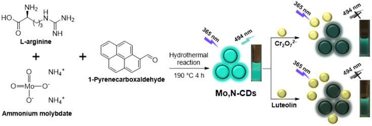
Synthesis of Mo, N-doped carbon dots for the fluorescence detection of Cr2O72− and luteolin and their application in anti-counterfeiting†
Received
14th March 2025
, Accepted 18th May 2025
First published on 10th June 2025
Abstract
In this work, Mo, N-doped carbon dots (Mo, N-CDs) were synthesized via a one-step hydrothermal method using L-arginine, ammonium molybdate and 1-pyrenecarboxaldehyde as precursors. The synthetic conditions, morphological structure, elemental composition and optical properties of Mo, N-CDs were carefully optimized and characterized. The as-synthesized Mo, N-CDs displayed cyan fluorescence emission at 494 nm with a high quantum yield (41.5%) and demonstrated superior pH stability, salt tolerance, thermostability and photobleaching resistance. In addition, Mo, N-CDs were employed as “on–off” sensors for the detection of Cr2O72− and luteolin with excellent linear correlation over a wide linear range (0–50 μM and 0–45 μM, respectively), low detection limits (0.96 μM and 0.64 μM, respectively) and short response time (<10 s). In addition, the practical detection of Cr2O72− and luteolin in actual samples demonstrated good recoveries (93–110%) with relative standard deviations less than 5%. Furthermore, the Mo, N-CD aqueous solution was directly used as a fluorescent ink and exhibited excellent anti-counterfeiting effects.
Introduction
Dichromate ion (Cr2O72−) is a dangerous oxygen-containing anion and is hazardous to the environment and organisms.1 With its wide use in chemical industries (electroplating, metallurgy, paint and leather tanning), Cr2O72− has become the most common heavy metal pollutant in water.2 Therefore, accurate detection of Cr2O72− in water is essential for environmental water monitoring and safeguarding biological health. Traditional methods for the detection of Cr2O72−, such as atomic absorption spectrometry,3 inductively coupled plasma mass spectrometry (ICP-MS),4 and colorimetric and electrochemical analyses,5 usually require expensive instruments and tedious operation procedures, which limit their application in the fast detection of Cr2O72−. Luteolin (3,4,5,7-tetrahydroxy-flavone) is a natural flavonoid compound with various pharmacological effects, such as antimicrobial, anti-inflammatory and antioxidant effects.6 Therefore, the detection of luteolin is of great practical importance in pharmaceutical analyses. Common methods for the detection of luteolin, including electrochemical methods,7 capillary zone electrophoresis (CZE),8 high-performance liquid chromatography (HPLC)9 and spectrophotometry (SP),10 have the drawbacks of high cost, low sensitivity and being time-consuming. Therefore, there is an urgent need for the development of new methods for fast and real-time detection of Cr2O72− and luteolin.
In order to address this issue, fluorescent detection strategies, especially carbon dot (CD)-based fluorescence strategies, stand out owing to their low-cost fabrication, low cytotoxicity, simple operation procedure, high sensitivity and selectivity and rapid response time.11,12 In the past years, numerous CD-based fluorescence sensors have been reported for the detection of Cr2O72− (for more details see Table S1†)13 and luteolin.14 However, these sensors often have the defects of low fluorescence quantum yield (FLQY), long response time and insufficient stability, which greatly limit their fluorescence detection applications. To solve these problems, researchers have found that doping CDs with heteroatoms can significantly improve their FLQY.15 Among them, non-metallic atom (N, B, P and S)16–19-doped CDs are frequently reported, while metal atom-doped CDs are relatively less reported and mainly focus on limited metal ions (Co, Cu, Fe and Zn).20,21 As an important transition metal element with high biocompatibility and good stability, molybdenum (Mo) has been reported for the synthesis of numerous functionalized molybdenum disulfide (MoS2) nanomaterials with different sulfur sources.22,23 However, Mo-doped CDs are rarely reported in literature.24
To enrich the scope of heteroatom-doped CDs and construct a fast detection platform for Cr2O72− and luteolin, we report a facile synthesis of Mo, N-doped CDs (Mo, N-CDs) using L-arginine,25 ammonium molybdate and 1-pyrenecarboxaldehyde as precursors via a simple one-step hydrothermal route (Scheme 1). The as-prepared Mo, N-CDs exhibited cyan fluorescence under a 365 nm UV light with a high FLQY of 41.5%. The morphology, structure and elemental composition of Mo, N-CDs were carefully characterized, indicating that molybdenum and nitrogen atoms were successfully doped. Further investigations showed that Mo, N-CDs exhibit good pH stability, salt tolerance, thermostability and photobleaching resistance. Moreover, Mo, N-CDs could be employed as “on–off” sensors for the sensitive detection of Cr2O72− and luteolin with low detection limits (0.96 and 0.64 μM) and wide linear range (0–50 μM, R2 = 0.9956 and 0–45 μM, R2 = 0.9992). Furthermore, the detection of Cr2O72− and luteolin in actual water samples and drugs was also investigated and showed acceptable recoveries (93–110%) with relative standard deviations of less than 5%. In addition, the application of Mo, N-CDs as fluorescent ink was explored and demonstrated excellent anti-counterfeiting effects.
 |
| | Scheme 1 Synthesis of Mo, N-CDs for Cr2O72− and luteolin detection. | |
Results and discussion
Synthesis and characterization
Initially, the hydrothermal reaction conditions and the molybdenum source for the preparation of Mo, N-CDs were optimized. As shown in Table S2,† 1-pyrenecarboxaldehyde and ammonium molybdate were indispensable (entries 1–4) and the highest fluorescence intensity of Mo, N-CDs was obtained with L-arginine (0.3 g), ammonium molybdate (0.3 g) and 1-pyrenecarboxaldehyde (0.02 g) under the optimal hydrothermal conditions (190 °C, 4 h as shown in entry 2 in Table S2†).
Then, the morphology structure of Mo, N-CDs was characterized by transmission electron microscopy (TEM), X-ray diffraction (XRD) and Raman spectroscopy (Fig. 1 and S2†). The TEM image showed that Mo, N-CDs were nearly spherical with a particle diameter of 1.7 ± 0.04 nm (Fig. 1a and b). High-resolution TEM (HRTEM) images showed Mo, N-CDs with a carbon core structure and lattice fringes of approximately 0.15 nm (Fig. 1c). Typical XRD pattern of Mo, N-CDs showed a broad peak at about 24.2°, corresponding to the peak of graphite (002) (Fig. 1d).26 Raman spectrum of Mo, N-CDs showed the main peak at around 1400 cm−1 (D band) and the shoulder peak at around 1600 cm−1 (G band), respectively, which should be assigned to disordered structures of the sp2 domains and in-plane vibrations of crystalline graphite (Fig. S2†).25d These results were compatible with the features of carbon dots.27
 |
| | Fig. 1 (a) TEM image; (b) particle size distribution histogram and curve; (c) HRTEM image; and (d) XRD pattern of Mo, N-CDs. | |
The surface functional groups of Mo, N-CDs were analyzed by Fourier transform infrared spectroscopy (FTIR). As shown in Fig. 2a, the broad absorption peaks at 3130 and 3010 cm−1 indicating the existence of N–H/O–H bonds.28 The peaks at 2916, 2848 and 1490 cm−1 indicated the C–H bonds.29 The two peaks at 1584 and 1400 cm−1 showed conjugated C![[double bond, length as m-dash]](https://www.rsc.org/images/entities/char_e001.gif) N, CO and CC bonds.30 The two peaks at 1435 and 1166 cm−1 indicated the existence of C–N and C–O bonds.31 The peak at 893 cm−1 should be the evidence of the existence of Mo–O bond.32 The elemental compositions of Mo, N-CDs were characterized by X-ray photoelectron spectroscopy (XPS). The full-scan XPS spectrum (Fig. 2b) shows distinct peaks at 232.23, 284.91, 399.55 and 530.56 eV, representing the presence of Mo3d (4.94%), C 1s (41.08%), N 1s (29.18%) and O 1s (24.79%), respectively. The high-resolution C 1s spectrum (Fig. 2c) showed three fitted peaks at 284.80, 286.05 and 288.37 eV, corresponding to C–C/CC, C–N/C–O and CO/CN bonds.31,33 The high-resolution O 1s spectrum (Fig. 2d) showed two fitted peaks at 533.69 and 534.85 eV, corresponding to the C–O and CO bonds.34 Three fitted peaks at 398.34, 399.93 and 401.37 eV in the high-resolution N 1 s spectrum (Fig. 2e) inferred the CN, C–N and N–H bonds.35 Four fitted peaks at 231.16, 232.25, 234.26 and 235.38 eV in the high-resolution Mo 3d spectrum (Fig. 2f) corresponded to Mo2+, Mo3+, Mo4+ and Mo6+.36 Furthermore, the presence of C, O, N and Mo in Mo,N-CDs was explored using the EDX spectrum (Fig. S3†).25d These results indicated that molybdenum and nitrogen were successfully doped in the as-synthesized carbon dots in this work.
N, CO and CC bonds.30 The two peaks at 1435 and 1166 cm−1 indicated the existence of C–N and C–O bonds.31 The peak at 893 cm−1 should be the evidence of the existence of Mo–O bond.32 The elemental compositions of Mo, N-CDs were characterized by X-ray photoelectron spectroscopy (XPS). The full-scan XPS spectrum (Fig. 2b) shows distinct peaks at 232.23, 284.91, 399.55 and 530.56 eV, representing the presence of Mo3d (4.94%), C 1s (41.08%), N 1s (29.18%) and O 1s (24.79%), respectively. The high-resolution C 1s spectrum (Fig. 2c) showed three fitted peaks at 284.80, 286.05 and 288.37 eV, corresponding to C–C/CC, C–N/C–O and CO/CN bonds.31,33 The high-resolution O 1s spectrum (Fig. 2d) showed two fitted peaks at 533.69 and 534.85 eV, corresponding to the C–O and CO bonds.34 Three fitted peaks at 398.34, 399.93 and 401.37 eV in the high-resolution N 1 s spectrum (Fig. 2e) inferred the CN, C–N and N–H bonds.35 Four fitted peaks at 231.16, 232.25, 234.26 and 235.38 eV in the high-resolution Mo 3d spectrum (Fig. 2f) corresponded to Mo2+, Mo3+, Mo4+ and Mo6+.36 Furthermore, the presence of C, O, N and Mo in Mo,N-CDs was explored using the EDX spectrum (Fig. S3†).25d These results indicated that molybdenum and nitrogen were successfully doped in the as-synthesized carbon dots in this work.
 |
| | Fig. 2 (a) FTIR spectrum of Mo, N-CDs and (b–f) XPS spectra of Mo, N-CDs: (b) full scan; (c–f) high-resolution spectrum of C 1s, O 1s, N 1s and Mo 3d. | |
Optical properties
The optical properties of Mo, N-CDs were investigated by UV-vis absorption and fluorescence spectroscopy. Mo, N-CDs showed strong absorption at 274 nm and weak absorption at 326 and 342 nm, which corresponded to the π–π* transition of CC bonds and n–π* transition of CN/CO bonds.37,38 Fluorescence spectra showed that the maximum excitation/emission wavelengths of Mo, N-CDs were 365/494 nm (Fig. 3a). Therefore, the Mo, N-CD aqueous solution appeared colourless under sunlight and cyan under UV irradiation (365 nm, insets of Fig. 3a). When the excitation wavelength was varied from 355 to 435 nm, the emission peaks of Mo, N-CDs remained almost unchanged, showing an excitation-wavelength-independent emission property (Fig. 3b). The fluorescence quantum yield (FLQY) of Mo, N-CDs was calculated to be 41.5% using quinine sulphate as a reference (Fig. S4†).
 |
| | Fig. 3 (a) UV-vis absorption and fluorescence spectra of Mo, N-CDs (insets: images of Mo, N-CDs under sunlight (left) and under a 365 nm UV light (right)); (b) fluorescence emission spectra of Mo, N-CDs under different excitation conditions; (c) fluorescence intensities of Mo, N-CDs at 494 nm at a continuous excitation of 365 nm; and (d–f) fluorescence intensity of Mo, N-CDs at different pH, NaCl concentration and incubation temperatures, respectively. | |
The stability of Mo, N-CDs was also investigated to evaluate their feasibility in natural environments (Fig. 3c–f). The fluorescence intensities of Mo, N-CDs did not show significant changes under continuous excitation irradiation at 365 nm for 90 min, different pH (3–12), NaCl concentrations (0–100 mM) and incubation temperatures (5–85 °C), demonstrating the excellent photobleaching resistance, pH stability, salt tolerance and thermostability of Mo, N-CDs.
Detection of Cr2O72-
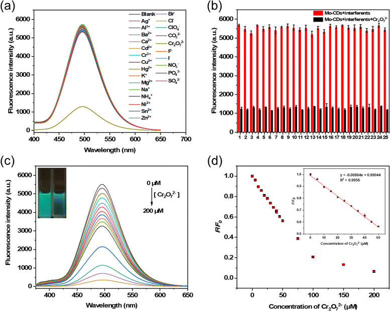
The recognition ability of Mo, N-CDs was studied by adding 25 common ions to the Mo, N-CD aqueous solution (Ag+, Al3+, Ba2+, Ca2+, Cd2+, Cr3+, Cu2+, Hg2+, K+, Mg2+, Na+, NH4+, Ni2+, Sn2+, Zn2+, Br−, Cl−, ClO4−, CO32−, Cr2O72−, F−, I−, NO3−, PO43− and SO42−). As shown in Fig. 4a, Cr2O72− quenched the fluorescence of Mo, N-CDs, whereas the other ions can hardly change the fluorescence of Mo, N-CDs. The anti-interference ability of Mo, N-CDs was studied through the addition of Cr2O72− to Mo, N-CD solutions containing other ions (Fig. 4b). The results showed that the selectivity of Mo, N-CDs for Cr2O72− was not affected. Response time studies showed that Cr2O72− could instantly (<10 s) quench the fluorescence of Mo, N-CDs (Fig. S5†). The sensitivity was explored by the measurements of fluorescence intensities of Mo, N-CDs with different amounts of Cr2O72−. As shown in Fig. 4c, the fluorescence intensity decreased with the gradual increase of Cr2O72−. The quenching efficiency was determined by F/F0, where F0 is the intensity of the blank Mo, N-CD solution and F is the intensity of the Mo,N-CDs with Cr2O72−. A linear curve was obtained by plotting the quenching efficiency vs. Cr2O72− (0–50 μM). As shown in Fig. 4d, the linear equation was calculated to be F/F0 = −0.0086 C(Cr2O72−) + 0.9904 with a correlation coefficient (R2) of 0.9956. The detection limit (LoD) was 0.96 μM (calculated using the (3σ/k) method, where σ is the standard deviation of the blank sample (n = 10) (Table S3†) and k is the slope of the linear calibration curve). Compared to previously published works for the detection of Cr2O72− (as listed in Table S1†), Mo, N-CDs in this work showed a fast response to Cr2O72− with a wide linear range and acceptable detection limit (<50 μg L−1 by WHO).37,39 In summary, Mo, N-CDs are promising for the rapid quantitative analysis of Cr2O72− in aqueous solution.
 |
| | Fig. 4 (a) Fluorescence spectra of Mo, N-CDs with different ions (100 μM); (b) fluorescence intensities of Mo, N-CDs with different ions (1–25: blank, Ag+, Al3+, Ba2+, Ca2+, Cd2+, Cr3+, Cu2+, Hg2+, K+, Mg2+, Na+, NH4+, Ni2+, Sn2+, Zn2+, Br−, Cl−, ClO4−, CO32−, F−, I−, NO3−, PO43− and SO42−) in the absence (red)/presence (black) of Cr2O72− (100 μM); (c) fluorescence emission spectra of Mo, N-CDs with different concentrations of Cr2O72− (inset: photograph of the Mo, N-CD solution without (left)/with (right) Cr2O72− (100 μM) under a 365 nm UV light); and (d) relationship between F/F0 and the concentration of Cr2O72−. | |
Detection of luteolin
In this experiment, 10 compounds (glutathione (GSH), glucose, L-arginine (L-Arg), L-cysteine (L-Cys), L-glutamic acid (L-Glu), luteolin, nicotinamide, ascorbic acid (AA), citric acid (CA) and urea) were selected to explore the selectivity and anti-interference capability of Mo, N-CDs. As shown in Fig. 5a–b, the fluorescence of Mo, N-CDs was significantly quenched by luteolin, whereas the other compounds did not influence the detection of luteolin. The exploration of response time showed that the fluorescence intensity of Mo, N-CDs was also instantly (<10 s) quenched by luteolin (Fig. S6†). The sensitivity of Mo,N-CDs to luteolin was explored using the same method for Cr2O72−. As shown in Fig. 5c and d, the quenching efficiency F/F0 vs. luteolin (0–45 μM) was plotted, and the linear equation was F/F0 = −0.0129 C (luteolin) + 0.9920 with a correlation coefficient (R2) of 0.9992. The detection limit is 0.64 μM, calculated using the (3σ/k) method. Compared with previous works,14 Mo, N-CDs in this work showed a fast response to luteolin with a low detection limit. These results show that Mo, N-CDs can be used for quantitative detection of luteolin in vitro.
 |
| | Fig. 5 (a) Fluorescence spectra of Mo, N-CDs with different compounds (luteolin (50 μM), other compounds (100 μM)); (b) fluorescence intensities of Mo, N-CDs with different compounds in the absence (red)/presence (black) of luteolin (50 μM); (c) fluorescence spectra of Mo, N-CDs with different concentrations of luteolin (inset: photographs of the Mo, N-CD solution without (left)/with (right) luteolin (50 μM) under a 365 nm UV light); and (d) relationship between F/F0 and the concentration of luteolin. | |
Quenching mechanism for Cr2O72−
The mechanism of fluorescence quenching was explored through UV-vis and fluorescence spectroscopy, zeta potential and fluorescence lifetime measurements. As shown in Fig. 6, compared to the theoretical UV-vis absorption spectra, the Mo, N-CDs/Cr2O72− mixtures did not give new absorption peaks, which ruled out the formation of non-fluorescent ground state complexes (Fig. 6a).40 Additionally, as illustrated in Fig. 6b, the UV-vis spectrum of Cr2O72− overlapped with the fluorescence spectra of Mo, N-CDs, indicating that the quenching mechanism might be inner-filter effect (IFE) and fluorescence resonance energy transfer (FRET).41 The negative zeta potential (−6.34 and −9.90 mV) indicated no electrostatic attraction between Mo, N-CDs and Cr2O72− (Fig. 6c), and unchanged fluorescence lifetime (6.24, 6.26, 6.31 and 6.31 ns) ruled out the FRET quenching mechanism (Fig. 6d).42 Therefore, an IFE mechanism was proposed for sensing Cr2O72−.
 |
| | Fig. 6 (a) UV-vis spectra of Mo, N-CDs, Cr2O72−, theoretical and experimental spectra of Mo, N-CDs/Cr2O72−; (b) UV-vis spectrum of Cr2O72− and fluorescence spectra of Mo, N-CDs; (c) zeta potentials of Mo, N-CDs, Cr2O72− and Mo, N-CDs/Cr2O72−; and (d) fluorescence lifetime of Mo, N-CDs (without/with Cr2O72−). | |
To calculate the IFE ratio, a series of Mo, N-CD solutions with different amounts of Cr2O72− were prepared, and their absorbance values (Aex and Aem) and fluorescence intensities (Fobsd) were measured. Then, the corrected fluorescence emission intensities (Fcor) were calculated according to Parker's equation (eqn (1).43 Thereafter, the correction factor (CF), observed fluorescence suppression efficiency (Eobsd) and corrected fluorescence suppression efficiency (Ecor) were calculated, as shown in Table 1 (CF < 3 to ensure the accuracy of the results). The IFE ratio was calculated as (Eobsd – Ecor)/Eobsd to be 95.34%.44 The ratio of the corrected fluorescence emission intensities without/with Cr2O72− (Fcor.o/Fcor) versus the concentration of Cr2O72− were plotted as shown in Fig. S8.† Fcor,0/Fcor was not linearly related with Cr2O72−, which further ruled out the static and dynamic quenching effects in this system according to the Stern–Volmer equation eqn (2).44
| |
 | (1) |
Fcor and
Fobsd are the corrected and measured fluorescence intensities at 494 nm;
d,
g and
s are the geometric parameters of the quartz cuvette (shown in Fig. S7,
† 1.00, 0.25 and 0.50 cm, respectively); and
Aex and
Aem are the absorbance at the excitation wavelength (365 nm) and the optimal emission wavelength (494 nm).
| | |
Fcor.0/Fcor = 1 + KSV[Q]
| (2) |
Fcor.0 and
Fcor are the corrected fluorescence intensities without and with Cr
2O
72−;
Ksv is the Stern–Volmer constant; [
Q] is the concentration of Cr
2O
72−.
Table 1 Calculation of the IFE ratio for Cr2O72−a
| Cr2O72− (μM) |
Aex |
Aem |
Fobsd |
Fcor |
CF |
Eobsd |
Ecor |
| CF = Fcor/Fobsd; Eobsd = 1 – Fobsd/Fobsd.0; Ecor = 1 – Fcor/Fcor.0 (Fobsd.0 and Fcor.0 are the measured and corrected fluorescence intensities, respectively, without Cr2O72−). |
| 0 |
0.52 |
0.025 |
5212 |
9198 |
1.76 |
0.00 |
0.000 |
| 5 |
0.56 |
0.020 |
5050 |
9158 |
1.82 |
0.03 |
0.001 |
| 10 |
0.62 |
0.024 |
4658 |
8991 |
1.93 |
0.11 |
0.022 |
| 15 |
0.66 |
0.019 |
4432 |
8804 |
1.99 |
0.15 |
0.043 |
| 20 |
0.72 |
0.016 |
4161 |
8651 |
2.08 |
0.20 |
0.059 |
| 25 |
0.80 |
0.042 |
3950 |
9075 |
2.30 |
0.24 |
0.013 |
Quenching mechanism of luteolin
The possible mechanism of luteolin detection was investigated using the same method described above. As shown in Fig. 7a, a new absorption band was formed at 379–390 nm in the experimental UV-vis absorption spectrum of the Mo, N-CDs/luteolin mixture, indicating the formation of non-fluorescent ground state complexes and a static quenching mechanism.45 Fig. 7b indicates that the quenching mechanism of Mo, N-CDs for luteolin might be IFE and FRET.46 Zeta potential measurements showed that no electrostatic interaction existed between Mo, N-CDs and luteolin (Fig. 7c), and fluorescence lifetime measurements ruled out FRET (Fig. 7d). In addition, the IFE ratio was calculated to be 80.55% (Table 2)43 and the presence of a static quenching effect was verified by the Stern–Volmer equation (Fig. S9†).47 In summary, the combination of IFE and static quenching may be the mechanism by which Mo, N-CDs detect luteolin.
 |
| | Fig. 7 (a) UV-vis spectra of Mo, N-CDs, luteolin, theoretical and experimental spectra of Mo, N-CDs/luteolin; (b) UV-vis spectrum of luteolin and fluorescence spectra of Mo, N-CDs; (c) zeta potentials of Mo, N-CDs, luteolin and Mo, N-CDs/luteolin; and (d) fluorescence lifetime of Mo, N-CDs (without/with luteolin). | |
Table 2 Calculation of the IFE ratio for luteolina
| Luteolin (μM) |
Aex |
Aem |
Fobsd |
Fcor |
CF |
Eobsd |
Ecor |
| CF = Fcor/Fobsd; Eobsd = 1 − Fobsd/Fobsd.0; Ecor = 1 – Fcor/Fcor.0 (Fobsd.0 and Fcor.0 are the measured and corrected fluorescence intensities, respectively, without luteolin). |
| 0 |
0.63 |
0.039 |
5786 |
11475 |
1.98 |
0.00 |
0.000 |
| 5 |
0.68 |
0.049 |
5390 |
11310 |
2.10 |
0.07 |
0.014 |
| 10 |
0.78 |
0.049 |
4865 |
11095 |
2.28 |
0.16 |
0.033 |
| 15 |
0.94 |
0.067 |
4119 |
10831 |
2.63 |
0.29 |
0.056 |
| 20 |
1.02 |
0.069 |
3809 |
10712 |
2.81 |
0.34 |
0.066 |
| 25 |
0.63 |
0.039 |
5786 |
11475 |
1.98 |
0.00 |
0.000 |
Testing of actual samples
The detection of Cr2O72− and luteolin in actual samples was further investigated to validate the reliability of the method. As shown in Tables 3 and 4, the recoveries of Cr2O72− in tap water, lake water and bottled drinking water and luteolin in Du Yi Wei capsule and Du Yi Wei tablet were 93–110% with relative standard deviations (RSD) less than 5%. Therefore, Mo, N-CDs can be used as “on-off” sensors for the detection of Cr2O72− and luteolin in the field of water quality monitoring and pharmaceutical analysis.
Table 3 Detection of Cr2O72− in actual water samples (n = 3)
| Samples |
Spiked (μM) |
Found (μM) |
Recovery (%) |
RSD (%) |
| Tap water |
15 |
14.67 |
97.78 |
0.46 |
| 25 |
25.17 |
100.68 |
0.23 |
| 35 |
34.51 |
98.61 |
1.27 |
| Lake water |
15 |
16.44 |
109.64 |
0.86 |
| 25 |
24.29 |
97.15 |
0.15 |
| 35 |
35.74 |
102.12 |
2.05 |
| Bottled drinking water |
15 |
14.51 |
96.73 |
0.76 |
| 25 |
25.52 |
102.10 |
0.22 |
| 35 |
36.19 |
103.41 |
0.28 |
Table 4 Detection of luteolin in actual samples (n = 3)
| Samples |
Spiked (μM) |
Found (μM) |
Recovery (%) |
RSD (%) |
| Du Yi Wei capsule |
0 |
5.26 |
— |
0.83 |
| 15 |
20.91 |
104.26 |
1.97 |
| 25 |
29.94 |
98.71 |
1.24 |
| 35 |
37.88 |
93.18 |
2.23 |
| Du Yi Wei tablet |
0 |
3.10 |
— |
3.39 |
| 15 |
18.53 |
102.85 |
0.25 |
| 25 |
28.48 |
101.50 |
1.08 |
| 35 |
36.67 |
95.92 |
2.71 |
Anti-counterfeiting application
Due to their stable optical property and relatively high quantum yields, Mo, N-CDs have potential application prospects in anti-counterfeiting. As shown in Fig. 8, two pictures were drawn by hand on test paper using a Mo, N-CD aqueous solution as fluorescent ink. The two pictures were almost invisible under sunlight but exhibited obvious cyan fluorescence under 365 nm UV light, showing good fluorescence anti-counterfeiting effects. With the removal of 365 nm UV light, the cyan fluorescence disappeared completely, demonstrating high-quality information encryption and decryption effects. The maintenance of fluorescence for several months indicates that the Mo, N-CD-based fluorescent ink exhibits better durability and reliability.
 |
| | Fig. 8 Photographs of pictures drawn by hand using Mo, N-CDs as fluorescent ink on test paper under sunlight (left) and under a 365 nm UV light (right). | |
Conclusions
In conclusion, a rarely reported Mo, N-doped cyan fluorescent carbon dots (Mo, N-CDs) was successfully synthesized by a one-step hydrothermal method using L-arginine, ammonium molybdate and indispensable 1-pyrenecarboxaldehyde as precursors. The results of morphological and compositional characterization showed that molybdenum and nitrogen atoms were successfully doped into carbon dots. Due to its high quantum yield, superior pH stability, salt tolerance and excellent photobleaching resistance, a fluorescence platform based on Mo, N-CDs was constructed for “on–off” detection of Cr2O72− and luteolin in aqueous solutions and actual samples. This method demonstrated high selectivity and sensitivity, a short response time and satisfactory recoveries. The fluorescence quenching mechanisms of Mo, N-CDs were explored and putatively identified as IFE (95.34%) for Cr2O72− and IFE (80.55%)/static quenching for luteolin. Furthermore, an aqueous solution of Mo, N-CDs can be directly used as a fluorescent ink and exhibits excellent anti-counterfeiting effects. The facial synthesis, high quantum yield, stable optical properties and rapid responsiveness of Mo, N-CDs make them an excellent alternative in environmental water monitoring and pharmaceutical analysis as well as anti-counterfeiting.
Data availability
The data supporting this article have been included as part of the ESI.†
Author contributions
Yang Geng and Ze-Yue Zhao: methodology, formal analysis, investigation, and writing – review and editing. Wei Zhang and Bai-Qiang Zhai: conceptualization and analysis. Ye Tian and Wen-Zhu Bi: validation and supervision.
Conflicts of interest
There are no conflicts to declare.
Acknowledgements
This work was supported by Henan Provincial Science and Technology International Cooperation Grant (242102521040), the Science and Technology Development Plan Project Henan Province (242102311178) and the Key Research Project of Zhengzhou Railway Vocational and Technical College (2025KY005).
Notes and references
-
(a) Z. Yu, C. Deng, S. Jiang, Y. Liu, C. Liu, F. Seidi, X. Zhang, Y. Huang, W. Wu, J. Han, Q. Yong and H. Xiao, J. Colloid Interface Sci., 2025, 679, 510 CrossRef CAS PubMed;
(b) T. Zheng, L. Qian, M. Li, Z. Wang, K. Li, Y. Zhang, B. Li and B. Wu, Dalton Trans., 2018, 47, 9103 RSC;
(c) B. Prathima, V. S. Raghvan, S. Soni, S. S. Gorthi and G. L. Sivakumar Babu, J. Water Process. Eng., 2024, 66, 105832 CrossRef.
- D. M. Hausladen, A. A. Ozinskas, C. N. McClain and S. Fendorf, Environ. Sci. Technol., 2018, 52, 8242 CrossRef CAS PubMed.
-
(a) F. A. S. Cunha, J. P. A. Fernandes, W. S. Lyra, A. G. G. Pessoa, J. C. C. Santos, M. C. U. Araújo and L. F. Almeida, J. Anal. At. Spectrom., 2024, 39, 1398 RSC;
(b) Z. Tahmasebi, S. S. H. Davarani, H. Ebrahimzadeh and A. A. Asgharinezhad, Microchem. J., 2018, 143, 212 CrossRef CAS.
- R. Pechancová, T. Pluháček, J. Gallo and D. Milde, Talanta, 2018, 185, 370 CrossRef PubMed.
-
(a) S. Liu, X. Wang, C. Zou, J. Zhou, M. Yang, S. Zhang, D. Huo and C. Hou, Anal. Chim. Acta, 2021, 1149, 238141 CrossRef CAS PubMed;
(b) F. Yao, M. Jia, Q. Yang, K. Luo, F. Chen, Y. Zhong, L. He, Z. Pi, K. Hou, D. Wang and X. Li, Chemosphere, 2020, 260, 127537 CrossRef CAS PubMed;
(c) J. Wan, M. Yu, W. Bi, Y. Sun, W. Wang, Y. Hou, S. Xie, T. Li and Y. Fan, J. Water Process. Eng., 2024, 68, 106447 CrossRef;
(d) I. Kabdaşli and O. Tünay, Molecules, 2023, 28, 2411 CrossRef PubMed.
-
(a) A. S. U. Mahamud, M. Ashrafudoulla, S. Nahar, M. A. H. Chowdhury, S. H. Park and S. D. Ha, Food Control, 2024, 166, 110734 CrossRef CAS;
(b) S. F. Nabavi, N. Braidy, O. Gortzi, E. S. Sanchez, M. Daglia, K. S. Woźniak and S. M. Nabavi, Brain Res. Bull., 2015, 119, 1 CrossRef CAS PubMed;
(c) N. Aziz, M. Y. Kim and J. Y. Cho, J. Ethnopharmacol., 2018, 225, 342 CrossRef CAS PubMed;
(d) M. Zhu, Y. Sun, Y. Su, W. Guan, Y. Wang, J. Han, S. Wang, B. Yang, Q. Wang and H. Kuang, Phytother. Res., 2024, 38, 3417 CrossRef PubMed.
-
(a) B. Wang, W. Zhao, L. Wang, K. Kang, X. Li, D. Zhang, J. Ren and X. Ji, Talanta, 2024, 273, 125836 CrossRef CAS PubMed;
(b) X. Hou, W. Wu, F. Zhao, W. Xie and Q. Yang, Microchim. Acta, 2021, 188, 86 CrossRef CAS PubMed;
(c) X. Li, R. Zou, Y. Niu, W. Sun, T. Shao and X. Chen, Sensors, 2018, 18, 2309 CrossRef PubMed.
- Y. Li, Q. Zhang, H. Sun, N. K. Cheung and H. Y. Cheung, Talanta, 2013, 105, 393 CrossRef CAS PubMed.
-
(a) S. H. Hwang, J. H. Paek and S. S. Lim, Molecules, 2016, 21, 1609 CrossRef PubMed;
(b) H. Y. Muti and S. Olimat, Orient. J. Chem., 2018, 34, 2721 CAS.
- G. Favaro, C. Clementi, A. Romani and V. Vickackaite, J. Fluoresc., 2007, 17, 707 CrossRef CAS PubMed.
-
(a) H. Liu, X. Zhong, Q. Pan, Y. Zhang, W. Deng, G. Zou, H. Hou and X. Ji, Coord. Chem. Rev., 2024, 498, 215468 CrossRef CAS;
(b) F. Liu, X. Lai, S. Zhao, Z. Lu, P. Han and L. Chen, Food Chem., 2023, 402, 134251 CrossRef CAS PubMed;
(c) C. Liu, Y. Mei, Q. Lei, X. Ma, X. Nan, Y. Zhu, J. Liao, Y. Xu, Y. Luo, H. Zhang, M. Yang, X. Lin and Q. Huang, Chem. Eng. J., 2024, 499, 156434 CrossRef CAS.
-
(a) E. K. Adotey, M. A. Torkmahalleh, L. Tastanova, A. Bekeshev, D. Shah, P. K. Hopke, W. Lee and M. P. Balanay, J. Hazard. Mater., 2024, 462, 132671 CrossRef CAS PubMed;
(b) X. Hu, J. Shi, Y. Shi, X. Zou, H. E. Tahir, M. Holmes, W. Zhang, X. Huang, Z. Li and Y. Xu, Meat Sci., 2019, 147, 127 CrossRef CAS PubMed;
(c) H. M. Junaid, A. R. Solangi and M. Batool, Analyst, 2021, 146, 2463 RSC.
- For selected examples see:
(a) R. Zhang, Y. Zheng, Q. Zhang, Z. Wu, L. Wang, J. Zhang, H. Ren and E. Duan, J. Environ. Chem. Eng., 2024, 12, 112391 CrossRef CAS;
(b) S. Liu, Z. Wu, N. Nian, P. Zhang and L. Ni, Water, Air, Soil Pollut., 2024, 235, 449 CrossRef CAS;
(c) S. Liu, Y. Zhang, Y. Zhang, J. Zhang, R. Tian and L. Zhao, Diamond Relat. Mater., 2024, 148, 111382 CrossRef CAS;
(d) M. He, X. Fu, G. Du, H. Li, P. Zhao and X. Liu, Packag. Technol. Sci., 2023, 36, 465 CrossRef CAS;
(e) X. Mei, D. Wang, S. Wang, J. Li and C. Dong, Anal. Bioanal. Chem., 2022, 414, 7253 CrossRef CAS PubMed;
(f) A. Tall, F. A. Cunha, B. Kaboré, C. A. E. S. Barbosa, U. Rocha, T. O. Sales, M. O. F. Goulart, I. Tapsoba and J. C. C. Santos, Microchem. J., 2021, 166, 106219 CrossRef CAS;
(g) H. Tian, G. Ju, M. Li, W. Fu, Y. Dai, Z. Liang, Y. Qiu, Z. Qin and X. Yin, RSC Adv., 2021, 11, 35946 RSC;
(h) Z. Ma, Y. Ma, M. Gu, X. Huo, S. Ma, Y. Lu, Y. Ning, X. Zhang, B. Tian and Z. Feng, Nanomaterials, 2020, 10, 1924 CrossRef CAS PubMed.
-
(a) Y. Wang, X. Hou, Y. Li, X. Sun, R. Hu, Y. Lv, R. Jia and L. Ding, Microchem. J., 2024, 206, 111562 CrossRef CAS;
(b) S. Zhang, Z. Wang, W. Yan and Y. Gou, Spectrochim. Acta, Part A, 2021, 259, 119887 CrossRef CAS PubMed;
(c) Q. Sun, Y. Long, S. Pan, H. Liu, J. Yang and X. Hu, Luminescence, 2018, 33, 1401 CrossRef CAS PubMed.
-
(a) S. Miao, K. Liang, J. Zhu, B. Yang, D. Zhao and B. Kong, Nano Today, 2020, 33, 100879 CrossRef CAS;
(b) Q. Xu, T. Kuang, Y. Liu, L. Cai, X. Peng, T. S. Sreeprasad, P. Zhao, Z. Yu and N. Li, J. Mater. Chem. B, 2016, 4, 7204 RSC.
-
(a) A. Saravanan, P. Das, M. Maruthapandi, S. Aryal, S. Michaeli, Y. Mastai, J. H. T. Luong and A. Gedanken, Surf. Interfaces, 2024, 46, 103857 CrossRef CAS;
(b) P. Das, S. Ganguly, S. Mondal, M. Bose, A. K. Das, S. Banerjee and N. C. Das, Sens. Actuators, B, 2018, 266, 583 CrossRef CAS.
- F. Wang, Y. Zhang, H. Li, W. Gong, J. Han, S. Jiang, D. Li and Z. Yao, Food Chem., 2025, 463, 141122 CrossRef CAS PubMed.
- P. Zhou, J. Xu, X. Hou, L. Dai, J. Zhang, X. Xiao and K. Huo, Int. J. Biol. Macromol., 2023, 253, 126714 CrossRef CAS PubMed.
-
(a) Z. Zhu, X. Li, M. Luo, M. Chen, W. Chen, P. Yang and X. Zhou, J. Colloid Interface Sci., 2022, 605, 330 CrossRef CAS PubMed;
(b) S. K. Park, H. Lee, M. S. Choi, D. H. Suh, P. Nakhanivej and H. S. Park, Energy Storage Mater., 2018, 12, 331 CrossRef.
-
(a) W. Lu, Y. Guo, J. Zhang, Y. Yue, L. Fan, F. Li, C. Dong and S. Shuang, ACS Appl. Mater. Interfaces, 2022, 14, 57206 CrossRef CAS PubMed;
(b) S. Huang, E. Yang, J. Yao, X. Chu, Y. Liu, Y. Zhang and Q. Xiao, ACS Omega, 2019, 4, 9333 CrossRef CAS PubMed;
(c) H. Behboudi, M. Pourmadadi, M. Omidi, M. Rahmandoust, S. O. R. Siadat and J. S. Shayeh, Surf. Interfaces, 2022, 29, 101710 CrossRef;
(d) M. Yang, J. Yao, B. Su, N. Xin, T. Zhou, M. Zeng, C. Wu, D. Wei, J. Sun and H. Fan, J. Mater. Chem. B, 2023, 11, 5898 RSC;
(e) P. Fan, C. Liu, C. Hu, F. Li, X. Lin, S. Yang and F. Xiao, New J. Chem., 2022, 46, 2526 RSC;
(f) X. Li, S. Ding, Z. Lyu, P. Tieu, M. Wang, Z. Feng, X. Pan, Y. Zhou, X. Niu, D. Du, W. Zhu and Y. Lin, Small, 2022, 18, 2203001 CrossRef CAS PubMed;
(g) W. Kong, C. Li, Z. Sun, F. Gao, J. Zheng and Y. Jiang, Molecules, 2023, 28, 5526 CrossRef CAS PubMed.
-
(a) Q. Miao, Y. Li, X. Fan, D. Deng, X. Yan, H. He and L. Luo, Analyst, 2021, 146, 6297 RSC;
(b) S. K. Tammina, Y. Wan, Y. Li and Y. Yang, J. Photochem. Photobiol., B, 2020, 202, 111734 CrossRef CAS PubMed;
(c) Q. Xu, W. Cai, M. Zhang, R. Su, Y. Ye, Y. Li, L. Zhang, Y. Guo, Z. Yu, S. Li, X. Lin, Y. Chen, Y. Luo, J. Street and M. Xu, RSC Adv., 2018, 8, 17254 RSC.
- L. Amaral and A. Daniel-da-Silva, Molecules, 2022, 27, 6782 CrossRef CAS PubMed.
- Z. Liang, R. Shen, Y. H. Ng, P. Zhang, Q. Xiang and X. Li, J. Mater. Sci. Technol., 2020, 56, 89 CrossRef CAS.
- X. Fu, J. Huang, X. Zhu, J. Rong, Z. Lin, Y. Dong and F. Fu, Chem. Commun., 2022, 58, 7180 RSC.
-
(a) P. Li, F. Han, W. Cao, G. Zhang, J. Li, J. Zhou, X. Gong, G. Turnbull, W. Shu, L. Xia, B. Fang, X. Xing and B. Li, Appl. Mater. Today, 2020, 19, 100601 CrossRef;
(b) F. Nemati, R. Z. Dorabei, M. Hosseini and M. R. Ganjali, Sens. Actuators, B, 2018, 255, 2078 CrossRef CAS.
- S. Kadian and G. Manik, Food Chem., 2020, 317, 126457 CrossRef CAS PubMed.
-
(a) B. Thangaraj, P. R. Solomon, S. Chuangchote, N. Wongyao and W. Surareungchai, ChemBioEng Rev., 2021, 8, 265 CrossRef CAS;
(b) J. Singh, S. Kaur, J. Lee, A. Mehta, S. Kumar, K. H. Kim, S. Basu and M. Rawat, Sci. Total Environ., 2020, 720, 137604 CrossRef CAS PubMed;
(c) S. Elkun, M. Ghali, T. Sharshar and M. M. Mosaad, Sci. Rep., 2024, 14, 27927 CrossRef CAS PubMed.
- W. Gao, H. Song, X. Wang, X. Liu, X. Pang, Y. Zhou, B. Gao and X. Peng, ACS Appl. Mater. Interfaces, 2017, 10, 1147 CrossRef PubMed.
- W. Lu, Y. Jiao, Y. Gao, J. Qiao, M. Mozneb, S. Shuang, C. Dong and C. Li, ACS Appl. Mater. Interfaces, 2018, 10, 42915 CrossRef CAS PubMed.
- C. Pan, T. Chen, L. Ma, X. Qin, Q. Wen and S. Feng, Chin. J. Anal. Chem., 2022, 50, 100069 Search PubMed.
- L. Dong, J. Wang, Y. Ma, Y. Ruan, Z. Hu and X. Ma, Arab. J. Chem., 2024, 17, 105660 CrossRef CAS.
- X. Lin, J. Chen, L. Zhang, T. Lan, Y. Yu, Z. Ke, L. Hu and Z. Zhang, Chin. J. Struct. Chem., 2010, 29, 437 CAS.
- W. Kong, C. Li, Z. Sun, F. Gao, J. Zheng and Y. Jiang, Molecules, 2023, 28, 5526 CrossRef CAS PubMed.
- J. Li, L. Zhang, P. Li, Y. Zhang and C. Dong, Sens. Actuators, B, 2018, 258, 580 CrossRef CAS.
-
(a) S. Huang, E. Yang, J. Yao, X. Chu, Y. Liu, Y. Zhang and Q. Xiao, ACS Omega, 2019, 4, 9333 CrossRef CAS PubMed;
(b) Q. Yang, W. Yang, Y. Zhang, W. Ge, X. Yang and P. Yang, Nanomaterials, 2020, 10, 460 CrossRef CAS PubMed.
- J. Wang, H. Wei, X. Chen, C. Chen and X. Chen, Int. J. Hydrogen Energy, 2020, 45, 595 CrossRef CAS.
- L. Zhu, D. Shen and K. Luo, J. Colloid Interface Sci., 2022, 617, 557 CrossRef CAS PubMed.
- Y. Yan, L. Li, H. Zhang, F. Du, Y. Meng, S. Shuang, R. Wang, S. Song and C. Dong, Spectrochim. Acta, Part A, 2021, 256, 119751 CrossRef CAS PubMed.
- Y. Mao, S. Gao, L. Yao, L. Wang, H. Qu, Y. Wu, Y. Chen and L. Zheng, J. Hazard. Mater., 2021, 408, 124898 CrossRef CAS PubMed.
- C. Ni, W. Zhang, W. Bi, M. Wu, S. Feng, X. Chen and L. Qu, RSC Adv., 2024, 14, 26667 RSC.
- X. Sun, M. Yuan, B. Liu and J. Shen, RSC Adv., 2018, 8, 19786 RSC.
- J. Xu, Y. Gao, L. Qin, X. Yue, Q. Zhang and L. Wang, Ceram. Int., 2023, 49, 7546 CrossRef CAS.
- W. He, Z. Huo, X. Sun and J. Shen, Microchem. J., 2020, 153, 104528 CrossRef CAS.
- M. Lu, C. Pan, X. Qin and M. Wu, ACS Omega, 2023, 8, 14499 CrossRef CAS PubMed.
- Y. Luo, C. Cui, X. Zhang, Y. Jiang, Z. Xiang, C. Ji and Z. Peng, Molecules, 2023, 28, 1566 CrossRef CAS PubMed.
- W. Chen, H. Lin, Y. Wu, M. Yang, X. Zhang, S. Zhu, M. He, J. Xie and Z. Shi, Adv. Compos. Hybrid Mater., 2022, 5, 2378 CrossRef CAS.
- H. Wu, L. Pang, M. Fu, X. Guo and H. Wang, J. Pharmaceut. Biomed. Anal., 2020, 180, 113052 CrossRef CAS PubMed.
|
| This journal is © The Royal Society of Chemistry 2025 |
Click here to see how this site uses Cookies. View our privacy policy here. 
 Open Access Article
 This Open Access Article is licensed under a Creative Commons Attribution-Non Commercial 3.0 Unported Licence
 *ab,
Ze-Yue Zhaoa,
Wei Zhanga,
Bai-Qiang Zhaia,
Ye Tian*a and
Wen-Zhu Bi
*ab,
Ze-Yue Zhaoa,
Wei Zhanga,
Bai-Qiang Zhaia,
Ye Tian*a and
Wen-Zhu Bi