
 Open Access Article
 This Open Access Article is licensed under a
 Creative Commons Attribution 3.0 Unported Licence
Recent advances of silver nanoparticle-based polymer nanocomposites for biomedical applications
Mohammad Harun-Ur-Rashid a,
Tahmina Foyezb,
Suresh Babu Naidu Krishna*c,
Sudhakar Podad and
Abu Bin Imran*e
a,
Tahmina Foyezb,
Suresh Babu Naidu Krishna*c,
Sudhakar Podad and
Abu Bin Imran*e
aDepartment of Chemistry, International University of Business Agriculture and Technology (IUBAT), Sector 10, Uttara Model Town, Dhaka 1230, Bangladesh. E-mail: mrashid@iubat.edu
bDepartment of Pharmacy, School of Life Sciences, United International University, United City, Madani Ave, Dhaka 1212, Bangladesh. E-mail: tahmina@pharmacy.uiu.ac.bd
cInstitute for Water and Wastewater Technology, Durban University of Technology, P. O. Box 1334, Durban 4000, South Africa. E-mail: sureshk@dut.ac.za
dDepartment of Biotechnology, Acharya Nagarjuna University, Andhra Pradesh, India. E-mail: sudhakarp@anu.ac.in
eDepartment of Chemistry, Bangladesh University of Engineering and Technology (BUET), Dhaka 1000, Bangladesh. E-mail: abimran@chem.buet.ac.bd
First published on 19th March 2025
Abstract
Silver nanoparticle–polymer nanocomposites (AgNP–PNCs) represent a transformative advancement in biomedical material science, integrating the potent antimicrobial properties of AgNPs with the structural versatility of polymer matrices. This synergy enables enhanced infection control, mechanical stability, and controlled drug delivery, making these nanocomposites highly suitable for applications such as wound healing, medical coatings, tissue engineering, and biosensors. Recent progress in synthesis and functionalization has led to greater control over particle morphology, dispersion, and stability, optimizing AgNP–PNCs for clinical and translational applications. However, challenges related to cytotoxicity, long-term stability, immune response, and scalability persist, necessitating systematic improvements in surface functionalization, hybridization strategies, and biocompatibility assessments. This review critically evaluates the latest advancements in AgNP–PNC development, focusing on their functionalization techniques, regulatory considerations, and emerging strategies to overcome biomedical challenges. Additionally, it discusses preclinical and translational aspects, including commercialization barriers and regulatory frameworks such as FDA and EMA guidelines, ensuring a comprehensive outlook on their clinical feasibility. By bridging the gap between innovation and practical application, this review investigates the transformative potential of AgNP–PNCs in advancing next-generation biomedical materials.
1. Introduction
In recent years, nanotechnology has revolutionized multiple scientific and clinical fields, enabling the development of advanced materials with unprecedented properties, which offer significant potential in addressing complex biomedical challenges.1 Among these materials, silver nanoparticles (AgNPs) have garnered considerable attention due to their powerful antimicrobial, optical, and electrical properties, making them particularly valuable in biomedical applications.2,3 However, stability, controlled release, and cytotoxicity challenges have limited their direct clinical use. To overcome these limitations, integrating AgNPs within polymer matrices to form AgNP–polymer nanocomposites (AgNP–PNCs) has emerged as a transformative strategy, combining the bioactive properties of silver with the versatility, stability, and biocompatibility of polymers.4 The AgNP–PNCs provide a controlled and sustained release of silver ions while mitigating the cytotoxic effects typically associated with free AgNPs.5 The polymer matrix is a stabilizing medium, reducing nanoparticle aggregation, improving biocompatibility, and allowing functional modifications to tailor the composite's mechanical, chemical, and biological properties for specific biomedical applications.6 AgNP–PNCs are, therefore, promising candidates for infection control, regenerative medicine, and targeted drug delivery, with applications from wound dressings and implantable medical devices to tissue scaffolds and biosensors.7 Despite these advantages, the widespread biomedical adoption of AgNP–PNCs remains hindered by several critical challenges. The biocompatibility and cytotoxicity of AgNPs remain a major concern, as their biological effects are strongly dependent on particle size, shape, surface chemistry, and silver ion release kinetics.8,9Additionally, achieving long-term stability and uniform dispersion of AgNPs within polymer matrices is a key challenge, as agglomeration can significantly reduce their functionality. The scalability and reproducibility of AgNP–PNC synthesis at an industrial level also present significant obstacles, requiring the development of cost-effective, eco-friendly, and standardized fabrication techniques. The regulatory approval for AgNP-based biomedical materials remains complex, with concerns over their long-term toxicity, environmental impact, and potential bacterial resistance. This review provides an in-depth analysis of AgNP–PNCs, focusing on their synthesis techniques, functionalization strategies, biomedical applications, and future perspectives. We critically examine the latest advancements in AgNP–PNC design, explore strategies for enhancing their stability, biocompatibility, and antimicrobial efficiency, and discuss the regulatory landscape governing their biomedical use. Finally, we highlight the current limitations and emerging innovations that will shape the future of AgNP–PNCs in clinical and commercial applications.
2. Background and scope of the review
AgNPs have attracted substantial interest in biomedical research due to their potent antimicrobial, anti-inflammatory, and regenerative properties, making them highly suitable for various medical applications such as wound care, surgical coatings, and drug delivery.10 Integrating AgNPs into polymer matrices to form AgNP–PNCs enhances stability, controls silver ion release, and reduces cytotoxicity while maintaining strong antimicrobial efficacy.11 However, despite the growing number of studies on AgNPs and polymer nanocomposites, a comprehensive review focusing specifically on AgNP–PNCs for biomedical applications remains limited.Existing literature has mainly focused on individual components-either AgNPs alone or polymer-based nanocomposites—but often lacks a systematic integration of advancements in AgNP–PNC synthesis, functionalization strategies, and application-specific modifications.12 While some studies have highlighted the use of AgNPs in commercial biomedical products, such as wound dressings, orthopedic implants, and biosensors, they often overlook critical challenges related to biocompatibility, long-term stability, cytotoxicity, and clinical translation.13 A key challenge in biomedical applications of AgNP–PNCs is biocompatibility and cytotoxicity, which depend on particle size, shape, concentration, and silver ion release kinetics, requiring optimized formulations for safe use. Stability and scalability also pose significant concerns, as nanoparticle agglomeration and degradation within polymer matrices can affect performance and safety. Fabrication techniques must be eco-friendly, cost-effective, and scalable to support large-scale production. Additionally, regulatory approval remains complex due to uncertainties regarding toxicity, environmental impact, and bacterial resistance, necessitating extensive preclinical and clinical studies.
This review aims to bridge existing gaps by providing a systematic and in-depth analysis of AgNP–PNCs, focusing on key aspects such as advancements in synthesis and functionalization, application-specific enhancements, and challenges in clinical translation. It critically evaluates various synthesis methods, including in situ polymerization, electrospinning, and solution blending, assessing their impact on biomedical performance. The review further explores the role of AgNP–PNCs in antimicrobial coatings, drug delivery, tissue engineering, and biosensing technologies, highlighting modifications that enhance their efficacy. Additionally, it discusses biocompatibility concerns, regulatory hurdles, and emerging innovations that could facilitate their safe and effective use in medical applications. A structured approach is adopted to systematically examine the fundamental properties of AgNPs, synthesis strategies, biomedical applications, and regulatory considerations. By integrating recent advancements with critical challenges, this review serves as a comprehensive resource for researchers, material scientists, and biomedical engineers, providing valuable insights into the development and clinical application of AgNP–PNCs.
3. Fundamentals of AgNPs
Silver has been valued for centuries due to its antimicrobial properties, conductivity, and chemical stability, making it an essential material in various applications, ranging from medicine and electronics to catalysis and water purification. The use of colloidal AgNPs in commercial applications dates back to 1897 with the introduction of Collargol, followed by stabilized forms such as Argyrol in the early 20th century. By 1954, colloidal silver was registered as a biocide under the U.S. Federal Insecticide, Fungicide, and Rodenticide Act (FIFRA), and it continues to be widely utilized in antimicrobial formulations.14 With the advent of nanotechnology, AgNPs have gained significant interest due to their unique size-dependent properties, enabling enhanced antimicrobial efficiency, tunable optical characteristics, and catalytic activity. Recent research has expanded AgNP applications beyond traditional uses to PNCs, leading to advanced biomedical solutions such as infection-resistant coatings, wound dressings, tissue scaffolds, biosensors, and targeted drug delivery systems.3.1. Unique properties of AgNPs
AgNPs exhibit distinct physicochemical properties that differ from bulk silver due to their high surface area-to-volume ratio, surface plasmon resonance (SPR), and quantum confinement effects. These properties contribute to their enhanced electrical conductivity, catalytic efficiency, and biological interactions, making them suitable for diverse biomedical applications.15• Cell membrane disruption – AgNPs bind to bacterial membranes, causing structural damage and increased permeability.
• Reactive Oxygen Species (ROS) generation – AgNPs induce ROS production, leading to oxidative stress, DNA fragmentation, and protein dysfunction in microbial cells.
• Protein and enzyme inhibition – silver ions interact with thiol (–SH) groups in bacterial proteins, disrupting essential enzymatic functions and preventing microbial growth.
These properties make AgNPs highly effective in antibacterial wound dressings, orthopedic implants, and medical coatings, providing long-term infection control without antibiotic resistance concerns.
3.2. Synthesis strategies for AgNPs
AgNP synthesis methods are broadly classified into top-down and bottom-up approaches, each offering distinct advantages for controlling particle size, shape, and stability.• Laser ablation – a high-energy laser pulse vaporizes bulk silver into nanosized particles, ensuring precise control over size and purity.17
• Ball milling – mechanical force reduces silver into nanoscale particles, often combined with surfactants to prevent agglomeration.
• Sputtering and lithography – used in microfabrication for thin-film deposition, creating patterned AgNP layers for biosensors and electronic devices.18
While top-down methods provide high purity nanoparticles, they require significant energy input and lack precise control over size uniformity.
• Chemical reduction – the most common method, utilizing reducing agents (e.g., sodium borohydride, citrate) to convert AgNO3 into AgNPs.
• Green synthesis – an eco-friendly approach using plant extracts, bacteria, or fungi as bioreducing agents, producing AgNPs with enhanced biocompatibility.
• Microbial and enzymatic synthesis – exploiting biological pathways in bacteria or fungi to create AgNPs with natural capping agents for stability.
Biogenic AgNP synthesis is gaining interest due to its low toxicity, sustainability, and tunable surface functionalization, making it highly suitable for biomedical and environmental applications.
4. Optimization of AgNPs for the fabrication of AgNP-based PNC
The AgNP-based PNC enhances their mechanical, electrical, and biological attributes, making them ideal for biomedical use. However, optimizing AgNPs for safe and effective application in PNCs requires precise control over nanoparticle size, shape, dispersion, stability, and biocompatibility. Additionally, ensuring scalability for industrial production is essential for their widespread clinical use.4.1. Optimization of synthetic methods
The synthesis method significantly influences the properties of AgNPs, impacting their integration into PNCs. Common techniques include chemical, electrochemical, photochemical, and green synthesis methods.20 Chemical reduction is widely preferred due to its cost-effectiveness, scalability, and ability to control particle size and shape by adjusting reducing agents, stabilizers, temperature, and reaction time. Electrochemical and photochemical reductions, although offering better precision, are less commonly employed due to their complexity and high costs. Recent advancements in green synthesis, using plant extracts, bacteria, or fungi, have emerged as eco-friendly alternatives, improving biocompatibility for medical applications. Table 1 provides a comparative overview of various synthesis methods for AgNPs and their integration into PNCs.| Synthesis method | Optimization parameters | Polymer type | Applications | Pros | Cons |
|---|---|---|---|---|---|
| Chemical reduction | Reducing agent type, stabilizer, temperature, reaction time, pH | Polyvinyl alcohol (PVA), poly(ethylene oxide) (PEO), polyethylene glycol (PEG), chitosan | Antimicrobial coatings, wound dressings, drug delivery | Cost-effective, scalable, easy size control | Requires toxic chemicals, stability issues |
| Electrochemical reduction | Voltage, electrolyte type, current density, reaction time | Polymethyl methacrylate (PMMA), poly(caprolactone) (PCL), PEG | Biosensors, orthopedic implants, drug carriers | High purity, precise control of shape/size | Expensive equipment, requires conductive polymer |
| Photochemical reduction | Light intensity, wavelength, irradiation time, solvent medium | PVP, polylactic acid (PLA), chitosan | Antimicrobial coatings, photodynamic therapy, wound dressings | Eco-friendly, avoids harsh chemicals | Requires precise light source, limited scalability |
| Laser ablation | Laser power, pulse duration, wavelength, target material | PLA, poly(dimethylsiloxane) PDMS, PU | Biosensors, dental materials, tissue engineering | No chemical reagents required, high purity | High cost, requires specialized equipment |
| Green synthesis (biogenic/plant-based) | Plant extract concentration, pH, temperature, reaction time | Alginate, chitosan, cellulose | Wound healing, tissue engineering, drug delivery | Environmentally friendly, non-toxic, biocompatible | Lack of reproducibility, longer reaction times |
| Microwave-assisted synthesis | Microwave power, reaction time, precursor concentration | PLA, PEG, PVA | Biosensors, antimicrobial coatings, drug delivery | Rapid synthesis, energy efficient, uniform particle distribution | Potential for overheating, polymer degradation |
| Sol–gel method | Solvent type, precursor concentration, hydrolysis time, drying temperature | PMMA, PEG, PEO | Bone tissue engineering, dental applications, implant coatings | High control over particle size, improved stability | Complex processing, long reaction time |
| Spray pyrolysis | Spray rate, solvent evaporation temperature, precursor concentration | PDMS, polyurethane (PU) | Antimicrobial coatings, biomedical sensors | Produces uniform nanoparticles, scalable | High-temperature process, risk of polymer degradation |
| Hydrothermal synthesis | Temperature, pressure, reaction time, precursor concentration | Chitosan, PVA, PCL | Tissue scaffolds, orthopedic implants, biosensors | High crystallinity, better biocompatibility | Requires high pressure, slow reaction time |
| In situ polymerization | Monomer ratio, reaction temperature, polymerization time | PEG, chitosan, PMMA | Drug delivery, tissue engineering, wound dressings | Direct nanoparticle–polymer integration, strong polymer–nanoparticle interactions | Requires precise reaction conditions, potential polymer degradation |
4.2. Optimization of particle size and shape
The size and shape of AgNPs significantly influence their antimicrobial activity, toxicity, and biocompatibility within PNCs. Factors such as reducing agent concentration, stabilizer type, reaction temperature, and synthesis duration determine the final nanoparticle morphology.21 The smaller AgNPs exhibit enhanced antimicrobial properties but may pose higher cytotoxicity risks. Additionally, anisotropic shapes (e.g., nanoplates, nanorods) offer unique plasmonic properties beneficial for biosensors and imaging applications. Precise control over these parameters ensures optimized AgNP performance in biomedical applications such as targeted drug delivery and wound healing.4.3. Optimization of particle dispersity in polymer matrix
Uniform dispersion of AgNPs within the polymer matrix ensures consistent properties and avoids aggregation, which can compromise the material's mechanical and biological performance. Techniques such as ultrasonication, high-shear mixing, and melt compounding are commonly employed to achieve homogeneity. Aguiar et al.22 successfully demonstrated uniform silver dispersion in a PVA matrix using mechanical alloying, producing nanocomposites with antibacterial and hydrophilic properties. Optimizing dispersion techniques is particularly important for ensuring the longevity and efficacy of AgNP–PNCs in medical devices and coatings.4.4. Optimization of particles' nanotoxicity and biocompatibility
Nanotoxicity remains a primary challenge in biomedical applications of AgNP–PNCs.23 The cytotoxicity of AgNPs varies depending on concentration, size, and surface chemistry. Surface functionalization strategies such as coating AgNPs with biocompatible polymers (e.g., PEG, chitosan) are employed to minimize adverse effects. Additionally, comprehensive in vitro and in vivo toxicity assessments help establish safe dosage levels for clinical applications. Recent studies have shown that AgNP concentrations below 10 ppm exhibit minimal cytotoxicity while retaining antimicrobial efficacy. Enhancing AgNP stability and optimizing release kinetics further ensure biocompatibility in biomedical applications.4.5. Optimization of particles' stability
AgNP stability is crucial, as aggregation and oxidation can degrade their antimicrobial and conductive properties over time. Surface functionalization and stabilizers such as surfactants or polymers can effectively prevent aggregation. Retout et al. developed a method using calix[4]arenetetradiazonium salt to enhance nanoparticle stability by preventing surface polymerization.24 Ensuring stable dispersion within the polymer matrix is vital for maintaining the long-term efficacy of AgNP–PNCs in medical applications.4.6. Optimization of mechanical characteristics
The mechanical properties of AgNP–PNCs must be optimized to enhance durability and flexibility for biomedical applications. Excessive AgNP loading can lead to particle aggregation, reducing mechanical strength. Deka et al. developed a stable PVA–guar gum hydrogel with AgNPs (10–20 nm), achieving a 74% increase in tensile strength and improved swelling capacity.25 The optimization of polymer compatibility and nanoparticle dispersion give superior mechanical performance, making these materials suitable for applications in tissue scaffolds and wound dressings.4.7. Optimization of production scalability
To ensure large-scale commercial viability, AgNP–PNC synthesis must be scalable, cost-effective, and eco-friendly. Various processing techniques such as melt blending, solution blending, and in situ polymerization allow efficient integration of AgNPs into polymer matrices while preserving nanoparticle properties. Nogueira et al. demonstrated a scalable AgNP production approach using a semi-batch reactor, ensuring consistent quality across reactor sizes and confirming antibacterial properties through UV-vis, TEM, and XRD analyses.26 These scalable methods are essential for transitioning AgNP–PNCs from laboratory research to industrial production for medical applications. By optimizing synthesis parameters, nanoparticle properties, dispersion techniques, biocompatibility, stability, mechanical characteristics, and scalability, AgNP–PNCs can be effectively tailored for biomedical applications. Future research should further refine these optimization strategies to improve safety, efficacy, and commercial feasibility in clinical settings.5. Prospective materials for fabricating AgNPs–PNCs
The selection of materials for AgNP–PNCs plays a crucial role in determining their biocompatibility, mechanical properties, antimicrobial efficacy, and functional stability for biomedical applications. Both natural and synthetic polymers, as well as inorganic and carbon-based materials, serve as effective matrices for incorporating AgNPs, enabling the development of advanced nanocomposites with tailored properties for applications such as wound dressings, implant coatings, biosensors, and drug delivery systems.5.1. Natural polymers
Natural polymers have been widely used in AgNP–PNC fabrication due to their biodegradability, biocompatibility, and ability to interact with biological systems. Chitosan, a polysaccharide derived from chitin, exhibits antimicrobial and anti-inflammatory properties, making it ideal for wound healing and drug delivery applications.27 Gelatin, a collagen-derived biopolymer, enhances the mechanical stability of nanocomposites and is commonly used in pharmaceuticals and tissue engineering.28 Alginate, a naturally occurring anionic polymer, is extensively utilized for wound dressings and tissue scaffolds due to its ability to form hydrogels in the presence of calcium ions.29 Cellulose-based materials, known for their strength and biodegradability, are stabilizing matrices in AgNP–PNCs, making them useful for coatings, films, and biomedical applications.30 Starch-based AgNP–PNCs have demonstrated promising results as biodegradable carriers for drug delivery, providing enhanced stability and controlled release properties.31 Silk fibroin, derived from silkworms, is increasingly being explored in regenerative medicine and tissue engineering, owing to its unique self-assembling nature and strong biocompatibility.32 Hyaluronic acid is essential in moisture retention and wound healing, making it highly effective for hydrogels and injectable drug carriers.33 Collagen, an essential structural protein, is widely employed in tissue scaffolding and regenerative medicine.34 Carrageenan, a sulfated polysaccharide obtained from red seaweed, has applications in biomedical coatings, antiviral formulations, and anti-inflammatory therapies.355.2. Synthetic polymers
Synthetic polymers such as PDMS, PEG, PVA, poly(lactic-co-glycolic acid) (PLGA), PEO, PLA, PU, PMMA, and PCL are widely used in AgNP–PNCs for biomedical applications due to their versatility, stability, and compatibility with medical uses. PDMS is highly flexible, biocompatible, and used in microfluidics, tissue engineering, and implantable devices.36 PEG is extensively applied in hydrogels and targeted imaging, enhancing drug solubility and controlled release properties37 provides excellent biocompatibility and mechanical properties, making it suitable for wound dressings and medical coatings.38 PLGA, also biodegradable, is commonly used in drug delivery and tissue engineering, while PEO's water solubility aids in similar applications.39 PU is frequently used in implantable medical devices and prosthetics due to its high elasticity and resistance to mechanical stress.40 PMMA, a well-established material in ophthalmology and dentistry, offers superior tissue compatibility and structural integrity, making it a preferred choice for intraocular lenses, bone cement, and dental implants.415.3. Inorganic materials
Inorganic materials serve as structural reinforcements in AgNP–PNCs, enhancing mechanical properties, stability, and bioactivity. Silica nanoparticles provide high surface area and excellent stability, making them suitable for biosensors, targeted drug delivery, and imaging applications.42 Titanium dioxide (TiO2), widely recognized for its biocompatibility and photocatalytic properties, is incorporated into AgNP–PNCs for antimicrobial coatings, dental materials, and orthopedic implants.43 Hydroxyapatite (HA), structurally similar to natural bone, is a key component in bone grafts, implants, and tissue engineering due to its ability to promote osseointegration and bioactivity.44 Zinc oxide (ZnO) exhibits antimicrobial, UV-blocking, and piezoelectric properties, making it useful for wound dressings, biosensors, and antimicrobial coatings.45 Gold nanoparticles (AuNPs) have been integrated into AgNP–PNCs to enhance plasmonic and photothermal properties, particularly for cancer therapy, imaging, and antimicrobial applications.465.4. Carbon-based materials
Carbon-based materials such as graphene, carbon nanotubes (CNTs), and carbon dots have gained significant interest in AgNP–PNC fabrication due to their high mechanical strength, electrical conductivity, and antimicrobial capabilities. Graphene provides a high surface area for AgNP immobilization, enhancing conductivity and stability in biosensors and drug delivery systems.47 CNTs, with their high aspect ratio and unique electronic properties, are being explored for targeted drug delivery and antimicrobial coatings. Carbon dots, due to their fluorescence properties and low cytotoxicity, have shown promise in bioimaging and cancer diagnostics.6. Methods for fabricating AgNP–PNCs
Fabricating AgNP–PNCs involves various techniques influencing particle distribution, stability, and overall composite performance. The choice of fabrication method determines nanocomposite morphology, mechanical strength, and suitability for biomedical applications.6.1. In situ polymerization
In situ polymerization allows for the direct synthesis of AgNPs within a polymer matrix, ensuring homogeneous nanoparticle distribution. This method provides enhanced mechanical properties and stability, making it suitable for implant coatings and drug carriers.486.2. Solution blending
Solution blending involves the physical mixing of AgNPs and polymers in a liquid solvent, ensuring uniform nanoparticle dispersion. This technique is commonly used for hydrogels, antimicrobial films, and biomedical coatings.496.3. Melt mixing
Melt mixing employs thermal and mechanical forces to disperse AgNPs into polymer matrices, resulting in strong interfacial interactions and improved mechanical strength. This approach is highly scalable, making it suitable for medical implants and prosthetic devices.506.4. Electrospinning
The electrospinning technique for creating AgNP–PNCs involves emitting a continuous jet of a charged polymer solution that is electrostatically drawn onto a collector.51 This method produces an ultrafine fibrous structure that embeds AgNPs within the polymer matrix. Electrospinning offers precise control over the dimensions and morphology of the fibers, which can be customized for specific biomedical applications.6.5. Layer-by-layer assembly
The creation of AgNP–PNCs via the layer-by-layer assembly technique involves the sequential deposition of alternating layers of a polymer and AgNPs onto a substrate.52 This method produces a multi-layered thin film nanocomposite characterized by precisely controlled thickness and uniform distribution of AgNPs within the polymer matrix. The layer-by-layer assembly provides a sophisticated means to fabricate nanocomposites with considerable structural and functional complexity, which can be specifically tailored for distinct biomedical applications.6.6. In situ reduction
The synthesis of AgNP–PNCs via in situ reduction involves the chemical reduction of a silver precursor in the presence of a polymer matrix to form AgNPs directly within the matrix.53 This method leverages chemical reduction to achieve the nanocomposite material. The in situ reduction process requires meticulous control over various parameters, including the concentration of the silver precursor and the reaction time, to ensure a well-dispersed and stable nanocomposite.6.7. Ultrasonication
Ultrasonication employs high-frequency sound waves to enhance AgNP dispersion within polymers, improving mechanical properties and antimicrobial performance in medical coatings and wound dressings.546.8. Spray pyrolysis
Spray pyrolysis produces AgNP–PNCs by depositing a mist of precursors onto a heated substrate, yielding uniform coatings with high thermal and chemical stability.556.9. Sol–gel method
The sol–gel method for fabricating AgNP–PNCs in biomedical applications involves preparing a colloidal AgNP solution and mixing it with a polymer precursor to form a gel. This gel is dried and heated to induce polymerization, creating a stable nanocomposite. Effective production requires precise control of precursor concentration and heating rate to ensure uniformity and stability, enhancing the composite's thermal and mechanical properties. Bagheri et al. demonstrated this method by synthesizing a silica–polydiphenylamine–AgNP nanocomposite (Ag–SiO2–PDPA) using butanethiol-capped AgNPs, where SiO2 spheres stabilized the AgNPs through surface adsorption.566.10. Microwave-assisted synthesis
Microwave-assisted synthesis of AgNP–PNCs for biomedical applications involves subjecting a polymer and AgNP precursor mix to microwave irradiation, rapidly reducing the precursor and embedding AgNPs within the polymer matrix. This efficient method yields high homogeneity and reproducibility, with enhanced thermal and electrical conductivity, making it ideal for biomedical use. Nicosia et al. developed antimicrobial AgNP–graphene nanocomposites (NanoHy-GPS) using microwave irradiation without reductants or surfactants, achieving controlled AgNP loading (5–87%) and uniform particle distribution (5–12 nm)57 (Fig. 1). To esterify graphene oxide (GO), PVA hydroxyl groups were used (Fig. 2, step 1), followed by a direct microwave-assisted silver ion reduction (Fig. 2, step 2). | ||
| Fig. 1 A diagram illustrating the process of polymer functionalization and AgNPs decoration. Reproduced with permission.57 Copyright 2020 by the authors. Licensee MDPI, Basel, Switzerland. | ||
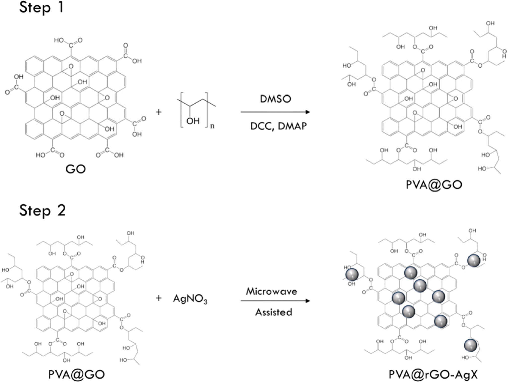 | ||
| Fig. 2 A diagram illustrating the process of creating hybrid systems made of PVA GO and silver denoted as PVA@rGO–AgX. Reproduced with permission.57 Copyright 2020 by the authors. Licensee MDPI, Basel, Switzerland. | ||
6.11. Hydrothermal synthesis
Hydrothermal synthesis generates well-dispersed AgNPs within polymer matrices under high-pressure conditions, ensuring enhanced biocompatibility and long-term stability.58 Table 2 provides a comparative overview of AgNP–PNC synthesis techniques, including their reaction conditions, polymer compatibility, advantages, disadvantages, and biomedical applications. It facilitates an informed selection of synthesis approaches based on the desired nanocomposite properties and specific medical applications.| Synthesis method | Reaction conditions | Polymer type | Advantages | Disadvantages | Applications |
|---|---|---|---|---|---|
| In situ polymerization | Controlled temperature, monomer–polymer interaction, catalyst-assisted reaction | PEG, PMMA, chitosan | Strong nanoparticle–polymer interaction, good dispersion, stable composite | Requires precise reaction conditions, potential polymer degradation | Drug delivery, tissue engineering, wound dressings |
| Solution blending | Polymer and AgNPs dissolved in solvent, mixed under controlled conditions | PVA, PEG, PU | Simple, cost-effective, scalable | Risk of AgNP aggregation, solvent removal required | Antimicrobial coatings, medical textiles, drug carriers |
| Melt mixing | High-temperature polymer melting, mechanical shear force for AgNP dispersion | PLA, PMMA, PDMS | No need for solvents, good dispersion, high mechanical properties | High temperature can degrade certain polymers and AgNPs | Orthopedic implants, antimicrobial coatings |
| Electrospinning | Polymer and AgNPs dissolved in a solution, electrostatic force used for nanofiber formation | PLA, PVA, chitosan | Produces ultrafine nanofibers, tunable porosity, high surface area | Requires optimized conditions, limited scalability | Wound dressings, scaffolds, filtration membranes |
| Layer-by-layer assembly | Sequential deposition of AgNPs and polymer layers on a substrate | PEO, PVP, chitosan | Precise thickness control, homogeneous nanoparticle distribution | Time-consuming, requires specialized equipment | Biosensors, antimicrobial coatings, tissue engineering |
| In situ reduction | Chemical reduction of silver precursor within polymer matrix | PVP, PEG, PMMA | Homogeneous nanoparticle distribution, good stability | Requires careful control of reaction parameters | Drug delivery, wound dressings, antibacterial coatings |
| Ultrasonication | High-frequency sound waves applied to AgNP–polymer mixture | PVA, chitosan, PEO | Good dispersion, prevents aggregation | Requires optimization to avoid polymer degradation | Biomedical coatings, antimicrobial films |
| Spray pyrolysis | Mist of AgNP–polymer precursor solution deposited on a heated substrate | PDMS, PU | Produces uniform AgNP–polymer coatings | High-temperature process, risk of polymer degradation | Antimicrobial medical devices, bioactive coatings |
| Sol–gel method | Hydrolysis and polymerization of precursors in solution | PMMA, PEG, PVA | High stability, precise control of AgNP size | Long processing time, requires careful pH and temperature control | Dental materials, tissue scaffolds, orthopedic applications |
| Microwave-assisted synthesis | Microwave irradiation applied to AgNP–polymer mixture | PLA, PEG, PVA | Rapid synthesis, energy efficient, uniform AgNP dispersion | Potential for overheating, polymer degradation | Drug delivery, antimicrobial textiles |
| Hydrothermal synthesis | High-pressure, high-temperature reaction in sealed vessel | Chitosan, PVA, PCL | Uniform AgNP distribution, biocompatible, good crystallinity | Requires specialized equipment, slow reaction time | Bone tissue engineering, biosensors, wound dressings |
7. AgNP-based PNC
A composite material combines substances with distinct physical and chemical properties at a macroscopic level, yielding unique properties unattainable by individual components. Composites consist of a matrix, typically the continuous phase, and reinforcements like particles, fibers, or layers. Recent advancements in nanocomposites, with features such as toughness nearly 1000 times greater than bulk counterparts, leverage nanoscale dimensions for high surface-to-volume ratios, enhancing interactions within the matrix and yielding multifunctional materials.59 PNCs containing 1–50 nm nanoparticles capitalize on the nanoscale's distinct physical and chemical properties. The polymer matrix in PNCs aids in binding, chemical protection, and orientation of nanofillers. AgNPs, known for conductivity, stability, catalytic activity, and optical properties, are commonly included in PNCs, enhancing performance and stability by acting as ion-capping agents.60The polymer matrix is an effective host for incorporating nanoparticles and controls nucleation to prevent excessive particle growth. Incorporating Ag nanoparticles significantly enhances the performance of the composite.61 AgNPs, which can be applied as coatings or additives, are used on various types of catheters, including PU ventricular catheters, commercially available under the brand names Silverline® and ON-Q SilverSoaker™.62 AgNPs are also present in hand gels63 and paints,64 providing durable antibacterial, antiviral, and antifungal properties. However, AgNPs alone face limitations due to high surface reactivity, which causes aggregation and reduces antimicrobial efficacy. To overcome this, researchers have focused on immobilizing AgNPs onto solid supports, with polymers as favored templates for easy incorporation, dispersion, and controlled synthesis. Silver–PNCs offer tailored antimicrobial coatings, sheets, or films that meet specific application needs, with natural polymers often preferred for their abundance, biocompatibility, and biodegradability.
7.1. AgNP-based natural PNC
AgNP-based biocidal coatings are crafted with biocompatible, non-toxic natural polymers like chitosan, alginate, and hyaluronic acid to tackle implant rejection, inflammation, and toxicity. AgNPs enhance chitosan's durability and antimicrobial properties. Nanotechnology increasingly favors eco-friendly natural polymers and nanoscale fillers to create bionanocomposites, especially AgNP composites. These natural polymer–AgNP composites improve mechanical, optical, and electrical properties, with polymers stabilizing AgNPs to prevent precipitation and agglomeration. AgNP-based natural PNCs can be synthesized using various methods, including mechanical mixing, in situ reduction of silver salts, and in situ polymerization.65 Alternative reduction methods, such as microwave and radiochemical techniques, are also employed.66 There have been numerous studies published on PNCs fabricated with a combination of AgNPs and natural polymers such as chitosan,67 gelatin 68, cellulose,69 collagen,70 alginate,71 starch,72 hyaluronic acid,73 silk fibroin,74 and carrageenans.757.2. AgNP-based synthetic PNC
Artificial synthetic polymers are nearly as diverse as natural ones and have rapidly gained use in medical applications, such as synthetic sutures made from polyesters and polyamides.76 These polymers are preferred for their customizable physical and chemical properties, often offering superior structural and mechanical strength compared to natural polymers. Their carbon-based composition allows them to closely mimic biological tissues, enhancing targeted interactions within the body. Reactive groups enable biofunctionalization, improving biocompatibility. Both biodegradable and non-biodegradable types are used, with biodegradable variants designed to degrade naturally after fulfilling their purpose, avoiding the need for surgical removal.77 Ideally, biodegradable synthetic polymers are designed to remain in the body only for their functional necessity and then degrade without further surgical intervention.788. Biomedical applications of AgNP–PNCs
AgNP–PNCs are promising materials in biomedical research due to their unique properties. They possess strong antimicrobial effects against bacteria, viruses, and fungi, making them ideal for wound dressings and infection control in medical devices. Additionally, their anti-inflammatory properties support potential use in treating inflammatory conditions. With biocompatibility and easy functionalization, AgNP–PNCs are also attractive for targeted drug delivery and imaging applications, offering significant potential for innovative biomedical technologies, as summarized in Fig. 3. | ||
| Fig. 3 Prospective and multipurpose biomedical applications of AgNP–PNCs. | ||
8.1. Antimicrobial coatings for medical devices
Antimicrobial refers to the capability to inhibit or eliminate microorganisms, including bacteria, viruses, fungi, and protozoans. Antimicrobial substances or agents are utilized to prevent or treat infections caused by these microorganisms. They are available in various forms, such as medications (including antibiotics, antivirals, and antifungals), disinfectants, sanitizers, and other products designed to control or eradicate harmful microorganisms from surfaces, objects, or living organisms. The antimicrobial properties of AgNP–PNCs play a crucial role in reducing the spread of infections and enhancing overall health and well-being. The process by which AgNPs are released from the polymer matrices and their antimicrobial action are depicted in Fig. 4 and 5, respectively. | ||
| Fig. 4 The schematic representation of the proposed release mechanism of AgNPs through polymeric matrices of AgNP–PNCs. Reproduced with permission.79 Copyright 2021 by Elsevier. | ||
 | ||
| Fig. 5 The schematic of antimicrobial actions of AgNPs–zeolite nanocomposite. Reproduced with permission.79 Copyright 2021 by Elsevier. | ||
Table 3 provides an overview of market-available AgNP–polymer nanocomposites used in wound care, surgical devices, orthopedic implants, dental hygiene, and infection control. It highlights the polymer type, manufacturer, application, and key functional features, offering insights into real-world implementations of AgNP–PNCs in biomedical fields.
| Product name | Manufacturer | Polymer type | Application | Key features |
|---|---|---|---|---|
| Acticoat™ | Smith & Nephew [https://www.smith-nephew.com/en/health-care-professionals/products/advanced-wound-management/acticoat-global] | Polyethylene (PE) | Antimicrobial wound dressing | Broad-spectrum antibacterial action, sustained silver release |
| SilvaSorb® | Medline [https://www.medline.com/product/SilvaSorb-Silver-Antimicrobial-Wound-Gel/Z05-PF00181] | PVA hydrogel | Wound healing, burn care | Moist wound healing environment, controlled silver release |
| Aquacel® Ag+ Extra | Convatec [https://www.convatec.com/advanced-wound-care/aquacel-family-of-dressings/] | Sodium carboxymethylcellulose | Chronic wounds, pressure ulcers | Enhanced absorption, silver-based antimicrobial action |
| Silverlon® | Argentum Medical [https://www.silverlon.com/products/catheter-dressings] | Nylon-based | Surgical wound dressing, burn care | Silver-coated antimicrobial fabric, promotes healing |
| SilvrSTAT® | AsepticMD [https://silvrstat.com/wp-content/uploads/2022/07/Product_Insert.pdf] | Chitosan-based hydrogel | Wound care, surgical incisions | Fast-acting antimicrobial gel, biocompatible |
| SilverShield® | Argentum Medical [https://www.microban.com/antimicrobial-solutions/technologies/microban-silver-technology] | Silicone-based | Catheters, medical device coatings | Prevents biofilm formation, extended silver ion release |
| SilverCare® Toothbrush | SilverCare [https://www.norwex.com/p/adult-silver-care-toothbrush-with-refill-light-blue?srsltid=AfmBOoot4qTh-IDjso-ehzXzbbX4zfpGATAheRrTwuQLqb1bEWnyCSc2] | Polypropylene (PP) | Dental hygiene | Antibacterial protection, silver-infused bristles |
| Silverlon® Catheter Wraps | Argentum Medical [https://www.silverlon.com/products/catheter-dressings] | Polyester | Central venous catheter protection | Reduces catheter-associated infections |
| Metal particles | Polymer matrix | Supporting materials | Purpose | Ref. |
|---|---|---|---|---|
| AgNP | Poly(hydroxymethyl 3,4-ethylenedioxythiophene) | Conjugated PNCs | Medical implant antimicrobial coating | 86 |
| Polystyrene | ||||
| (3-Aminopropyl)triethoxysilane | ||||
| AgNP | Chitosan | Antifungal | 87 | |
| AgNP | Chitosan | Antibacterial, antibiofilm, antifungal, antioxidant and wound-healing activities | 88 | |
| AgNP | Polysulfone (PSF), polyimide (PI) or polyetherimide (PEI) | Zeolite crystals | Antifungal | 89 |
| AgNP | Montmorillonite | Antifungal | 90 | |
| AgNP | Acrylic latexes | Antifungal | 91 | |
| AgNP | PDMS | Lecithin and montmorillonite | Antibacterial | 92 |
| AgNP | PLA | Polyethyleneimine (PEI) | Antimicrobial and antiviral | 93 |
| AgNP | GO | Antimicrobial | 94 |
8.2. Tissue engineering
AgNP–PNCs show strong potential in tissue engineering, offering antimicrobial properties that prevent implant infections while enhancing mechanical strength and electrical conductivity. Their polymer matrices add biocompatibility and controlled degradation, useful for applications like wound healing, bone regeneration, and implants.95 Hosseini et al. synthesized polyaniline (PANI) in bacterial cellulose/AgNP hydrogels with rose-like PANI morphologies, achieving high porosity (80%) and enhanced modulus (G′) under specific synthesis conditions.96 Zulkifli's team developed hydroxyethyl cellulose/AgNP scaffolds for skin tissue, showing high porosity, moderate degradation, and low cytotoxicity with hFB cells, supporting their potential as skin substitutes.97 Marsich engineered antibacterial alginate/hydroxyapatite scaffolds with AgNPs for bone grafts, achieving a 341.5 μm pore size and effective osteoblast proliferation with antimicrobial activity, highlighting their promise for tissue engineering.98 Table 5 summarizes the recent applications of AgNP–PNCs for tissue engineering purposes.| Metal particles | Polymer matrix | Supporting materials | Purpose | Ref. |
|---|---|---|---|---|
| Ag | Hydroxyethyl cellulose (HEC) | Implant and skin tissue engineering | 99 | |
| Ag | Chitosan, carboxymethyl cellulose | Cellulose nanowhiskers | Bone tissue engineering | 95 |
| Ag | Starch and gelatin | Bioactive glass particles (BG) | Bone tissue engineering and antibacterial | 100 |
| Ag | Bacterial cellulose, polyaniline, PEG | Nanocomposite aerogel | Soft tissue engineering and antibacterial | 96 |
| Ag | Agarose, chitosan | Soft tissue engineering and antibacterial | 101 |
8.3. Wound dressings and tissue scaffolds
AgNPs are highly valued in wound dressings and tissue scaffolds for their antibacterial, biocompatible, and tissue-regenerative properties. Integrating AgNPs into PNCs has shown significant efficacy in reducing bacterial growth and accelerating wound healing, with promising applications in tissue regeneration.51 However, further research is needed to confirm long-term biocompatibility and safety. Mehrabani et al. created silk fibroin/chitin nanocomposite scaffolds with AgNPs (0.001–0.1%), achieving strong antimicrobial activity, porosity, and biocompatibility, suitable for wound dressings.102 Additionally, cellulose nanofiber (CNF) films with AgNPs derived from Cassia alata demonstrated mechanical strength, water absorption, and potential anti-diabetic effects, suggesting a cost-effective wound-healing material.103 Ediyilyam et al. fabricated biogenic AgNP–chitosan scaffolds via lyophilization, using Mussaenda frondosa for synthesis. These biocompatible scaffolds exhibited enhanced biological and physical properties, showing potential for broader biomedical applications.104 Table 6 summarizes the recent applications of AgNP–PNCs for wound dressing purposes.| Metal particles | Polymer matrix | Supporting materials | Purpose | Ref. |
|---|---|---|---|---|
| Ag | Collagen | Catechin | Second degree infected burns | 105 |
| Angiogenic | ||||
| Antibacterial | ||||
| AgNP | Chitosan | Pectin | Wound healing | 106 |
| Ag | Starch | Nanocomposites hydrogel membranes | Wound dressing | 107 |
| PVA | Antimicrobial | |||
| Ag | Chitosan | Antibacterial antibiofilm and wound healing | 88 | |
| Ag | Sodium alginate | Nanocomposites hydrogel | 108 | |
| Ethylene glycol | ||||
| Acrylic acid | ||||
| Ag | Cellulose acetate | GO modified nanofibrous mats | Antimicrobial performance and cutaneous wound healing | 109 |
| PU | ||||
| AgNP | PVA | Xanthan gum | Dermal dressings antibacterial wound healing | 110 |
| Sodium carboxymethyl cellulose | Hypromellose | |||
| AgO | Carboxymethyl chitosan | Tragacanthin gum | Wound dressing | 111 |
| TiO2 | ||||
| Ag2O | Cellulose acetate | GO | Antibacterial wound healing | 112 |
| ZnS |
8.4. Drug delivery systems
AgNP–PNCs show strong potential in drug delivery systems, combining a polymer matrix with AgNPs. The high surface area and antimicrobial properties of AgNPs enhance drug solubility, controlled release, and targeted delivery to specific cells or tissues. The polymer matrix provides stability, while AgNPs improve drug loading and release efficiency.113 These nanocomposites promise advancements in drug delivery by offering enhanced solubility, controlled release, and precise targeting, though further research is needed to confirm their clinical safety. The proposed mechanism, including three main processes, is illustrated in Fig. 6. | ||
| Fig. 6 The proposed mechanism of predominant three processes involved in polymer nanocomposite-based drug delivery system. Reproduced with permission.114 Copyright 2021 by the authors. Licensee MDPI Basel Switzerland. | ||
Su et al. developed a chitosan–AgNP nanocomposite with a raspberry-like morphology combined with GO, using a microplasma process at room temperature and atmospheric pressure for efficient AgNP incorporation (Fig. 7).115 This pH-sensitive composite enables single or dual drug release: at pH 7.4, methyl blue released quickly in the first 10 hours, reaching 70–80% after 40–50 hours, while fluorescein sodium achieved a 45% release over 240 hours. The dual system slowed methyl blue release, with both rates accelerating at pH 4. The composite also showed strong antibacterial effects against E. coli and S. aureus, along with efficient photothermal conversion under near-infrared lasers, making it suitable for applications in healthcare, water treatment, and antimicrobial use.
 | ||
| Fig. 7 (a) The schematic diagram of the atmospheric pressure microplasma setup for the preparation of CS/AgNPs (b) CS + AgNO3 solution before and after RT-APM treatment and (c) the preparation of CS/RB-AgNPs/GO nanocomposite using a freeze-drying process. Reproduced with permission.115 Copyright 2021 by the authors. Licensee MDPI Basel Switzerland. | ||
Yadollahi et al. explored chitosan hydrogel beads crosslinked with sodium tripolyphosphate to encapsulate AgNPs for drug delivery.116 X-ray diffraction and SEM confirmed AgNP integration, with the beads exhibiting strong antibacterial effects against E. coli and S. aureus and enhanced swelling capacity. In vitro studies showed that AgNPs facilitated sustained drug release, increasing with higher AgNP concentrations, positioning these PNC beads as a promising drug delivery system. Neri et al. examined the release of silibinin, a liver-protective drug, from a PEG–PLA@Ag composite, where AgNPs responded to specific laser wavelengths via Surface Plasmon Resonance (SPR), generating localized heat for controlled drug release. This system's photothermal properties show potential for targeted drug delivery and photothermal therapies.117 Yang et al. developed C-β-CD-modified AgNPs with quaternary ammonium and amino groups for drug delivery.118 Thymol was loaded into this cyclodextrin–AgNP composite, with C-β-CD acting as both a reducing agent and stabilizer. The successful synthesis, confirmed by XRD, UV-vis, FT-IR, and 1H-NMR, showed effective dispersion and aggregation resistance. HS-SPME-GC-MS and UV-vis spectroscopy verified drug-loading efficiency, with a noted increase in particle size after drug loading. Table 7 summarizes the recent applications of AgNP–PNCs for drug delivery system.
| Metal particles | Polymer matrix | Supporting materials | Purpose | Ref. |
|---|---|---|---|---|
| Ag | Chitosan | Nanochitosan polymer composite | Drug delivery | 114 |
| Ag | Carboxymethylcellulose, poly(acrylamide) | Nanocomposite hydrogel | pH sensitive drug delivery | 119 |
| AgNP | Tragacanth gum | Nanocomposite hydrogel | Drug delivery and inactivation of multidrug-resistant bacteria | 120 |
8.5. Biosensors for disease diagnosis
AgNP–PNCs hold great potential for disease diagnosis biosensors due to AgNPs' high surface area and signal amplification, making them ideal for biosensing.121,122 As shown in Fig. 8, PNCs with metallic nanoparticles like Ag and Au are well-suited for these applications. The polymer matrix in these composites provides stability, while AgNPs enhance sensitivity and specificity, which are useful for detecting cancer biomarkers, infectious diseases, and cardiovascular markers, as well as small molecules like drugs and toxins. AgNP–PNCs can be tailored to specific biosensing needs, with applications for electrochemical and optical sensors. Ananth et al. developed AgNP biosensors coated with BSA and PVA, showing wavelength shifts and no behavioral toxicity in a mouse model, indicating potential in examining cellular processes.121 Yazdanparast et al. introduced a biosensor for detecting MCF-7 breast cancer cells using an aptamer-based AgNP assay, achieving high selectivity and a detection limit of 25 cells, suitable for early breast cancer detection.123 | ||
| Fig. 8 A graphical representation to depict the various forms of MNPs (such as AuNPs or AgNPs) and the use of ligands to stabilize them. The optical and electrochemical characteristics of the MNPs were examined using techniques for optical and electrochemical sensing confirming their properties. Reproduced with permission.122 Copyright 2021 by the authors. Licensee MDPI Basel Switzerland. | ||
8.6. Anticancer agents and cancer therapy
AgNP–PNCs show promise as anticancer agents, leveraging AgNPs' cell-killing and antimicrobial properties, with the polymer matrix providing stability and biocompatibility. These composites enable targeted drug delivery and can act as photothermal agents, where AgNPs generate heat upon near-infrared activation to destroy cancer cells. AgNP–PNCs also enhance chemotherapy by boosting drug uptake and reducing side effects, potentially preventing drug resistance.124 Senthilkumar et al. developed a PDMA/Ag nanocomposite using a chemical oxidative method, achieving a high AgNPs content (42.1%) with spherical particles around 25 nm.125 The PDMA/Ag composite showed strong antibacterial properties and significantly reduced cell viability in HeLa cancer cells compared to PDMA alone. Haghparasti et al. synthesized AgNPs with white tea extract, forming a Wt/Ag@Mt nanocomposite with antioxidant properties. The Wt/Ag@Mt-NCs displayed a crystalline structure and effective cytotoxicity against MOLT-4 cells, with an IC50 of 0.0039 μM, outperforming drugs like doxorubicin and cisplatin. These bio-synthesized nanoparticles hold promise for pharmaceutical applications, offering an environmentally sustainable approach.1268.7. Dental materials for caries and periodontal disease prevention
AgNPs have been widely explored for their antimicrobial potential and applications in dentistry. Integrating AgNPs into PNCs enhances the mechanical, physical, and antibacterial properties of dental materials, aiding in dental imaging, where their optical properties improve contrast in X-rays, aiding cavity and periodontal disease detection.127–129 AgNPs are particularly effective against bacteria causing dental caries and periodontal disease; when embedded in dental composites and coatings, they inhibit bacterial growth more effectively than traditional materials. This efficacy is due to AgNPs' small size, allowing them to penetrate bacterial cells and disrupt functions, while the polymer matrix ensures even distribution throughout the material. To address the need for low-cost, user-friendly detection of dental caries and periodontal diseases, Ma et al. developed a wearable mouthguard with Au@Ag nanorods and PDMS, which detects lesion sites by a color change due to H2S gas from bacterial decay.127 The mouthguard is mechanically robust, chemically stable, biocompatible, and accurately locates lesion sites, showing promise for self-monitoring oral health and targeted treatment. Table 8 summarizes the recent applications of AgNP–PNCs for dental materials and treatment.| Metal particles | Polymer matrix | Supporting materials | Purpose | Ref. |
|---|---|---|---|---|
| AgNPs | Polydopamine (PDA) | Hydroxyapatite nanowires | Long-term clinical restorations in dental treatment | 128 |
| AgNPs | Resin | Halloysite nanotubes (HNT) | Restorative dental material for patients with high risk of dental caries | 129 |
| AgNP | Acrylic resin | Sodium citrate | Antimicrobial effects against C. albicans biofilm | 130 |
| Dental treatment | ||||
| AgNP | Gelatin | Halloysite nanotubes (HNT) | Dental prostheses | 131 |
| PMMA | ||||
| AgNP | PMMA | Boron nitride nanosheets (h-BNNs) | Oral denture bases | 132 |
8.8. Other biomedical applications
AgNP–PNCs have emerged as promising candidates for various biomedical applications, including optical materials such as contact lenses, orthopedic implants, neural interfaces, medical textile coatings, skin protection, cosmetics, and anti-inflammatory products. The diverse biomedical uses of AgNP–PNCs are comprehensively summarized in Table 9.| Metal particles | Polymer matrix | Supporting materials | Purpose | Reference |
|---|---|---|---|---|
| AgNPs | PVA | PNC hydrogel | Sensing devices cosmetic | 133 |
| Poly(vinyl pyrrolidone) | Medical contact lenses | |||
| AgNPs | Polyvinyl-pyrrolidone (PVP) | Poly(2-hydroxyethyl methacrylate) (pHEMA) | Contact lens | 134 |
| AuNPs | ||||
| Spherical AgNPs | pHEMA | Contact lens blue-yellow color vision deficiency | 135 | |
| AgNP | Arabinoxylan-co-acrylic acid | GO | Orthopedic implant | 136 |
| Al2O3NP | Nano-hydroxyapatite (nHAp) | |||
| AgNP Ti | Amorphous hydrocarbon | Nanocomposite coating | Orthopedic implant | 137 |
| AgNP Ti | Nanocomposite Ag layer | Orthopedic implant | 138 | |
| Ag nanowires | Poly(ε-decalactone) (EDL) | Electrically conducting neural interface biomaterial | 139 | |
| Ag/Au nanowires | Poly(3-methoxypropyl acrylate) (PMC3A) | Long-term implantable flexible and transparent neural interface | 140 | |
| Au nanoshell | PDMS | Tough stretchable and self-healing polymer | Neuroprosthetics for in vivo bidirectional signaling | 141 |
| AgNP | Methylenebis(phenyl urea) (MPU) | |||
| Isophorone bisurea units (IU) | ||||
| Silver nanowires (AgNW) | Poly(ε-decalactone) | CNT | Neural interface materials | 142 |
| Poly(hydroxymethyl 3,4-ethylenedioxythiophene) microspheres (MSP) | ||||
| AgNP | Carboxymethyl chitosan | Cotton fabric | Antibacterial and antifungal textile coating | 143 |
| AgNP | Chitosan | One-pot microwave synthesized PNC | Antibacterial antioxidant wound healing | 144 |
| Polyethylene oxide | ||||
| Photothermal AgNPs | Hyaluronic acid | Tannic acids (TA) | Antibacterial and antioxidant | 145 |
| Au, Ag and ZnO NPs | PVA | MMT clay | Skin protection cosmetics UV protection coating | 146 |
| Starch | ||||
| AgNP | Poly(amino acid) | Nanocomposite fibrous mesh | Anti-inflammatory drug | 147 |
9. Challenges and solutions for successful biomedical application of AgNP–PNCs
The successful translation of AgNP–PNCs into biomedical applications requires addressing key biocompatibility, stability, cost, standardization, and regulatory hurdles. While AgNP–PNCs hold great promise in infection control, drug delivery, and implantable medical devices, challenges related to toxicity, stability, large-scale production, and regulatory compliance must be resolved to facilitate clinical adoption. This section presents current challenges along with strategic solutions to enhance the real-world application of AgNP–PNCs in biomedical fields.9.1. Biocompatibility and cytotoxicity concerns
AgNPs exhibit size- and dose-dependent toxicity, which can induce oxidative stress, DNA damage, mitochondrial dysfunction, and inflammatory responses in human cells, limiting their safe use in biomedical applications.148 The mechanism of toxicity primarily involves the generation of ROS, disruption of cellular membranes, and interference with protein function. The immune response triggered by AgNP exposure remains a critical issue, requiring further investigation into inflammatory pathways and potential long-term effects. To enhance biocompatibility, AgNPs can be functionalized with biocompatible coatings, such as PEG, chitosan, or albumin, which minimize direct AgNP–cell interactions and reduce inflammatory responses.149 Hybrid nanostructures, such as AgNPs embedded in lipid or polymeric nanocarriers, provide a controlled release mechanism that mitigates acute toxicity while preserving therapeutic effects. Surface engineering techniques, including biomimetic coatings, can improve AgNP compatibility with biological environments. Preclinical evaluation through advanced organ-on-a-chip models and 3D cell cultures will provide more accurate predictions of AgNP–PNC biocompatibility before in vivo testing.9.2. Stability challenges in biological environments
AgNP–PNC stability is a critical factor influencing long-term efficacy and safety in biomedical applications. The tendency of AgNPs to aggregate, oxidize, and release excess silver ions in physiological conditions can significantly impact their effectiveness.150 Key factors affecting stability include pH variations, ionic strength, temperature fluctuations, and interactions with biological macromolecules. To counteract these challenges, surface modifications using stabilizing agents such as PEG, surfactants (SDS, CTAB, PVP), and natural polymers (chitosan, alginate, or dextran) have been shown to prevent aggregation and enhance dispersion in biological media. Incorporating AgNPs into a crosslinked polymeric network improves nanoparticle stability by restricting their movement and controlling silver ion release. Hybrid AgNP-based systems, such as AgNP-loaded mesoporous silica nanoparticles or graphene-based nanocomposites, offer improved chemical stability and prolonged antimicrobial activity, making them promising candidates for long-term medical implants and wound dressings.9.3. Cost constraints and scalable production
The large-scale production of AgNP–PNCs remains expensive due to high silver consumption, energy-intensive synthesis processes, and the need for advanced functionalization techniques. Traditional chemical synthesis methods require toxic reducing agents, which complicate purification steps and increase production costs. To make AgNP–PNCs more commercially viable, cost-effective and sustainable synthesis approaches have been explored. Green synthesis techniques, utilizing plant extracts, microbial cultures, or biomolecules as reducing and stabilizing agents, have demonstrated promise in producing AgNPs with minimal environmental impact and reduced production costs.151 Microwave-assisted synthesis has emerged as a rapid and energy-efficient method that ensures controlled AgNP growth with minimal precursor wastage. Adopting scalable microfluidic reactors allows for continuous synthesis, ensuring batch-to-batch consistency and optimized nanoparticle characteristics. Advances in automated in-line characterization will enhance production efficiency, enabling real-time monitoring of AgNP properties during synthesis.9.4. Standardization issues in AgNP–PNC fabrication
The absence of standardized protocols in AgNP–PNC synthesis leads to inconsistencies in particle size, shape, surface charge, and stability, affecting reproducibility and clinical reliability. Variability in nanoparticle formulations presents significant challenges for regulatory approval and large-scale manufacturing. To address these challenges, it is essential to establish standardized synthesis and characterization protocols that define optimal nanoparticle physicochemical properties for biomedical applications. Characterization techniques such as dynamic light scattering (DLS), transmission electron microscopy (TEM), zeta potential analysis, and X-ray photoelectron spectroscopy (XPS) should be employed systematically to ensure batch consistency. Regulatory bodies such as the International Organization for Standardization (ISO) and the National Institute of Standards and Technology (NIST) are working toward defining standard measurement frameworks for nanomaterials. Establishing reference AgNP materials for biomedical applications will facilitate cross-laboratory comparisons and regulatory compliance.9.5. Regulatory approval and clinical translation
AgNP-based materials face regulatory barriers due to concerns over nanoparticle toxicity, environmental persistence, and potential long-term effects on human health. Regulatory agencies such as the U.S. Food and Drug Administration (FDA), European Medicines Agency (EMA), and World Health Organization (WHO) require comprehensive preclinical and clinical evaluations before approving AgNP–PNCs for medical applications. Currently, FDA-approved AgNP-based products are limited, with most applications restricted to topical antimicrobials, wound dressings, and medical coatings. For systemic applications, including drug delivery and injectable nanomedicines, extensive pharmacokinetic and toxicological assessments are required to evaluate silver biodistribution, clearance rates, and immunogenicity. Compliance with Good Manufacturing Practices (GMP) and ISO nanotechnology standards will be essential for commercializing AgNP–PNCs in clinical settings. To facilitate regulatory approval, collaborative efforts between academia, industry, and regulatory agencies are needed to develop standardized safety assessment frameworks. Risk-benefit analyses, focusing on the therapeutic advantages of AgNPs versus potential long-term risks, will be vital in shaping future regulatory guidelines.9.6. Comparison with alternative antimicrobial nanomaterials
While AgNPs are among the most effective antimicrobial agents, concerns about toxicity, environmental impact, and potential bacterial resistance have led researchers to explore alternative nanomaterials with comparable efficacy and lower cytotoxicity. ZnO, copper nanoparticles (CuNPs), TiO2, and carbon-based nanomaterials such as GO have demonstrated broad-spectrum antimicrobial properties with minimal adverse effects on mammalian cells. ZnO nanoparticles exhibit photoactivated antimicrobial properties, generating ROS under UV exposure, making them ideal for antimicrobial coatings and wound dressings. CuNPs provide effective bacterial inhibition, though their higher cytotoxicity and oxidative stress potential require careful dose optimization. TiO2, often combined with AgNPs, enhances photodynamic antibacterial therapy, offering a synergistic approach to infection control. Carbon-based materials, including GO and CNTs, exhibit membrane-disrupting antibacterial mechanisms, reducing the risk of resistance development. A comparative evaluation of AgNP–PNCs with alternative antimicrobial nanomaterials will provide deeper insights into material selection for specific biomedical applications, ensuring optimal efficacy with minimal side effects. Future research should explore hybrid nanostructures, integrating AgNPs with other nanomaterials to create multifunctional antimicrobial coatings with controlled release capabilities.10. Future perspective of AgNP–PNCs for biomedical applications
The potential of AgNP–PNCs in biomedical applications continues to be a promising field of study. The unique properties of AgNPs, including antimicrobial activity, high thermal and electrical conductivity, and enhanced mechanical properties, when integrated with polymer matrices, create innovative materials suited for various medical applications. However, the full realization of these materials in clinical settings will depend on advancements across several fronts. As research progresses, the biocompatibility of AgNP–PNCs remains a primary concern. Innovations in surface modification and functionalization of nanoparticles to reduce cytotoxicity while maintaining antimicrobial efficacy will be crucial. Future studies must establish robust biocompatibility profiles through comprehensive in vitro and in vivo assessments.152Developing cost-effective, scalable manufacturing processes that can produce high-quality AgNP–PNCs consistently is essential. Techniques like 3D printing and automated synthesis could revolutionize the production of these nanocomposites, making them more accessible for widespread medical use.153,154 Darbandi et al. reported significant advancements in the fabrication of 3D-printed dental restorations using resins modified with zirconium dioxide (ZrO2) nanoparticles and AgNP-immobilized halloysite nanotubes (HNC/Ag).154 Their study highlights the potential of these nanoparticles to enhance the mechanical properties of 3D-printed resins, which is crucial for biomedical applications. They evaluated the impact of different mass fractions of ZrO2 and HNC/Ag on the flexural strength, flexural modulus, fracture toughness, and microhardness of the resins. The optimal ratios identified were ZrO2 at 4% and HNC/Ag at 5%, which significantly increased the flexural strength, flexural modulus, and fracture toughness of the resin. Moreover, higher concentrations of ZrO2 (up to 16%) and HNC/Ag (up to 7.5%) were found to improve the hardness of the resin. The study underscores the potential of advanced manufacturing techniques in developing high-performance, biocompatible materials for biomedical applications. By leveraging the properties of ZrO2 and HNC/Ag nanoparticles, it is possible to create dental restorations with superior mechanical and physical characteristics, paving the way for more durable and practical biomedical devices. Future research could explore the integration of these nanoparticles into other types of resins and their applications in various biomedical fields beyond dentistry. The use of 3D printing technology, combined with advanced nanoparticle modifications, holds promise for developing innovative and highly functional biomedical materials.
AgNP–PNCs show promise in targeted drug delivery applications. The ability to engineer nanoparticles that can navigate to specific tissue sites and release therapeutic agents in a controlled manner is an area ripe for development. Future research could focus on enhancing the targeting capabilities of these systems, possibly using ligand-directed strategies.155 The optical properties of AgNPs can be exploited to improve diagnostic imaging technologies. AgNP–PNCs could be designed to enhance contrast in imaging modalities such as MRI or ultrasound, providing clearer, more precise images that could aid in the early detection of diseases. As with any emerging medical technology, regulatory challenges must be navigated to ensure safety and efficacy before clinical adoption. Establishing standardized protocols for the testing, use, and disposal of AgNP–PNCs will be necessary. Additionally, ethical considerations, particularly concerning long-term effects and environmental impact, will require ongoing dialogue among scientists, regulators, and the public.
While current applications are promising, exploring AgNP–PNCs in regenerative medicine, neural interfaces, and wearable health monitors could provide new avenues for treatment and patient care. Research into these applications could open up novel therapeutic avenues and potentially revolutionize aspects of healthcare. In the face of rising antibiotic resistance, AgNP–PNCs could offer a potent alternative due to their broad-spectrum antimicrobial properties. Future research might optimize these properties to create surfaces and devices resistant to colonization by pathogenic microbes, thus reducing the incidence of hospital-acquired infections. The future of AgNP–PNCs in biomedical applications looks promising, with potential breakthroughs spanning various medical fields. The interdisciplinary nature of this research, involving materials science, nanotechnology, medicine, and engineering, suggests a collaborative approach will be pivotal. Continuous evaluation and adaptation to emerging challenges and innovations will be key to its success as technology matures.
11. Conclusion
AgNP–PNCs represent a promising frontier in biomedical materials, combining the antimicrobial, optical, and conductive properties of AgNPs with the versatility, stability, and biocompatibility of polymers. These nanocomposites have demonstrated exceptional potential across various medical applications, from antimicrobial coatings for medical devices to advanced wound dressings, tissue engineering scaffolds, and drug delivery systems. The development and optimization of AgNP–PNCs have significantly enhanced the functionality of these materials, making them highly suitable for addressing critical health challenges and improving patient outcomes. Despite these advances, challenges remain. The biocompatibility and cytotoxicity of AgNPs, dependent on particle size, shape, and concentration, are essential to ensure safe clinical applications. Moreover, standardizing synthesis methods and improving scalability are vital for adopting AgNP–PNCs widely in commercial and clinical settings. Addressing these factors will facilitate regulatory approvals and streamline the integration of AgNP–PNCs into various biomedical fields.This review has comprehensively analyzed the current landscape, covering recent advancements in synthesis and functionalization, innovative applications, and future research directions for AgNP–PNCs. By fostering interdisciplinary research and collaboration, the field is poised to overcome present limitations and unlock the full potential of AgNP–PNCs. As research progresses, these nanocomposites could revolutionize medical materials, offering safer, more effective solutions for various biomedical applications.
Data availability
No primary research results, software or code have been included and no new data were generated or analyzed as part of this review.Author contributions
Mohammad Harun-Ur-Rashid: conceptualization, data curation, formal analysis, writing original draft, and review. Tahmina Foyez: data curation, formal analysis, writing original draft, and review. Suresh Babu Naidu Krishna: formal analysis, data curation, resources, visualization, writing-review, and editing. Sudhakar Poda: data curation, formal analysis, validation. Abu Bin Imran: conceptualization, supervision, data curation, visualization, writing original draft, writing review and editing.Conflicts of interest
All authors certify that they have no affiliations with or involvement in any organization or entity with any financial interest or non-financial interest in the subject matter or materials discussed in this manuscript.Acknowledgements
M. Harun-Ur-Rashid would like to thank the International University of Business Agriculture and Technology (IUBAT) for providing the necessary resources and support for this research. A. B. Imran is thankful to the Committee for Advanced Studies and Research (CASR) in BUET for funding. KSBN would like to thank Durban University of Technology for the research fellowship.References
- A. Ullah, M. Ullah and S. I. Lim, Recent advancements in nanotechnology based drug delivery for the management of cardiovascular disease, Curr. Probl. Cardiol., 2024, 49(4), 102396, DOI:10.1016/j.cpcardiol.2024.102396.
- A. S. Monem, et al., Assessment of the Effect of Surface Modification of Metal Oxides on Silver Nanoparticles: Optical Properties and Potential Toxicity, Cell Biochem. Biophys., 2024, 82(2), 1213–1224, DOI:10.1007/s12013-024-01272-2.
- H. Jangid, et al., Advancing biomedical applications: an in-depth analysis of silver nanoparticles in antimicrobial, anticancer, and wound healing roles, Front. Pharmacol, 2024, 15, 1438227, DOI:10.3389/fphar.2024.1438227.
- S. Raza, et al., Biosynthesis of silver nanoparticles for the fabrication of non cytotoxic and antibacterial metallic polymer based nanocomposite system, Sci. Rep., 2021, 11(1), 10500, DOI:10.1038/s41598-021-90016-w.
- S. M. Nasef, E. E. Khozemy and G. A. Mahmoud, pH-responsive chitosan/acrylamide/gold/nanocomposite supported with silver nanoparticles for controlled release of anticancer drug, Sci. Rep., 2023, 13(1), 7818, DOI:10.1038/s41598-023-34870-w.
- N. K. Dhiman, S. Agnihotri and R. Shukla, Silver-Based Polymeric Nanocomposites as Antimicrobial Coatings for Biomedical Applications, in Nanotechnology in Modern Animal Biotechnology: Recent Trends and Future Perspectives, ed. S. Singh and P. K. Maurya, Springer, Singapore, 2019, pp. 115–171, DOI:10.1007/978-981-13-6004-6_4.
- S. Renu, K. S. Shivashangari and V. Ravikumar, Incorporated plant extract fabricated silver/poly-D,l-lactide-co-glycolide nanocomposites for antimicrobial based wound healing, Spectrochim. Acta, Part A, 2020, 228, 117673, DOI:10.1016/j.saa.2019.117673.
- A. Woźniak, et al., Size and shape-dependent cytotoxicity profile of gold nanoparticles for biomedical applications, J. Mater. Sci.: Mater. Med., 2017, 28(6), 92, DOI:10.1007/s10856-017-5902-y.
- B. Reidy, et al., Mechanisms of Silver Nanoparticle Release, Transformation and Toxicity: A Critical Review of Current Knowledge and Recommendations for Future Studies and Applications, Materials, 2013, 6, 2295–2350, DOI:10.3390/ma6062295.
- O. Gherasim, et al., An Updated Review on Silver Nanoparticles in Biomedicine, Nanomaterials, 2020, 10(11), 2318, DOI:10.3390/nano10112318.
- A.-C. Burduşel, et al., Biomedical Applications of Silver Nanoparticles: An Up-to-Date Overview, Nanomaterials, 2018, 8(9), 681, DOI:10.3390/nano8090681.
- S. S. D. Kumar, et al., Recent advances on silver nanoparticle and biopolymer-based biomaterials for wound healing applications, Int. J. Biol. Macromol., 2018, 115, 165–175, DOI:10.1016/j.ijbiomac.2018.04.003.
- M. A. Wahab, et al., Silver Nanoparticle-Based Nanocomposites for Combating Infectious Pathogens: Recent Advances and Future Prospects, Nanomaterials, 2021, 11(3), 581, DOI:10.3390/nano11030581.
- A. Uthaman, H. M. Lal and S. Thomas, Fundamentals of silver nanoparticles and their toxicological aspects, in Polymer Nanocomposites Based on Silver Nanoparticles: Synthesis, Characterization Applications, ed. H. M. Lal, et al., Springer, Gewerbestrasse 11, 6330 Cham, Switzerland, 2021, pp. 1–24, DOI:10.1007/978-3-030-44259-0_1.
- O. A. Yeshchenko, et al., Surface plasmon as a probe for melting of silver nanoparticles, Nanotechnology, 2009, 21(4), 045203, DOI:10.1088/0957-4484/21/4/045203.
- N. Karak, R. Konwarh and B. Voit, Catalytically active vegetable-oil-based thermoplastic hyperbranched polyurethane/silver nanocomposites, Macromol. Mater. Eng., 2010, 295(2), 159–169, DOI:10.1002/mame.200900211.
- A. Menazea, Femtosecond laser ablation-assisted synthesis of silver nanoparticles in organic and inorganic liquids medium and their antibacterial efficiency, Radiat. Phys. Chem., 2020, 168, 108616, DOI:10.1016/j.radphyschem.2019.108616.
- K. M. Kabusure, et al., Optical characterization of DNA origami-shaped silver nanoparticles created through biotemplated lithography, Nanoscale, 2022, 14(27), 9648–9654, 10.1039/D1NR06256E.
- A. Gubarev, et al., Ag (0) nanoparticles stabilized with Poly (Ethylene Glycol) s modified with amino groups: formation and properties in solutions, Colloid J., 2019, 81, 226–234 CrossRef CAS.
- S. Iravani, et al., Synthesis of silver nanoparticles: chemical, physical and biological methods, Res. Pharm. Sci., 2014, 9(6), 385–406 CAS.
- S. Ibrahim, et al., Optimization for biogenic microbial synthesis of silver nanoparticles through response surface methodology, characterization, their antimicrobial, antioxidant, and catalytic potential, Sci. Rep., 2021, 11(1), 770, DOI:10.1038/s41598-020-80805-0.
- D. B. Aguiar, et al., Obtaining Ultrafine Dispersions of Silver Particles in Poly(vinyl Alcohol) Matrix Using Mechanical Alloying, Polymers, 2022, 14(17), 3588, DOI:10.3390/polym14173588.
- S. Das, et al., Biocompatible silver nanoparticles: An investigation into their protein binding efficacies, anti-bacterial effects and cell cytotoxicity studies, J. Pharm. Anal., 2021, 11(4), 422–434, DOI:10.1016/j.jpha.2020.12.003.
- M. Retout, I. Jabin and G. Bruylants, Synthesis of Ultrastable and Bioconjugable Ag, Au, and Bimetallic Ag_Au Nanoparticles Coated with Calix[4]arenes, ACS Omega, 2021, 6(30), 19675–19684, DOI:10.1021/acsomega.1c02327.
- R. Deka, et al., Highly stable silver nanoparticles containing guar gum modified dual network hydrogel for catalytic and biomedical applications, Carbohydr. Polym., 2020, 248, 116786, DOI:10.1016/j.carbpol.2020.116786.
- A. L. Nogueira, et al., Influence of process parameters and scalability of the semi-batch production of functionalized silver nanoparticles, Can. J. Chem. Eng., 2016, 94(8), 1472–1485, DOI:10.1002/cjce.22530.
- A. J. Friedman, et al., Antimicrobial and Anti-Inflammatory Activity of Chitosan–Alginate Nanoparticles: A Targeted Therapy for Cutaneous Pathogens, J. Invest. Dermatol., 2013, 133(5), 1231–1239, DOI:10.1038/jid.2012.399.
- K. A. Milakin, et al., Biocompatible and antibacterial gelatin-based polypyrrole cryogels, Polymer, 2020, 197, 122491, DOI:10.1016/j.polymer.2020.122491.
- E. G. Arafa, et al., Eco-friendly and biodegradable sodium alginate/quaternized chitosan hydrogel for controlled release of urea and its antimicrobial activity, Carbohydr. Polym., 2022, 291, 119555, DOI:10.1016/j.carbpol.2022.119555.
- T. Aziz, et al., Developing multifunctional cellulose derivatives for environmental and biomedical applications: Insights into modification processes and advanced material properties, Int. J. Biol. Macromol., 2024, 278, 134695, DOI:10.1016/j.ijbiomac.2024.134695.
- E. Ojogbo, E. O. Ogunsona and T. H. Mekonnen, Chemical and physical modifications of starch for renewable polymeric materials, Materials Today Sustainability, 2020, 7–8, 100028, DOI:10.1016/j.mtsust.2019.100028.
- B. Kundu, et al., Silk fibroin biomaterials for tissue regenerations, Adv. Drug Delivery Rev., 2013, 65(4), 457–470, DOI:10.1016/j.addr.2012.09.043.
- S. Tiwari and P. Bahadur, Modified hyaluronic acid based materials for biomedical applications, Int. J. Biol. Macromol., 2019, 121, 556–571, DOI:10.1016/j.ijbiomac.2018.10.049.
- A. Irastorza, et al., The versatility of collagen and chitosan: From food to biomedical applications, Food Hydrocolloids, 2021, 116, 106633, DOI:10.1016/j.foodhyd.2021.106633.
- I. J. Das and T. Bal, Exploring carrageenan: From seaweed to biomedicine—A comprehensive review, Int. J. Biol. Macromol., 2024, 268, 131822, DOI:10.1016/j.ijbiomac.2024.131822.
- I. Miranda, et al., Properties and Applications of PDMS for Biomedical Engineering: A Review, J. Funct. Biomater., 2022, 13(1), 2, DOI:10.3390/jfb13010002.
- A. Alexander, et al., Polyethylene glycol (PEG)–Poly(N-isopropylacrylamide) (PNIPAAm) based thermosensitive injectable hydrogels for biomedical applications, Eur. J. Pharm. Biopharm., 2014, 88(3), 575–585, DOI:10.1016/j.ejpb.2014.07.005.
- R. A. Rebia, et al., Biodegradable PHBH/PVA blend nanofibers: Fabrication, characterization, in vitro degradation, and in vitro biocompatibility, Polym. Degrad. Stab., 2018, 154, 124–136, DOI:10.1016/j.polymdegradstab.2018.05.018.
- A. M. Maadani and E. Salahinejad, Performance comparison of PLA- and PLGA-coated porous bioceramic scaffolds: Mechanical, biodegradability, bioactivity, delivery and biocompatibility assessments, J. Controlled Release, 2022, 351, 1–7, DOI:10.1016/j.jconrel.2022.09.022.
- B. Naureen, et al., Recent advances in tissue engineering scaffolds based on polyurethane and modified polyurethane, Mater. Sci. Eng., C, 2021, 118, 111228, DOI:10.1016/j.msec.2020.111228.
- M. Wekwejt, et al., Influence of several biodegradable components added to pure and nanosilver-doped PMMA bone cements on its biological and mechanical properties, Mater. Sci. Eng., C, 2020, 117, 111286, DOI:10.1016/j.msec.2020.111286.
- Q. Lei, et al., Sol–gel-based advanced porous silica materials for biomedical applications, Adv. Funct. Mater., 2020, 30(41), 1909539, DOI:10.1002/adfm.201909539.
- H. B. Dias, et al., Titanium dioxide and modified titanium dioxide by silver nanoparticles as an anti biofilm filler content for composite resins, Dent. Mater., 2019, 35(2), e36–e46, DOI:10.1016/j.dental.2018.11.002.
- A. Szcześ, L. Hołysz and E. Chibowski, Synthesis of hydroxyapatite for biomedical applications, Adv. Colloid Interface Sci., 2017, 249, 321–330, DOI:10.1016/j.cis.2017.04.007.
- R. Gopikrishnan, et al., Synthesis, characterization and biocompatibility studies of zinc oxide (ZnO) nanorods for biomedical application, Nano-Micro Lett., 2010, 2(1), 31–36, DOI:10.1007/BF03353614.
- M. A. Dheyab, et al., Synthesis and coating methods of biocompatible iron oxide/gold nanoparticle and nanocomposite for biomedical applications, Chin. J. Phys., 2020, 64, 305–325, DOI:10.1016/j.cjph.2019.11.014.
- P. Sarkar, et al., Chapter 20 – Biocompatibility and biomedical applications of various carbon-based materials, in Handbook of Carbon-Based Nanomaterials, ed. S. Thomas, et al., Elsevier, 2021, pp. 829–875, DOI:10.1016/B978-0-12-821996-6.00015-4.
- P. A. Zapata, et al., Nanocomposites based on polyethylene and nanosilver particles produced by metallocenic “in situ” polymerization: synthesis, characterization, and antimicrobial behavior, Eur. Polym. J., 2011, 47(8), 1541–1549, DOI:10.1016/j.eurpolymj.2011.05.008.
- S. B. Aziz, et al., Polymer blending as a novel approach for tuning the SPR peaks of silver nanoparticles, Polymers, 2017, 9(10), 486, DOI:10.3390/polym9100486.
- N. Vidakis, et al., Three-dimensional printed antimicrobial objects of polylactic acid (PLA)-silver nanoparticle nanocomposite filaments produced by an in-situ reduction reactive melt mixing process, Biomimetics, 2020, 5(3), 42, DOI:10.3390/biomimetics5030042.
- R. Thomas, et al., Electrospun polycaprolactone membrane incorporated with biosynthesized silver nanoparticles as effective wound dressing material, Appl. Biochem. Biotechnol., 2015, 176, 2213–2224, DOI:10.1007/s12010-015-1709-9.
- X. Liu, et al., Synthesis and characterization of novel antibacterial silver nanocomposite nanofiltration and forward osmosis membranes based on layer-by-layer assembly, Water Res., 2013, 47(9), 3081–3092, DOI:10.1016/j.watres.2013.03.018.
- J. Upadhyay, et al., Antibacterial and hemolysis activity of polypyrrole nanotubes decorated with silver nanoparticles by an in-situ reduction process, Mater. Sci., 2015, 54, 8–13 CAS.
- S. Jaworski, et al., Graphene oxide-based nanocomposites decorated with silver nanoparticles as an antibacterial agent, Nanoscale Res. Lett., 2018, 13, 1–17, DOI:10.1186/s11671-018-2533-2.
- M. Keskar, et al., Synthesis and characterization of silver nanoparticle-loaded amorphous calcium phosphate microspheres for dental applications, Nanoscale Adv., 2019, 1(2), 627–635, 10.1039/C8NA00281A.
- H. Bagheri and S. Banihashemi, Sol–gel-based silver nanoparticles-doped silica–Polydiphenylamine nanocomposite for micro-solid-phase extraction, Anal. Chim. Acta, 2015, 886, 56–65, DOI:10.1016/j.aca.2015.06.012.
- A. Nicosia, et al., Polymer-based graphene derivatives and microwave-assisted silver nanoparticles decoration as a potential antibacterial agent, Nanomaterials, 2020, 10(11), 2269, DOI:10.3390/nano10112269.
- N.-T. Nguyen and J.-H. Liu, A green method for in situ synthesis of poly (vinyl alcohol)/chitosan hydrogel thin films with entrapped silver nanoparticles, J. Taiwan Inst. Chem. Eng., 2014, 45(5), 2827–2833, DOI:10.1016/j.jtice.2014.06.017.
- K. K. Sadasivuni, et al., Silver nanoparticles and its polymer nanocomposites—Synthesis, optimization, biomedical usage, and its various applications, in Polymer Nanocomposites in Biomedical Engineering, ed. K. Sadasivuni, et al., Springer, Gewerbestrasse 11, 6330 Cham, Switzerland, 2019, pp. 331–373, DOI:10.1007/978-3-030-04741-2_11.
- P. Khanna, et al., Synthesis and characterization of Ag/PVA nanocomposite by chemical reduction method, Mater. Chem. Phys., 2005, 93(1), 117–121, DOI:10.1016/j.matchemphys.2005.02.029.
- H.-J. Li, et al., Large-scale synthesis and self-organization of silver nanoparticles with Tween 80 as a reductant and stabilizer, Nanoscale Res. Lett., 2012, 7(612), 1–13 Search PubMed .DOI: https://www.nanoscalereslett.com/content/7/1/612.
- U. Samuel and J. Guggenbichler, Prevention of catheter-related infections: the potential of a new nano-silver impregnated catheter, Int. J. Antimicrob. Agents, 2004, 23, 75–78, DOI:10.1016/j.ijantimicag.2003.12.004.
- J. Jain, et al., Silver nanoparticles in therapeutics: development of an antimicrobial gel formulation for topical use, Mol. Pharm., 2009, 6(5), 1388–1401, DOI:10.1021/mp900056g.
- A. Kumar, et al., Silver-nanoparticle-embedded antimicrobial paints based on vegetable oil, Nat. Mater., 2008, 7(3), 236–241, DOI:10.1038/nmat2099.
- S. Jadoun and K. Anna Dilfi, Silver nanoparticles with natural polymers, in Polymer Nanocomposites Based on Silver Nanoparticles: Synthesis, Characterization Applications, ed. H. M. Lal, et al., Springer, Gewerbestrasse 11, 6330 Cham, Switzerland, 2021, pp. 139–157, DOI:10.1007/978-3-030-44259-0_6.
- M. Zahran and A. H. Marei, Innovative natural polymer metal nanocomposites and their antimicrobial activity, Int. J. Biol. Macromol., 2019, 136, 586–596, DOI:10.1016/j.ijbiomac.2019.06.114.
- R. Arora and U. Issar, Biomedical Application of Chitosan-Based Nanocomposites as Antifungal Agents, in Chitosan-Based Nanocomposite Materials: Fabrication, Characterization and Biomedical Applications, Springer, 2022, pp. 251–271 Search PubMed.
- M. Yang, et al., Silver nanoparticle-loaded gelatin-based nanocomposite films toward enhanced mechanical properties and antibacterial activity, ACS Appl. Bio Mater., 2022, 5(5), 2193–2201, DOI:10.1021/acsabm.2c00039.
- T. Yuan, et al., Silver nanoparticles immobilized on cellulose nanofibrils for starch-based nanocomposites with high antibacterial, biocompatible, and mechanical properties, Cellulose, 2021, 28, 855–869, DOI:10.1007/s10570-020-03567-y.
- P. E. Antezana, et al., Collagen hydrogels loaded with silver nanoparticles and Cannabis sativa oil, Antibiotics, 2021, 10(11), 1420, DOI:10.3390/antibiotics10111420.
- A. M. Adel, et al., Fabrication of packaging paper sheets decorated with alginate/oxidized nanocellulose-silver nanoparticles bio-nanocomposite, Int. J. Biol. Macromol., 2021, 181, 612–620, DOI:10.1016/j.ijbiomac.2021.03.182.
- F. Ortega, V. B. Arce and M. A. Garcia, Nanocomposite starch-based films containing silver nanoparticles synthesized with lemon juice as reducing and stabilizing agent, Carbohydr. Polym., 2021, 252, 117208, DOI:10.1016/j.carbpol.2020.117208.
- A. Martorana, et al., Fabrication of silver nanoparticles by a diethylene triamine-hyaluronic acid derivative and use as antibacterial coating, Carbohydr. Polym., 2022, 295, 119861, DOI:10.1016/j.carbpol.2022.119861.
- K. Wang, et al., Multifunctional silk fibroin/PVA bio-nanocomposite films containing TEMPO-oxidized bacterial cellulose nanofibers and silver nanoparticles, Cellulose, 2022, 29(3), 1647–1666, DOI:10.1007/s10570-021-04369-6.
- J. Liu, et al., Soluble Soybean Polysaccharide/Carrageenan Antibacterial Nanocomposite Films Containing Green Synthesized Silver Nanoparticles, ACS Appl. Polym. Mater., 2022, 4(8), 5608–5618, DOI:10.1021/acsapm.2c00635.
- M. F. Maitz, Applications of synthetic polymers in clinical medicine, Biosurface and Biotribology, 2015, 1(3), 161–176 CrossRef.
- G. Hofmann, Biodegradable implants in orthopaedic surgery—a review on the state-of-the-art, Clin. Mater., 1992, 10(1–2), 75–80 CrossRef CAS PubMed.
- E. Bat, et al., Biodegradable elastomers for biomedical applications and regenerative medicine, Regener. Med., 2014, 9(3), 385–398 CrossRef CAS PubMed.
- M. Azizi-Lalabadi, F. Garavand and S. M. Jafari, Incorporation of silver nanoparticles into active antimicrobial nanocomposites: Release behavior, analyzing techniques, applications and safety issues, Adv. Colloid Interface Sci., 2021, 293, 102440, DOI:10.1016/j.cis.2021.102440.
- N. K. Dhiman, S. Agnihotri and R. Shukla, Silver-based polymeric nanocomposites as antimicrobial coatings for biomedical applications, in Nanotechnology in Modern Animal Biotechnology: Recent Trends Future Perspectives, ed. S. Singh and P. Maurya, Springer, Singapore, 2019, pp. 115–171, DOI:10.1007/978-981-13-6004-6_4.
- H. R. Bakhsheshi-Rad, et al., Co-incorporation of graphene oxide/silver nanoparticle into poly-L-lactic acid fibrous: A route toward the development of cytocompatible and antibacterial coating layer on magnesium implants, Mater. Sci. Eng., C, 2020, 111, 110812, DOI:10.1016/j.msec.2020.110812.
- J. Jeevanandam, et al., Synthesis approach-dependent antiviral properties of silver nanoparticles and nanocomposites, J. Nanostruct. Chem., 2022, 12, 809–831, DOI:10.1007/s40097-021-00465-y.
- M. Hasanin, et al., Ecofriendly preparation of silver nanoparticles-based nanocomposite stabilized by polysaccharides with antibacterial, antifungal and antiviral activities, BioMetals, 2021, 34, 1313–1328, DOI:10.1007/s10534-021-00344-7.
- V. Demchenko, et al., Nanocomposites based on polylactide and silver nanoparticles and their antimicrobial and antiviral applications, React. Funct. Polym., 2022, 170, 105096, DOI:10.1016/j.reactfunctpolym.2021.105096.
- F. V. Ferreira, et al., Silver nanoparticles coated with dodecanethiol used as fillers in non-cytotoxic and antifungal PBAT surface based on nanocomposites, Mater. Sci. Eng., C, 2019, 98, 800–807, DOI:10.1016/j.msec.2019.01.044.
- S. Gomez-Carretero, R. Nybom and A. Richter-Dahlfors, Electroenhanced Antimicrobial Coating Based on Conjugated Polymers with Covalently Coupled Silver Nanoparticles Prevents Staphylococcus aureus Biofilm Formation, Adv. Healthcare Mater., 2017, 6(20), 1700435, DOI:10.1002/adhm.201700435.
- V. T. Le, et al., Synthesis and antifungal activity of chitosan-silver nanocomposite synergize fungicide against Phytophthora capsici, J. Macromol. Sci., Part A:Pure Appl. Chem., 2019, 56(6), 522–528, DOI:10.1080/10601325.2019.1586439.
- A. M. Shehabeldine, et al., Multifunctional Silver Nanoparticles Based on Chitosan: Antibacterial, Antibiofilm, Antifungal, Antioxidant, and Wound-Healing Activities, J. Fungi, 2022, 8(6), 612, DOI:10.3390/jof8060612.
- C. A. Chiericatti, L. A. Lozano and J. M. Zamaro, Composite Films Based on Silver-Zeolite/Polymer as Efficient Antifungals, ChemistrySelect, 2020, 5(33), 10391–10400, DOI:10.1002/slct.202001391.
- S. L. Iconaru, et al., Preparations of Silver/Montmorillonite Biocomposite Multilayers and Their Antifungal Activity, Coatings, 2019, 9(12), 817, DOI:10.3390/coatings9120817.
- G. Boivin, A. M. Ritcey and V. Landry, Silver Nanoparticles as Antifungal Agents in Acrylic Latexes: Influence of the Initiator Type on Nanoparticle Incorporation and Aureobasidium pullulans Resistance, Polymers, 2023, 15(6), 1586, DOI:10.3390/polym15061586.
- X. Huang, et al., Functional modification of polydimethylsiloxane nanocomposite with silver nanoparticles-based montmorillonite for antibacterial applications, Colloids Surf., A, 2022, 642, 128666, DOI:10.1016/j.colsurfa.2022.128666.
- V. Demchenko, et al., Nanocomposites based on polylactide and silver nanoparticles and their antimicrobial and antiviral applications, React. Funct. Polym., 2022, 170, 105096, DOI:10.1016/j.reactfunctpolym.2021.105096.
- M. Cobos, et al., Graphene Oxide–Silver Nanoparticle Nanohybrids: Synthesis, Characterization, and Antimicrobial Properties, Nanomaterials, 2020, 10(2), 376, DOI:10.3390/nano10020376.
- A. Hasan, et al., Nano-biocomposite scaffolds of chitosan, carboxymethyl cellulose and silver nanoparticle modified cellulose nanowhiskers for bone tissue engineering applications, Int. J. Biol. Macromol., 2018, 111, 923–934, DOI:10.1016/j.ijbiomac.2018.01.089.
- H. Hosseini, et al., Lightweight aerogels based on bacterial cellulose/silver nanoparticles/polyaniline with tuning morphology of polyaniline and application in soft tissue engineering, Int. J. Biol. Macromol., 2020, 152, 57–67, DOI:10.1016/j.ijbiomac.2020.02.095.
- F. H. Zulkifli, et al., A facile synthesis method of hydroxyethyl cellulose-silver nanoparticle scaffolds for skin tissue engineering applications, Mater. Sci. Eng., C, 2017, 79, 151–160, DOI:10.1016/j.msec.2017.05.028.
- E. Marsich, et al., Nano-composite scaffolds for bone tissue engineering containing silver nanoparticles: preparation, characterization and biological properties, J. Mater. Sci.: Mater. Med., 2013, 24, 1799–1807, DOI:10.1007/s10856-013-4923-4.
- F. H. Zulkifli, et al., A facile synthesis method of hydroxyethyl cellulose-silver nanoparticle scaffolds for skin tissue engineering applications, Mater. Sci. Eng., C, 2017, 79, 151–160, DOI:10.1016/j.msec.2017.05.028.
- A. Akturk, M. Erol Taygun and G. Goller, Optimization of the electrospinning process variables for gelatin/silver nanoparticles/bioactive glass nanocomposites for bone tissue engineering, Polym. Compos., 2020, 41(6), 2411–2425, DOI:10.1002/pc.25545.
- N. Kumar, et al., Biocompatible agarose-chitosan coated silver nanoparticle composite for soft tissue engineering applications, Artif. Cells, Nanomed., Biotechnol., 2018, 46(3), 637–649, DOI:10.1080/21691401.2017.1337021.
- M. G. Mehrabani, et al., Preparation of biocompatible and biodegradable silk fibroin/chitin/silver nanoparticles 3D scaffolds as a bandage for antimicrobial wound dressing, Int. J. Biol. Macromol., 2018, 114, 961–971, DOI:10.1016/j.ijbiomac.2018.03.128.
- V. Subha, et al., Functionalization of spray coated cellulose nanofiber sheet with montmorillonite (MMT) and silver nanoparticles (AgNPs) to biomedical nanocomposite as wound regeneration scaffold, Prog. Org. Coat., 2022, 166, 106782, DOI:10.1016/j.porgcoat.2022.106782.
- S. Ediyilyam, et al., Synthesis, characterization and physicochemical properties of biogenic silver nanoparticle-encapsulated chitosan bionanocomposites, Polymers, 2022, 14(3), 463, DOI:10.3390/polym14030463.
- C. Kalirajan and T. Palanisamy, Bioengineered Hybrid Collagen Scaffold Tethered with Silver-Catechin Nanocomposite Modulates Angiogenesis and TGF-β Toward Scarless Healing in Chronic Deep Second Degree Infected Burns, Adv. Healthcare Mater., 2020, 9(12), 2000247, DOI:10.1002/adhm.202000247.
- T. T. T. Nguyen, et al., Passion fruit peel pectin/chitosan based antibacterial films incorporated with biosynthesized silver nanoparticles for wound healing application, Alexandria Eng. J., 2023, 69, 419–430, DOI:10.1016/j.aej.2023.01.066.
- S. Batool, et al., Biogenic synthesis of silver nanoparticles and evaluation of physical and antimicrobial properties of Ag/PVA/starch nanocomposites hydrogel membranes for wound dressing application, J. Drug Delivery Sci. Technol., 2019, 52, 403–414, DOI:10.1016/j.jddst.2019.05.016.
- S. Sudarsan, et al., Nontoxic pH-sensitive silver nanocomposite hydrogels for potential wound healing applications, Polym.-Plast. Technol. Mater., 2021, 60(1), 84–104, DOI:10.1080/25740881.2020.1786584.
- E. Esmaeili, et al., The biomedical potential of cellulose acetate/polyurethane nanofibrous mats containing reduced graphene oxide/silver nanocomposites and curcumin: Antimicrobial performance and cutaneous wound healing, Int. J. Biol. Macromol., 2020, 152, 418–427, DOI:10.1016/j.ijbiomac.2020.02.295.
- S. Singh, et al., Poly (vinyl alcohol) copolymerized with xanthan gum/hypromellose/sodium carboxymethyl cellulose dermal dressings functionalized with biogenic nanostructured materials for antibacterial and wound healing application, Int. J. Biol. Macromol., 2022, 216, 235–250, DOI:10.1016/j.ijbiomac.2022.06.172.
- H. Liang, et al., Fabrication of tragacanthin gum-carboxymethyl chitosan bio-nanocomposite wound dressing with silver-titanium nanoparticles using freeze-drying method, Mater. Chem. Phys., 2022, 279, 125770, DOI:10.1016/j.matchemphys.2022.125770.
- A. F. Alharthi, et al., Cellulose-Acetate-Based Films Modified with Ag2O and ZnS as Nanocomposites for Highly Controlling Biological Behavior for Wound Healing Applications, Materials, 2023, 16(2), 777, DOI:10.3390/ma16020777.
- I. Gholamali, M. Asnaashariisfahani and E. Alipour, Silver nanoparticles incorporated in pH-sensitive nanocomposite hydrogels based on carboxymethyl chitosan-poly (vinyl alcohol) for use in a drug delivery system, Regener. Eng. Transl. Med., 2020, 6, 138–153, DOI:10.1007/s40883-019-00120-7.
- I. Sivanesan, et al., Green Synthesized Chitosan/Chitosan Nanoforms/Nanocomposites for Drug Delivery Applications, Polymers, 2021, 13(14), 2256, DOI:10.3390/polym13142256.
- Z. Su, et al., Chitosan/silver nanoparticle/graphene oxide nanocomposites with multi-drug release, antimicrobial, and photothermal conversion functions, Mater. Sci., 2021, 14(9), 2351, DOI:10.3390/ma14092351.
- M. Yadollahi, S. Farhoudian and H. Namazi, One-pot synthesis of antibacterial chitosan/silver bio-nanocomposite hydrogel beads as drug delivery systems, Int. J. Biol. Macromol., 2015, 79, 37–43, DOI:10.1016/j.ijbiomac.2015.04.032.
- F. Neri, et al., Biocompatible silver nanoparticles embedded in a PEG–PLA polymeric matrix for stimulated laser light drug release, J. Nanopart. Res., 2016, 18, 1–14 CrossRef CAS.
- K. Yang, et al., Synthesis of cationic β-cyclodextrin functionalized silver nanoparticles and their drug-loading applications, RSC Adv., 2023, 13(11), 7250–7256 RSC.
- M. Gulsonbi, et al., Green synthesis, characterization and drug delivery applications of a novel silver/carboxymethylcellulose – poly(acrylamide) hydrogel nanocomposite, Ecotoxicol. Environ. Saf., 2016, 134, 421–426, DOI:10.1016/j.ecoenv.2015.10.031.
- K. Nagaraja, et al., Tragacanth gum-based multifunctional hydrogels and green synthesis of their silver nanocomposites for drug delivery and inactivation of multidrug resistant bacteria, Int. J. Biol. Macromol., 2021, 174, 502–511, DOI:10.1016/j.ijbiomac.2021.01.203.
- A. N. Ananth, et al., PVA and BSA stabilized silver nanoparticles based surface–enhanced plasmon resonance probes for protein detection, Colloids Surf., B, 2011, 85(2), 138–144, DOI:10.1016/j.colsurfb.2011.02.012.
- N. Ibrahim, et al., A review on the development of gold and silver nanoparticles-based biosensor as a detection strategy of emerging and pathogenic RNA virus, Sensors, 2021, 21(15), 5114, DOI:10.3390/s21155114.
- S. Yazdanparast, et al., Dual-aptamer based electrochemical sandwich biosensor for MCF-7 human breast cancer cells using silver nanoparticle labels and a poly (glutamic acid)/MWNT nanocomposite, Microchim. Acta, 2018, 185, 1–10, DOI:10.1007/s00604-018-2918-z.
- F. M. Zahed, et al., Silver nanoparticles decorated polyaniline nanocomposite based electrochemical sensor for the determination of anticancer drug 5-fluorouracil, J. Pharm. Biomed. Anal., 2018, 161, 12–19, DOI:10.1016/j.jpba.2018.08.004.
- M. Senthilkumar, et al., Facile synthesis of metal nanoparticle-loaded polymer nanocomposite with highly efficient an optically enhanced biocidal and anticancer agents, J. Biomater. Sci., Polym. Ed., 2021, 32(17), 2210–2226, DOI:10.1080/09205063.2021.1962057.
- Z. Haghparasti and M. M. Shahri, Green synthesis of water-soluble nontoxic inorganic polymer nanocomposites containing silver nanoparticles using white tea extract and assessment of their in vitro antioxidant and cytotoxicity activities, Mater. Sci. Eng., C, 2018, 87, 139–148, DOI:10.1016/j.msec.2018.02.026.
- D. Ma, et al., Au@Ag Nanorods-PDMS Wearable Mouthguard as a Visualized Detection Platform for Screening Dental Caries and Periodontal Diseases, Adv. Healthcare Mater., 2022, 11(10), 2102682, DOI:10.1002/adhm.202102682.
- M. Ai, et al., Composite resin reinforced with silver nanoparticles–laden hydroxyapatite nanowires for dental application, Dent. Mater., 2017, 33(1), 12–22, DOI:10.1016/j.dental.2016.09.038.
- T. Barot, D. Rawtani and P. Kulkarni, Physicochemical and biological assessment of silver nanoparticles immobilized Halloysite nanotubes-based resin composite for dental applications, Heliyon, 2020, 6(3), e03601, DOI:10.1016/j.heliyon.2020.e03601.
- A. S. Takamiya, et al., Biocompatible silver nanoparticles incorporated in acrylic resin for dental application inhibit Candida albicans biofilm, Mater. Sci. Eng., C, 2021, 118, 111341, DOI:10.1016/j.msec.2020.111341.
- K. Gawdzinska, et al., Preparation and Characterization of Hybrid Nanocomposites for Dental Applications, Appl. Sci., 2019, 9(7), 1381, DOI:10.3390/app9071381.
- M. Li, et al., The Mechanical and Antibacterial Properties of Boron Nitride/Silver Nanocomposite Enhanced Polymethyl Methacrylate Resin for Application in Oral Denture Bases, Biomimetics, 2022, 7(3), 138, DOI:10.3390/biomimetics7030138.
- L. M. Shaker, et al., Highly transparent antibacterial hydrogel-polymeric contact lenses doped with silver nanoparticles, J. Vinyl Addit. Technol., 2023, 29(6), 1023–1035, DOI:10.1002/vnl.21995.
- A. E. Salih, et al., Syntheses of Gold and Silver Nanocomposite Contact Lenses via Chemical Volumetric Modulation of Hydrogels, ACS Biomater. Sci. Eng., 2022, 8(5), 2111–2120, DOI:10.1021/acsbiomaterials.2c00174.
- A. E. Salih, et al., Silver Nanoparticle-Loaded Contact Lenses for Blue-Yellow Color Vision Deficiency, Phys. Status Solidi A, 2022, 219(1), 2100294, DOI:10.1002/pssa.202100294.
- M. U. A. Khan, et al., Synthesis and Characterization of Silver-Coated Polymeric Scaffolds for Bone Tissue Engineering: Antibacterial and In Vitro Evaluation of Cytotoxicity and Biocompatibility, ACS Omega, 2021, 6(6), 4335–4346, DOI:10.1021/acsomega.0c05596.
- M. Thukkaram, et al., Investigation of Ag/a-C:H Nanocomposite Coatings on Titanium for Orthopedic Applications, ACS Appl. Mater. Interfaces, 2020, 12(21), 23655–23666, DOI:10.1021/acsami.9b23237.
- A. Devlin-Mullin, et al., Atomic Layer Deposition of a Silver Nanolayer on Advanced Titanium Orthopedic Implants Inhibits Bacterial Colonization and Supports Vascularized de Novo Bone Ingrowth, Adv. Healthcare Mater., 2017, 6(11), 1700033, DOI:10.1002/adhm.201700033.
- K. Krukiewicz, et al., Analysis of a poly(ε-decalactone)/silver nanowire composite as an electrically conducting neural interface biomaterial, BMC Biomed. Eng., 2019, 1(1), 9, DOI:10.1186/s42490-019-0010-3.
- T. Araki, et al., Long-Term Implantable, Flexible, and Transparent Neural Interface Based on Ag/Au Core–Shell Nanowires, Adv. Healthcare Mater., 2019, 8(10), 1900130, DOI:10.1002/adhm.201900130.
- H. Seo, et al., Durable and Fatigue-Resistant Soft Peripheral Neuroprosthetics for In Vivo Bidirectional Signaling, Adv. Mater., 2021, 33(20), 2007346, DOI:10.1002/adma.202007346.
- K. Krukiewicz, et al., Electrical percolation in extrinsically conducting, poly(ε-decalactone) composite neural interface materials, Sci. Rep., 2021, 11(1), 1295, DOI:10.1038/s41598-020-80361-7.
- C. A. Arenas-Chávez, et al., Antibacterial and Antifungal Activity of Functionalized Cotton Fabric with Nanocomposite Based on Silver Nanoparticles and Carboxymethyl Chitosan, Processes, 2022, 10(6), 1088, DOI:10.3390/pr10061088.
- M. M. Aljohani, et al., One-pot microwave synthesis of chitosan-stabilized silver nanoparticles entrapped polyethylene oxide nanofibers, with their intrinsic antibacterial and antioxidant potency for wound healing, Int. J. Biol. Macromol., 2023, 235, 123704, DOI:10.1016/j.ijbiomac.2023.123704.
- R. Chang, et al., Nanocomposite multifunctional hyaluronic acid hydrogel with photothermal antibacterial and antioxidant properties for infected wound healing, Int. J. Biol. Macromol., 2023, 226, 870–884, DOI:10.1016/j.ijbiomac.2022.12.116.
- S. Chakraborty, et al., Physicochemical stability evaluation of cosmetic formulations of PVA, starch and MMT clay nanocomposites, SN Appl. Sci., 2019, 1(6), 581, DOI:10.1007/s42452-019-0607-0.
- D. Barczikai, et al., Interaction of silver nanoparticle and commonly used anti-inflammatory drug within a poly(amino acid) derivative fibrous mesh, J. Mol. Liq., 2021, 322, 114575, DOI:10.1016/j.molliq.2020.114575.
- C. A. Dos Santos, et al., Silver Nanoparticles: Therapeutical Uses, Toxicity, and Safety Issues, J. Pharm. Sci., 2014, 103(7), 1931–1944, DOI:10.1002/jps.24001.
- H. M. Fahmy, et al., Coated silver nanoparticles: Synthesis, cytotoxicity, and optical properties, RSC Adv., 2019, 9(35), 20118–20136, 10.1039/C9RA02907A.
- A. A. Farah, R. A. Alvarez-Puebla and H. Fenniri, Chemically stable silver nanoparticle-crosslinked polymer microspheres, J. Colloid Interface Sci., 2008, 319(2), 572–576, DOI:10.1016/j.jcis.2007.11.030.
- R. Zhang, et al., A simple, low-cost approach to prepare flexible highly conductive polymer composites by in situ reduction of silver carboxylate for flexible electronic applications, Compos. Sci. Technol., 2011, 71(4), 528–534, DOI:10.1016/j.compscitech.2011.01.001.
- F. Ciftci, Bioadhesion, antimicrobial activity, and biocompatibility evaluation bacterial cellulose based silver nanoparticle bioactive composite films, Process Biochem., 2024, 137, 99–110, DOI:10.1016/j.procbio.2023.12.021.
- C. S. Shivananda, et al., Silver nanoparticles reinforced on silk fibroin/carboxymethylcellulose composite films for electrical applications, Mater. Sci. Eng., B, 2024, 304, 117392, DOI:10.1016/j.mseb.2024.117392.
- K. R. Darbandi and B. K. Amin, Innovation and Evaluations of 3D Printing Resins Modified with Zirconia Nanoparticles and Silver Nanoparticle-Immobilized Halloysite Nanotubes for Dental Restoration, Coatings, 2024, 14(3), 310, DOI:10.3390/coatings14030310.
- X. Chen, et al., A Ligand-Directed Spatial Regulation to Structural and Functional Tunability in Aggregation-Induced Emission Luminogen-Functionalized Organic–Inorganic Nanoassemblies, Adv. Mater., 2024, 2313381, DOI:10.1002/adma.202313381.
| This journal is © The Royal Society of Chemistry 2025 |