DOI:
10.1039/D5NR00201J
(Paper)
Nanoscale, 2025,
17, 12350-12360
Fabrication of an In2O3 NP-based high-performance low-operating voltage phototransistor and tuning of its photosensitivity from UV to blue region†
Received
15th January 2025
, Accepted 12th April 2025
First published on 14th April 2025
Abstract
In this work, a visible-blind low-operating voltage phototransistor was fabricated using colloidal In2O3 nanoparticles (NPs) via a solution process technique, and its photosensitivity was tuned to the blue region by adding a PbI2 layer to the channel. The low-voltage operation of this thin-film transistor (TFT) was achieved by employing an LiInSnO4 gate dielectric with high areal capacitance, which originated from the mobile Li+ ions inside the dielectric thin film. Furthermore, the photosensitivity of the low-voltage TFT was improved through the implementation of an asymmetric source–drain (S–D) electrode of TFT with different work functions, which worked as a driving voltage for photo-generated carriers. Specifically, LiF/Al and MoO3/Ag were used as source and drain electrodes, respectively, which exhibited a work-function difference of ∼−1.16 eV. Incorporating these asymmetric S–D electrodes markedly improved the performance of the In2O3 NP TFT, reducing the subthreshold swing (SS) from 682 to 160 mV per decade, representing a fourfold decrease, and enhancing the on/off current ratio by an order of magnitude. As the band gap of In2O3 NP was ∼3.7 eV, the device was sensitive only towards deep UV region, making it a visible-blind device. The photosensitivity of the device under UV illumination was enhanced by twenty times using the asymmetric S–D electrodes. The photo-response band of this TFT was further tuned to the blue region by adding a PbI2 layer on the In2O3 channel of the TFT. The photosensitivity of the asymmetric electrode-based PbI2/In2O3 heterojunction TFT in the deep UV (∼395 nm) and blue (∼445 nm) regions was 492 and 152, respectively.
1. Introduction
In the last two decades, a large variety of photodetector (PD) devices have been reported owing to their various applications in image sensing, optical fiber communication, wireless communication, and bio-medical applications.1,2 These PDs are made up of various materials, including different conventional semiconductors, inorganic nanostructure materials, metal oxide semiconductors, and organic semiconductors, with different device structures such as photoconductors, photodiodes and phototransistors.3–5 Among these devices, phototransistors have various advantages including their high gain with excellent sensitivity.6,7 Besides, the semiconductor channel of a phototransistor can be directly illuminated with an optical signal that can avoid loss of optical signal, making them highly suitable for low-light detection and imaging applications. This is serious concern particularly for UV- and blue-sensitive photodetectors because of their poor penetration depth.8–10 Moreover, during photocurrent generation of a phototransistor, multiple device parameters change, enabling one to pick up the most sensitive parameter to calibrate the device with light intensity.8,11
Among the various semiconductors, Si is the most widely used material for photodetector application because of its unique band gap of 1.1 eV that facilitates fabricating a device which is sensitive from the UV to the NIR region. Moreover, the synthesis method of Si is a highly advanced one, which enables growing highly crystalline Si with extremely high purity. Si can be doped either with p- or n-type dopants, which commonly require tuning of its charge transport properties in different microelectronics and optoelectronics applications. However, the Si PD exhibit poor response to the UV and blue regions for its indirect semiconducting nature, which requires at least several tens of micrometer widths to absorb photons efficiently.12 However, UV and blue light have a poor penetration depth to the crystalline Si. Therefore, for UV/blue photodetector fabrication, wide band gap semiconductors such as SiC, GaN, halide perovskite, and metal oxide semiconductors are widely used.13–17 Among them, metal oxide semiconductors are also commonly used for the fabrication of n-channel TFTs because of their high electron mobility, and easy and low cost fabrication method.18–21 Again, the fabrication of these TFTs using a high-κ dielectric can reduce the operating voltage of the TFT, which is required for portable PDs.22–24 However, UV/blue responsivity and detectivity of these devices need to be improved with more cost-effective fabrication techniques for their realistic applications.
In this work, a low-voltage In2O3 nanoparticle (NP)-based phototransistor was fabricated using an ion-conducting LiInSnO4 dielectric via a solution process. The performance of the TFT was improved by employing an asymmetric work function source–drain (S–D) electrode. These two different electrode combinations were chosen to collect photo-generated electrons and holes selectively from the semiconductor channel to the electrodes at a faster rate. Additionally, an LiInSnO4 thin film was used as a gate insulator that has high areal capacitance due to the mobile Li+ ions inside the dielectric thin film, which can reduce the operating voltage of these TFTs to 2.0 V.25 This asymmetric S–D electrode In2O3 NP TFT exhibited a substantial improvement in the on/off ratio and subthreshold swing reduction. Importantly, ∼two order of improvement of photosensitivity of the device was observed with respect to the symmetric electrode TFT, which is a very necessary requirement to overcome the limitations of earlier efforts on UV/blue photodetector works.13,26 This improvement was attributed to the selective and efficient collection of electrons and holes by the electrodes, driven by their work function difference of the S–D electrodes. The photosensitivity of this TFT is restricted only in the UV region due to the wide band gap of In2O3 NP, making it a visible-blind UV photodetector.27 Further, a PbI2 layer was added on the channel of this TFT to tune the photo-response band of the device from the UV to the blue region without reducing its device performance.
2. Experimental section
2.1. Material synthesis
The synthesis of the precursor solution for LiInSnO4 thin films involved the equimolar integration of lithium acetate [C2H3LiO2] (purity > 98.0%, TCI), anhydrous indium chloride [InCl3] (purity > 99.99%, Alfa Aesar), and tin(II) chloride [SnCl2] (purity > 99.99%, SIGMA-ALDRICH). These three individual components are dissolved in 2-methoxy ethanol (2MEA) separately to prepare a solution of concentration 200 mM each via rigorous stirring for one hour under ambient conditions. Then these solutions are mixed in a volume ratio of 1/1/1 to prepare a 200 mM precursor solution for LiInSnO4 (LITO). Further, this precursor solution was filtered through a PVDF syringe (0.45 μm) filter to remove unwanted microparticles. For the semiconductor channel deposition, a commercial indium(III) oxide nanoparticle (In2O3 nano powder <100 nm particle size, 99.99%, Sigma Aldrich) was dispersed in 2-methoxy ethanol (2MEA) in definite quantity and subjected to ultrasonic sonication. Lead(II) iodide (PbI2), purchased from Sigma Aldrich (99%), was dissolved in dimethyl formamide (DMF), to prepare a solution of concentration 5 mg ml−1, which was used as an additional photosensitive layer to tune the photo-response of the TFT.
2.2. Device fabrication
The heavily p-doped silicon substrate (p+-Si) with dimensions of 15 mm × 15 mm was used as a substrate and cleaned with an extran solution (MA02, Merck) to remove macroscopic contamination. Subsequently, the substrates were subjected to a wet cleaning process in an ultrasonic bath using DI water, acetone, and isopropyl alcohol for 20 minutes each in succession.28 To abolish the effect of organic substances, the wet-cleaned substrates were treated with an oxygen plasma for 10 minutes. After cleaning, the precursor solution of LiInSnO4 (200 mM) was spin-coated onto the substrates at 5000 rpm for 50 seconds, placed on a preheated hot plate at 90 °C for 5 minutes and subsequently transferred to a pre-heated (350 °C) furnace where it annealed for 30 minutes. This dielectric coating process was repeated three times followed by final annealing at 500 °C for 1 hour. Then the colloidal solution of In2O3 NPs is spin coated onto the dielectric film at a speed of 4000 rpm for 45 s followed by an ambient atmosphere annealing process at 125 °C for 30 minutes. The thickness of the prepared LiInSnO4 and In2O3 tin films are 90 and 30 nm, respectively, which are also shown in the cross-sectional SEM data in the ‘Surface morphology and thickness analysis’ section. Finally, S–D electrodes with a ‘width-to-length’ ratio (W/L) of 118 (23.6 mm/0.2 mm) were deposited by a thermal evaporation method. Aluminum S–D electrodes were used for symmetric electrode devices (Device-1), whereas LiF (4.5 nm)/Al (60 nm) and MoO3 (4.5 nm)/Ag (60 nm) were used as source and drain electrodes, respectively, for the asymmetric electrode device (Device-2). In case of asymmetric electrodes, LiF and MoO3 were used as interface materials of Al and Ag electrodes, respectively.29 After the electrical and optical characterization of these devices, the PbI2 (Lead(II) iodide) precursor solution was spin-coated on top of these TFTs at a speed of 2500 rpm for 1 minute, and subsequently, kept on the preheated hotplate at 60 °C for 20 minutes to remove the solvent. Besides, a set of devices were fabricated on a p+-Si substrate following exactly the same procedure by using an Al top electrode, but excluding the In2O3 layer (Al/LiInSnO4/p+-Si). This metal–insulator–metal (MIM) geometry device was used for the electrical characterization of the LiInSnO4 dielectric thin film. The real and SEM images of Device-1 and Device-2 with labelled source (S), Drain (D) and Gate (G) electrodes are shown in Fig. SI 1.†
2.3. Material characterization
The structural examination of LiInSnO4 and In2O3 NP thin films were carried out using an X-ray diffractometer (Rigaku) with monochromatic Cu Kα radiation (λ = 0.15405 nm). An Atomic Force Microscope (AFM) (NT-MDT NTEGRA-prima) was used to investigate the surface morphology of various layers comprising the device structure. The UV–Visible spectrophotometer (JASCO V-650) assisted us in determining the absorption spectra of the semiconducting material, which was the focal point of this research. Capacitance vs. frequency (C–f) measurements of thin-film dielectric samples were carried out using an LCR meter (Keysight Technology Model E4990A). Electrical characterization of Thin-Film Transistors (TFTs) and leakage current measurements of the dielectric material were performed using a semiconductor parameter analyzer (Keysight B1500 A). The External Quantum efficiency (EQE%) data were investigated using a quantum efficiency measurement set-up (ENLITECH QE-R). Electrical connections with the devices were established using a micromanipulator of a manual probe station. All electrical measurements were conducted under open atmospheric conditions.
3. Results and discussion
3.1. Structural analysis of LiInSnO4 and In2O3 NPs
The Grazing Incidence X-ray Diffraction (GIXRD) patterns of LiInSnO4 and In2O3 NP thin films are shown in Fig. 1(a) and (b), respectively. In Fig. 1(a), no clear peaks are observed in the GIXRD pattern of the LiInSnO4 dielectric thin film, revealing an amorphous phase of the film.4Fig. 1(b) shows the XRD data of In2O3 nanoparticles, indicating its diffraction peaks at 2θ angles of 20.9°, 30°, 34.8° and 50.4°, which correspond to the (112), (222), (400), and (440) reflection planes, respectively. The heightened intensity observed from the (222) plane serves as clear evidence of the cubic phase (JCPDS No. 06-0416) of the In2O3 NPs.30,31
 |
| | Fig. 1 (a) GIXRD pattern of LiInSnO4 thin film, indicating its amorphous phase and (b) GIXRD pattern of In2O3 nanoparticles thin film, indicating the crystalline nature of the nanoparticle. | |
3.2. Optical properties of LiInSnO4 and In2O3 NPs
The optical properties of the LiInSnO4 gate dielectric and In2O3 NPs thin films were analyzed using a UV-Vis spectrophotometer, and the results are shown in Fig. 2. For this optical study, the LiInSnO4 thin film was deposited on a quartz substrate, which demonstrates an average transmittance above 85% in the visible range (400–800 nm), indicating minimal impurities and defects (Fig. 2(a)). Besides, from Fig. 2(a), it is clear that the LiInSnO4 thin film has absorption in the deep UV region only. Tauc plot of these absorption data reveals an optical band gap of 5.3 eV of the LiInSnO4 thin film (Fig. 2(b)), which is suitable as the gate dielectric of a TFT. Similarly, UV-Vis measurements of an In2O3 thin film show an absorbance peak at 330 nm, (Fig. 2(c)). These findings indicate the optical band gap of In2O3 NPs to be ∼3.75 eV. Additionally, the UV-Vis absorption spectra of the PbI2 thin film on a quartz substrate are shown in Fig. 2(d), displaying a prominent peak across 445 nm wavelength, which can allow the device to respond under the blue region (445 nm) of the electromagnetic spectra.
 |
| | Fig. 2 (a) Optical transmittance/absorbance spectra of the solution-processed LiInSnO4 dielectric thin film annealed at 550 °C for LiInSnO4/quartz. (b) Tauc plot of LiInSnO4; the band gap is 5.3 eV. Absorbance spectra of (c) In2O3 NPs and (d) PbI2 thin films. | |
3.3. Surface morphology and thickness analysis
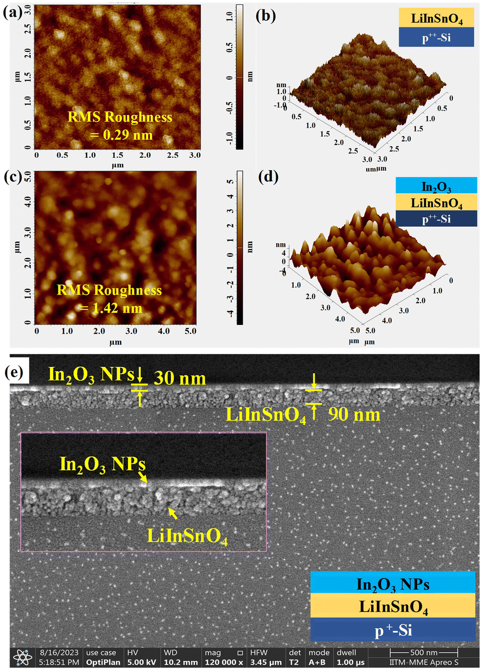
The surface roughness of individual films plays a vital role in the device's performance. For this assessment, atomic force microscopic (AFM) study of the LiInSnO4 and In2O3 NPs was performed. Fig. 3(a) and (b) show the 2D and 3D AFM images of the LiInSnO4 thin film. The root mean square (RMS) surface roughness of this film is 0.29 nm, which is quite low for its use as a gate dielectric of a TFT.32 In addition, the AFM analysis was also performed for the In2O3 NP-coated LiInSnO4 dielectric thin film, i.e. on the surface of p+-Si/LiInSnO4/In2O3. The 2D and 3D AFM images of that surface are shown in Fig. 3(c) and (d), respectively, and indicate its RMS roughness of ∼1.42 nm, which is quite low. For the thickness measurement of LiInSnO4 and In2O3 NP thin films, a cross-sectional FESEM analysis of p+-Si/LiInSnO4/In2O3 was performed, and the image is shown in Fig. 3(e). The measured thickness of the LiInSnO4 and In2O3 NP thin film, as obtained from this study, are 90 nm and 30 nm, respectively.
 |
| | Fig. 3 AFM images: (a) 2D and (b) 3D images of LiInSnO4 dielectric thin film (p+-Si/LiInSnO4) with Rrms ∼ 0.29 nm, (c) 2D and (d) 3D images of In2O3 NPs thin film (p+-Si/LiInSnO4/In2O3) Rrms ∼ 1.42 nm, and (e) cross-sectional SEM image of the p+-Si/LiInSnO4/In2O3 NP film. | |
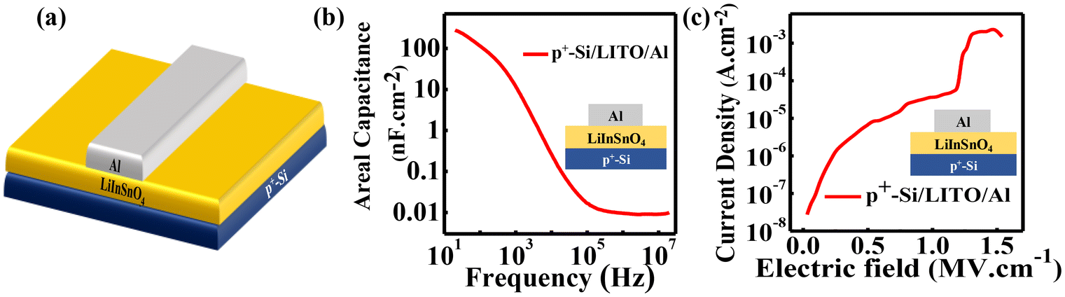
3.4. Dielectric measurement of LiInSnO4 through an MIM device
Capacitance vs. frequency (C–f) and current vs. voltage (I–V) characterizations were performed in a metal–insulator–metal (MIM) device structure (Fig. 4(a)) to understand the dielectric quality of the LiInSnO4 thin film. The frequency–dependent capacitance (C–f) of the proposed LiInSnO4 dielectric was studied within the frequency range of 20 Hz–20 MHz, as shown in Fig. 4(b). These data indicate that the areal capacitance of LiInSnO4 is ∼190 nF cm−2 at a frequency of 50 Hz and decreases ∼15% of its initial value at 500 Hz. Although it drops faster above 103 Hz, which is due to the mobile Li+ ions that fail to contribute to its ionic polarization at higher frequencies. Since this LiInSnO4 dielectric film has a high areal capacitance (≥190 nF cm−2) at lower frequencies, making it suitable for TFTs with low operating voltages.33 Moreover, the I–V characteristics (Fig. 4(c)) show that the LiInSnO4 thin film has a low current density, which is due to its high compactness and a large optical band gap of 5.3 eV. Under 1 MV cm−1 applied electric field, the current density is ∼10−6 A cm−2, which can be low enough for fabricating a high on/off ratio TFT. Besides, the device remains stable up to an external field of 1.25 MV cm−1. This high breakdown field of the LiInSnO4 thin film suggests the existence of minimal pinholes with good uniformity, which is required for choosing it as a gate dielectric for a TFT.
 |
| | Fig. 4 (a) Schematic of the MIM device structure of p+-Si/LiInSnO4/Al. Electrical characterization of LiInSnO4 dielectric film with MIM device. (b) Variation in capacitance with frequency and (c) variation in current density with applied field. | |
3.4. TFT characterization
The electrical characteristics of TFTs were evaluated under dark and ambient conditions. For the output characteristics (ID–VD) of Device-1(Fig. 5(a)), the drain voltage (VD) varied from 0 V to 2 V with a constant gate voltage (VG) ranging from −0.5 V to 2 V with a step of 0.63 V (Fig. 5(b)). In the transfer characteristics (ID–VG) of Device-1, the gate voltage varied from −2 V to 2 V at constant drain voltage (1 V) (Fig. 5(c)). Similarly, the output characteristics of Device-2 (Fig. 5(d)) were investigated under the same applied voltages as for Device-1, as shown in Fig. 5(e). It can be noted that the accumulation mode drain current (ID) of Device-2 is significantly lower (∼1/20 times) than that of Device-1, which originated from the Schottky contact of the drain electrode (MoO3/Ag). Besides, the transfer characteristics of Device-2 were investigated by varying VG from −1.5 to 1.5 V at a constant drain voltage of 1 V (Fig. 5(f)). The parameters such as saturation mobility (μsat), subthreshold swing (SS), and maximum number of the trap states (NMaxSS) of the TFTs were calculated from the transfer characteristics of the devices using the following equations:34,35| |  | (1) |
| |  | (2) |
| |  | (3) |
 |
| | Fig. 5 (a) Schematic of Device-1 (symmetric electrodes), and its (b) output and (c) transfer characteristics. (d) Schematic of Device-2 (asymmetric electrodes), and its (e) output and (f) transfer characteristics. | |
The on/off ratios of Device-1 and Device-2 are 3.1 × 10² and 2.2 × 10³, respectively. This enhancement of the on/off ratio of Device-2 is due to its Schottky contact in the drain electrode that reduces the off current of the device significantly. The effective carrier mobility (μ) of these Device-1 and Device-2 calculated using eqn (1) is 4.37 cm2 V−1 s−1 and 0.13 cm2 V−1 s−1, respectively. It is worth noting that the subthreshold swing (SS) of Device-2 is 160 mV dec−1, which is reduced by six times compared to Device-1 (931 mV dec−1). Besides, the dielectric/semiconductor interface trap states of Device-2, calculated using eqn (3), are approximately one order lower than that of Device-1. All these parameters of Device-1 and Device-2 are summarized and compared in Table 1.
Table 1 TFT parameters of Device-1 and Device-2 without PbI2
| Device |
On–off ratio |
Threshold voltage (Vth) |
Carrier mobility (cm2 V−1 s−1) |
Subthreshold swing (mV per decade) |
Interface trap states density (cm−2) |
| Device-1 |
6.5 × 10² |
−1.10 |
4.37 |
682 |
1.24 × 10¹³ |
| Device-2 |
2.9 × 10³ |
−0.19 |
0.13 |
160 |
2.0 × 10¹² |
As mentioned earlier, after the electrical characterization of Device-1 and Device-2, a PbI2 layer is over coating on both the devices. The respective output and transfer characteristics of PbI2-coated Device-1 (Fig. 6(a)) under dark conditions are shown in Fig. 6(b) and (c), respectively, whereas these characteristics for PbI2-coated Device-2 (Fig. 6(d)) are shown in Fig. 6(e) and (f), respectively.
 |
| | Fig. 6 (a) Schematic of PbI2-coated Device-1, and its (b) output characteristics and (c) transfer characteristics. (d) Schematic of PbI2-coated Device-2, and its (e) output characteristics and (f) transfer characteristics. | |
All electrical characterizations of these devices were performed in the same range of applied voltages as off uncoated TFTs. From Fig. 6(b), it is clear that the accumulation mode current of the PbI2 coated Device-1 is reducing by half with respect to the uncoated device. Moreover, from Fig. 6(c), it can be noted that the off current of PbI2-coated Device-1 also decreased by one order of magnitude with respect to the uncoated device that effectively increased the on/off ratio (∼2.7 × 10³) and decreased the sub-threshold voltage of the device to 160 mV per decade. However, the effective electron mobility is reduced to 3.2 V cm−2, which is due to the reduction of on state current. Almost similar behaviors were observed in the PbI2-coated Device-2 as well. The summary of device parameters of PbI2-coated Device-1 and Device-2 is given in Table 2.
Table 2 TFT parameters of Device-1 and Device-2 with an additional PbI2 layer
| Device |
On–off ratio |
Threshold voltage (Vth) |
Carrier mobility (cm2 V−1 s−1) |
Subthreshold swing (mV per decade) |
Interface trap states density (cm−2) |
| Device-1 (with PbI2) |
2.7 × 10³ |
−0.22 |
3.2 |
357 |
5.9 × 10¹² |
| Device-2 (with PbI2) |
7.4 × 10³ |
−0.14 |
0.14 |
78 |
3.6 × 10¹¹ |
3.6. Optical response of the devices
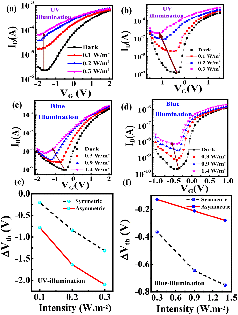
To investigate the photoresponse of the In2O3 NPs thin-film transistor (TFT), a UV LED source of peak intensity at ∼395 nm (Fig. SI 2(a)†) was used and the transfer characteristics of Device-1 and Device-2 are illustrated in Fig. 7(a) and (b), respectively. The In2O3 TFT demonstrated an observable photocurrent response under UV illumination, due to the higher energy of UV photons with respect to the band gap of the In2O3 NPs. The photoresponse of the devices can be estimated in terms of the enhancement in the depletion mode drain current (off current) and the shifting in the threshold voltage. The off current and threshold voltage of Device-1 were changing by more than one order and by a difference of 2.15 V under 0.3 W m−2 of UV illumination, while for Device-2, these changes are more than two orders and 1.37 V, respectively. Moreover, both of the devices are illuminated with a blue LED with a peak intensity of ∼445 nm (Fig. SI 2(b)†), and exhibit negligible photoresponse, as shown in Fig. SI 3(a) and SI 3(b).† The photoresponse of Device-1 was also realized through the output characteristics under UV illumination (Fig. SI 4(a)†). Therefore, it can be concluded that under UV illumination, the asymmetric S–D electrode device (Device-2) has a significantly larger variation of ‘off current’ and ‘threshold voltage shifting’ with respect to the symmetric S–D electrode device (Device-1), indicating a larger sensitivity of Device-2 under UV illumination. The shifting in the threshold voltage of the Device-1 and Device-2 is from −1.10 V to −3.2 V and −0.19 V to −1.53 V under 0.3 W m−2 UV illumination, respectively, as presented in Fig. 7(e).
 |
| | Fig. 7 Photoresponse observed in transfer characteristics under UV illumination for (a) symmetric Device-1 and (b) asymmetric Device-2, and under the blue light illumination for (c) symmetric and (d) asymmetric devices. The change in threshold voltage vs. intensity for symmetric and asymmetric electrode devices: (e) UV-illumination and (f) blue light illumination after PbI2 coating. | |
Similar studies were performed with PbI2-coated Device-1 and Device-2 by illuminating blue light, which are shown in Fig. 7(c) and (d), respectively. From these data, it is clear that the asymmetric electrode device has a larger variation of off current than that of the symmetric electrode devices. Moreover, it can be noted that the threshold voltage of PbI2-coated Device-1 shifted from −0.22 V to −1.73 V (Fig. 7(f)), whereas, in the case of the PbI2-coated Device-2, this shifting is prominently lesser but is following the trend of shift towards a negative voltage axis, i.e. from −0.14 V to −0.39 V (Fig. 7(f)). However, the depletion mode photocurrent of PbI2-coated Device-2 is seven times higher than that of PbI2-coated Device-1. In Fig. SI 4(b),† the photoresponse of PbI2-coated Device-1 is presented with the output characteristics under Blue light illumination.
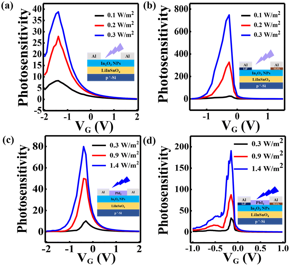
The gate bias-dependent photosensitivity of the devices under UV and blue light illumination was calculated using eqn (4),36 and is shown in Fig. 8. The photosensitivity of Device-1 and Device-2 under UV illumination is shown in Fig. 8(a) and (b), respectively, and it can be noted that the peak photosensitivity of Device-2 (750) is almost 18 times higher than that of Device-1 (40) at 0.3 W m−2 in the depletion region. The enhancement in the UV photosensitivity of Device-2 is due to the carrier selective absorption of the LiF/Al and MoO3/Ag electrodes in the TFT. Similarly, the peak blue light photosensitivity of the PbI2-coated Device-2 is around two times of the PbI2-coated Device-1 in the flat-band region, as shown in Fig. 8(c) and (d).
| |  | (4) |
 |
| | Fig. 8 Photosensitivity plot of TFTs under UV illumination with (a) symmetric electrode and (b) asymmetric electrode. Photosensitivity plot of PbI2-coated TFTs under blue illumination with (c) symmetric electrode and (d) asymmetric electrode. | |
Moreover, the detectivity of Device-1 and Device-2 with and without the PbI2-coated TFT was calculated using eqn (5) and (6):6,37
| |  | (5) |
| |  | (6) |
where
IPh,
Popt,
S, and
Idark are the photocurrent, power of incident light, active area, and dark current of the device, respectively.
q,
Ilight, and
h are the electronic charge (1.6 × 10
−19 Coulomb), device current under light conditions, and Planck's constant (6.62 × 10
−34 m
2 kg s
−1).
The detectivity plots of TFTs under UV illumination for Device-1 and Device-2 are presented in Fig. S5(a) and S5(b),† respectively. Meanwhile, Fig. S5(c) and S5(d)† correspond to PbI2-coated Device-1 and Device-2, respectively. The values of detectivity of all devices are presented in Table 3. Furthermore, symmetric electrode Device-1 exhibits a higher detectivity than that of asymmetric S–D electrode Device-2, primarily because Device-1 generates a higher photocurrent due to the formation of a good Ohmic junction at the In2O3 and Al interface.
Table 3 Summary of the transient photoresponses of devices under blue light illumination
| In2O3 TFT under UV light illumination |
| Device |
EQE (%) |
Photosensitivity at VG = −0.5 V |
Detectivity (Jones) |
τ
rise (sec) |
τ
fall (sec) |
| UV at 0.3 W m−2 |
| Device-1 |
4.46 |
8 |
1.05 × 10¹¹ |
3.1 |
24.8 |
| Device-2 |
5.69 |
492 |
4.63 × 10¹⁰ |
1.9 |
14.6 |
| With PbI2 layer under blue light illumination |
| Device |
EQE (%) |
Photosensitivity at VG = −0.1 V (depletion mode) |
Detectivity (Jones) |
τ
rise (sec) |
τ
fall (sec) |
| Blue at 1.4 W m−2 |
| Device-1 (with PbI2) |
5.29 |
55 |
4.71 × 10¹⁰ |
3.7 |
24 |
| Device-2 (with PbI2) |
12.25 |
152 |
1.9 × 10⁹ |
4.6 |
40.8 |
3.7. Transient response of the devices
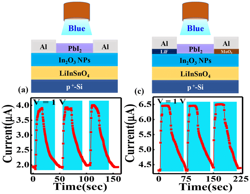
The multiple cycle transient photoresponse of Device-1 and Device-2 under UV illumination at 0.3 W m−2 with a gate bias of 0 V is shown in Fig. 9(a) and (b), respectively. The rise time (τRise) and fall time (τFall) of Device-1 are 3.1 seconds and 24.8 s, respectively. It was observed that the rise time and fall time of Device-2 are much faster than Device-1 with values of 1.9 s and 14.6 s, respectively. In addition, the transient response of Device-1 and Device-2 with a PbI2 layer was measured under blue light illumination at 1.4 W m−2 at 0 V gate biasing, as shown in Fig. 10(a) and (b), respectively. This study gives the response time of Device-1 as rise time and fall time of 3.7 and 24 s., respectively, whereas these values are 4.6 and 40.8 s., respectively, for Device-2. A summary of the photoresponse time of the devices is given in Table 3.
 |
| | Fig. 9 Transient photoresponse of the TFT under multiple cycle UV light illumination: (a) Device-1 (with symmetric S-D electrodes) and (b) Device-2 (with asymmetric S-D electrodes). | |
 |
| | Fig. 10 Transient photoresponse of PbI2-coated TFT under multiple cycle blue light illumination: (a) Device-1 (with symmetric S-D electrodes) and (b) Device-2 (with asymmetric S-D electrodes). | |
3.8. External quantum efficiency of device
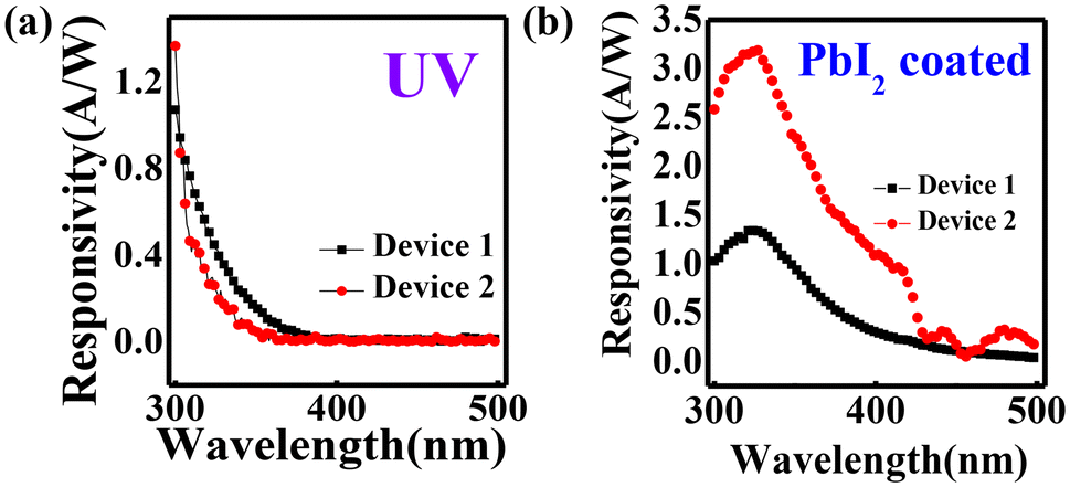
The external quantum efficiency (EQE) quantifies the relationship between the “quantity of generated charge carriers per incident photon” within a photodetector. The EQE data of Device-1 and Device-2 at 0 V (Black) and 2 V (Red) drain bias and 0 V gate biasing are shown in Fig. 11(a) and (b), respectively. The EQE data of both of the devices reveal its photosensitivity in the UV region only. At 2 V drain biasing, the EQE values of both the devices increase very significantly. Moreover, the values of EQE of these two devices are almost the same. However, it can be noted that the Device-2 shows a reasonable EQE at the 0-volt drain biasing (inset Fig. 11(a)), which is not observed in Device-1 (inset Fig. 11(b)). It can also be noted that no significant photocurrent is generated in the region > 365 nm in both of these two devices, even after applying 2 V drain bias, depicting the visible-blind nature of the device. The EQE spectra of PbI2-coated Device-1 and Device-2 at 2 V drain and 0 V gate biasing are shown in Fig. 11(c) and (d), respectively. The percentage EQE of Device-2 (∼12) is higher than that of Device-1 (∼5), which may be due to the additional bias originating from the asymmetric S–D electrodes of Device-2. Moreover, it is observed that with the incorporation of an additional layer of blue-sensitive PbI2 material, there are notable extensions in the EQE spectra towards the visible region (450–495 nm) in the devices, confirming the blue light sensitivity of the device. Therefore, it can be concluded that these visible-blind phototransistors may become a particular color or spectrum-sensitive device based on the materials coated on top of these In2O3 TFTs without compromising device performance. Again photocurrent extraction in the external circuit can be improved by incorporating asymmetric work-function S-D electrodes. The responsivity data of Device-1 and Device-2 are shown in Fig. 12(a), signifying the comparable responsivity in the UV region of both the device. In contrast with PbI2-coated Device-1 and Device-2, the responsivity of Device-2 exceeds that of Device-1 in the visible blue region of the electromagnetic spectra (Fig. 12(b)). Overall, the devices are showing nice photoresponse under UV as well as blue light illumination compared to the earlier reports, as presented in Table SI 1.†
 |
| | Fig. 11 External quantum efficiency at 0 V and 2 V without any additional PbI2 layer: (a) symmetric electrode TFT and (b) asymmetric electrode TFT. External Quantum efficiency with the PbI2-coated device at 2 V with (c) symmetric electrode and (d) asymmetric electrode TFTs. | |
 |
| | Fig. 12 Responsivity of Device-1 and Device-2: (a) without PbI2 layer and (b) with PbI2 layer (plotted from the EQE data). | |
3.9. Working mechanism of the device
The working principle of the photocurrent generation of these devices is explained based on the energy band alignment of the channel semiconductor with their source and drain electrodes, which is shown in Fig. 13. In the equilibrium condition, i.e. when the gate electrode and drain electrodes are electrically unbiased, the energy band diagrams of Device-1 and Device-2 are shown in Fig. 13(a) and (b), respectively. It can be noted that, in the case of the symmetric electrode (Al), the potential barrier for carrier in the In2O3 NPs at the S and D electrodes remains the same because of the similar work function (Φ) of the Al (ΦAl ∼ 4.09 eV).38 Therefore, there is no driving force that allows carriers (e−) to move from the source to the drain electrode. However, in Device-2, due to substantial work function difference between the LiF/Al (ΦLiF/Al ∼ 3.79 eV) and MoO3/Ag (ΦMoO3/Ag ∼ 4.88 eV) electrodes,38 it manifests band bending with a downward slope towards the drain electrode, which originated due to the thin interface layers of Al and Ag electrodes, which modulate the work function of the electrodes.29,39 Specifically, LiF decreases the work function of the Al electrode that diminishes the barrier height (ΦB) of electrons, dictating electron transfer from the source electrode to In2O3 NPs. However, the MoO3 interface layer attains an optimal thickness before silver (Ag) deposition, increasing the work function of MoO3/Ag beyond that of pristine Ag.40,41 Hence, it forms a Schottky barrier for electron transport at the drain electrode and reduces the barrier potential for hole injection into the In2O3 NP channel simultaneously.42 The electron transport of this Schottky junction can arise due to the thermionic emission (TE)- and thermionic field emission (TFE)-related tunneling process that effectively reduce the sub-threshold swing of the device.43 This tuning in the work function of original Al and Ag metals creates a reasonable potential difference across the (S–D) electrodes (between LiF/Al and MoO3/Ag). Moreover, this potential difference across the S–D electrodes provides a driving force from the source to the drain, even at zero S–D biasing in asymmetric electrodes (Device-2), which is the origin of photocurrent generation in the EQE spectra of Device-2.44
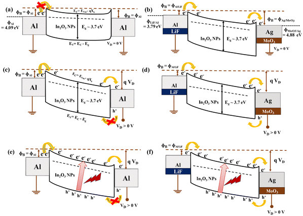 |
| | Fig. 13 Band diagram of (a) Device-1 and (b) Device-2 at equilibria with zero biasing (VD = 0 V), (c) Device-1 and (d) Device-2 under electrical biasing (VD > 0 V), and (e) Device-1 and (f) Device-2 under electrical (VD > 0 V) and optical illumination. | |
Under applied positive drain biasing in the device, electrons are conducted from S to D in the In2O3 NP channel in both devices. However, the hole injection is strictly blocked in the Al electrode of Device-1. In contrast, there is a chance of hole conduction in Device-2, because of a lower barrier for holes at the MoO3/Ag electrode, as shown in Fig. 13(d). The imposition of an additional positive drain bias results in an augmented barrier potential gradient between the source and drain electrodes is realized, as shown in Fig. 13(e) and (f), respectively. This heightened potential disparity prompts electron injection from the source, supplementing the population of photogenerated electrons. In both classes, electrons are drifted toward the drain electrode and concurrently, photogenerated holes move towards the source electrode under the influence of positive drain bias. This concerted effect serves to substantially reduce the rate of electron–hole recombination. Thus, Device-2 manifests an amplified photoresponse under positive drain bias with respect to Device-1. After the addition of PbI2 layer in the channel, the absorption spectra of the transistor channel are enhanced, resulting in photo-generated carrier formation inside the PbI2 layer due to blue light illumination. Again, photo-generated electrons can transfer to the In2O3 layer due to the favorable energy band and transport through this oxide channel, resulting in the blue sensitivity of the device.
4. Conclusion
In conclusion, an asymmetric work-function S–D electrode In2O3 NP TFT has been fabricated using an LiInSnO4 thin film as a gate dielectric. This solution-processed TFT can be operated within 2 V external bias due to high areal capacitance of the LiInSnO4 thin film. The on/off ratio of this asymmetric S–D electrode is ∼10 times higher than that of the corresponding symmetric S–D reference device due to the elevated sheet resistance of the In2O3 NP channel, originating from the Schottky contact of the drain electrode. In addition, the subthreshold swing of the asymmetric S–D electrode device (Device-2) showed a depleted value compared to the symmetric electrode device (Device-1). Both Device-1 and Device-2 showed visible-blind UV sensitivity, which originated from the wide band gap of In2O3 NPs. Additionally, Device-2 showed enhanced photosensitivity with a value by 20 times under UV illumination due to the additional driving force between S–D electrodes, originating from their work-function differences. Besides, the carrier-selective charge collection to the source and drain electrodes also enhanced this effective photocurrent generation. This visible-blind UV sensitivity was further tuned to the blue region by adding PbI2 in the channel. Therefore, this study paves the way to fabricate a phototransistor by adjusting the optical window for photosensitivity and its enhancement by choosing an additional coating or different combinations of source–drain electrodes.
Data availability
URL for Data availability_ In2O3 NP TFT: https://drive.google.com/drive/folders/1ED-6xHgO6fAPaHiegWWuR-clksVCI03H?usp=drive_link.
Conflicts of interest
There are no conflicts to declare.
Acknowledgements
Bhola Nath Pal graciously acknowledges the financial support received from SPARCS, India (SPARC/2019-2020/P2588/SL), and DST, India (DST/INT/SWD/VR/P-12/2019). The authors extend their appreciation to the Central Instrument Facility Centre, IIT (BHU), for their invaluable assistance with instrument support for AFM. Authors thanks DST-FIST (SR/FST/ET-II/2018/277(C)) for providing instrument facility for XRD study. Akhilesh Kumar Yadav and Utkarsh Pandey are grateful to IIT (BHU) for the provision of an SRF fellowship. Pijush Kanti Aich expresses gratitude to the UGC for the SRF fellowship provided.
References
- R. Saran and R. J. Curry, Nat. Photonics, 2016, 10, 81–92 CrossRef CAS
 .
.
- M. Long, P. Wang, H. Fang and W. Hu, Adv. Funct. Mater., 2019, 29, 1803807 CrossRef .
- M. Ahmadi, T. Wu and B. Hu, Adv. Mater., 2017, 29, 1605242 CrossRef PubMed .
- D. Yang and D. Ma, Adv. Opt. Mater., 2019, 7, 1800522 CrossRef .
- S. Dahiya, S. V. Singh, U. Pandey, S. Hazra and B. N. Pal, ACS Appl. Opt. Mater., 2024, 2, 776–783 CrossRef CAS .
- H. Yoo, I. S. Lee, S. Jung, S. M. Rho, B. H. Kang and H. J. Kim, Adv. Mater., 2021, 33, 2006091 CrossRef CAS PubMed .
- N. Li, Z. Lan, L. Cai and F. Zhu, J. Mater. Chem. C, 2019, 7, 3711–3729 RSC .
- J. Guo, S. Jiang, M. Pei, Y. Xiao, B. Zhang, Q. Wang, Y. Zhu, H. Wang, J. Jie, X. Wang, Y. Shi and Y. Li, Adv. Electron. Mater., 2020, 6, 2000062 CrossRef CAS .
- S. Kim, S. Oh and J. Kim, ACS Photonics, 2019, 6, 1026–1032 CrossRef CAS .
- Z. Lian, J. Wei, Z. Liu, G. Chen, H.-C. Kuo, Y. Dan, C.-C. Tu and R. Yang, ACS Photonics, 2024, 11(10), 4224–4234 CAS .
- D. Li, Y. Chen, Y. Tang, K. Liang, H. Ren, F. Li, Y. Wang, G. Liu, C. Song, L. Meng and B. Zhu, ACS Appl. Electron. Mater., 2023, 5, 578–592 CrossRef CAS .
- L. Shi and S. Nihtianov, IEEE Sens. J., 2012, 12, 2453–2459 CAS .
- J. Zheng, H. Chong, L. Wang, S. Chen, W. Yang, G. Wei and F. Gao, J. Mater. Chem. C, 2020, 8, 6072–6078 RSC .
- Y. Yun, G. S. Han, G. N. Park, J. Kim, J. Park, D. Vidyasagar, J. Jung, W. C. Choi, Y. J. Choi, K. Heo, J. Kang, J.-S. Park, H. S. Jung and S. Lee, Adv. Mater., 2022, 34, 2270351 CrossRef .
- L. Liu, C. Yang, A. Patanè, Z. Yu, F. Yan, K. Wang, H. Lu, J. Li and L. Zhao, Nanoscale, 2017, 9, 8142–8148 RSC .
- N. K. R. Nallabala, S. S. Kushvaha, S. Sangaraju and V. K. Kummara, Mater. Sci. Semicond. Process., 2024, 170, 107954 CrossRef .
- N. K. R. Nallabala, S. Godavarthi, V. K. Kummara, M. K. Kesarla, C. Yuvaraj, S. Kumar, N. Ravi, G. K. Guntupalli, S. Jilani and S. P. Vattikuti, Sol. Energy Mater. Sol. Cells, 2021, 225, 111033 Search PubMed .
- Y. Qin, S. Long, Q. He, H. Dong, G. Jian, Y. Zhang, X. Hou, P. Tan, Z. Zhang, Y. Lu, C. Shan, J. Wang, W. Hu, H. Lv, Q. Liu and M. Liu, Adv. Electron. Mater., 2019, 5, 1900389 Search PubMed .
- K.-J. Zhou, P.-H. Chen, Y.-Z. Zheng, M.-C. Tai, Y.-X. Wang, Y.-T. Chien, P.-J. Sun, H.-C. Huang, T.-C. Chang and S. M. Sze, J. Mater. Chem. C, 2022, 10, 9192–9197 RSC .
- B. N. Pal, P. Trottman, J. Sun and H. E. Katz, Adv. Funct. Mater., 2008, 18, 1832–1839 CrossRef CAS .
- N. K. R. Nallabala, V. R. M. Reddy, V. Singh, K. R. Bakash, S. Kumar, D. Saha, V. Mahendran, V. K. Kummara, G. K. Guntupalli and S. P. Vattikuti, Sens. Actuators, A, 2022, 339, 113502 CrossRef CAS .
- B. N. Pal, B. M. Dhar, K. C. See and H. E. Katz, Nat. Mater., 2009, 8, 898–903 CrossRef CAS PubMed .
- A. Sharma, N. K. Chourasia, A. Sugathan, Y. Kumar, S. Jit, S.-W. Liu, A. Pandey, S. Biring and B. N. Pal, J. Mater. Chem. C, 2018, 6, 790–798 RSC .
- U. Pandey, N. Pal, A. Ghosh, S. Suman, S. Biring and B. N. Pal, Nanoscale, 2024, 16, 8504–8513 RSC .
- U. Pandey, N. K. Chourasia, N. Pal, S. Biring and B. N. Pal, IEEE Trans. Electron Devices, 2022, 69, 1077–1082 CAS .
- S. Yin, Y. Cheng, Y. Li, W. Liang, T. Li, J. Ma, D. Wu, Z. Shi and X. Li, J. Mater. Sci., 2021, 56, 13633–13645 CrossRef CAS .
- C. Chen, J. Moir, N. Soheilnia, B. Mahler, L. Hoch, K. Liao, V. Hoepfner, P. O'Brien, C. Qian, L. He and G. A. Ozin, Nanoscale, 2015, 7, 3683–3693 RSC .
- N. Pal, R. Chakraborty, A. Sharma, U. Pandey, V. Acharya, K. Prajapati, A. Gupta, S. Suman, P. Swaminathan, A. K. Singh, P. K. Roy and B. N. Pal, J. Alloys Compd., 2023, 960, 170691 CrossRef CAS .
- P. K. Aich, Z. Genene, U. Pandey, A. K. Yadav, E. Wang and B. N. Pal, ACS Photonics, 2024, 11, 3704–3712 CrossRef CAS .
- B. K. Yap, Z. Zhang, G. S. H. Thien, K.-Y. Chan and C. Y. Tan, Appl. Surf. Sci. Adv., 2023, 16, 100423 CrossRef .
- H. Ullah, Z. H. Yamani, A. Qurashi, J. Iqbal and K. Safeen, J. Mater. Sci.: Mater. Electron., 2020, 31, 17474–17481 CrossRef CAS .
- N. Pal, B. Thakurta, R. Chakraborty, U. Pandey, V. Acharya, S. Biring, M. Pal and B. N. Pal, J. Mater. Chem. C, 2022, 10, 14905–14914 RSC .
- A. Sharma, N. K. Chourasia, A. Sugathan, Y. Kumar, S. Jit, S.-W. Liu, A. Pandey, S. Biring and B. N. Pal, J. Mater. Chem. C, 2018, 6, 790–798 Search PubMed .
- R. Chen and L. Lan, Nanotechnology, 2019, 30, 312001 CrossRef CAS PubMed .
- R. Shyam, P. K. Aich, U. Pandey, B. N. Pal and R. Prakash, IEEE Sens. J., 2024, 24(20), 32004–32010 Search PubMed .
- R. Shyam, T. Manaka and R. Prakash, J. Mater. Chem. C, 2024, 12, 18514–18525 Search PubMed .
- S. Suman, U. Pandey, N. Pal, P. Swaminathan and B. N. Pal, ACS Photonics, 2023, 11, 93–101 Search PubMed .
- S. Dahiya, U. Pandey, S. Hazra, R. Chakraborty, S. Pramanik, P. P. Maurya and B. N. Pal, Adv. Mater. Technol., 2025, 10(7), 2401532 Search PubMed .
- T. Brown, R. Friend, I. Millard, D. Lacey, J. Burroughes and F. Cacialli, Appl. Phys. Lett., 2000, 77, 3096–3098 CrossRef CAS .
- U. Pandey, A. K. Yadav, N. Pal, P. K. Aich and B. N. Pal, J. Mater. Chem. C, 2023, 11, 15276–15287 RSC .
- S. Dahiya, S. Hazra, U. Pandey, S. Pramanik, P. Dahiya, S. V. Singh, N. Kumari and B. N. Pal, Opt. Mater., 2024, 157, 116182 CrossRef CAS .
- A. V. Polotai, T.-H. Jeong, G.-Y. Yang, E. C. Dickey, C. A. Randall, P. Pinceloup and A. S. Gurav, J. Electroceram., 2009, 23, 6–12 Search PubMed .
- S. Lee and A. Nathan, Science, 2016, 354, 302–304 Search PubMed .
- N. K. R. Nallabala, S. P. Vattikuti, V. Verma, V. Singh, S. Alhammadi, V. K. Kummara, V. Manjunath, M. Dhanalakshmi and V. R. M. Reddy, Mater. Sci. Semicond. Process., 2022, 138, 106297 CrossRef .
Footnote |
| † Electronic supplementary information (ESI) available: The emission spectra of (a) ultra-violet (UV) and (b) blue light sources; photoresponse of the transfer characteristics under blue illumination of (a) Device-1 and (b) Device-2, (without PbI2 coat); photoresponse of the output characteristics (a) under UV illumination and (b) under blue illumination (with PbI2 coating). See DOI: https://doi.org/10.1039/d5nr00201j |
|
| This journal is © The Royal Society of Chemistry 2025 |
Click here to see how this site uses Cookies. View our privacy policy here. 
 Open Access Article
 This Open Access Article is licensed under a Creative Commons Attribution-Non Commercial 3.0 Unported Licence
 a,
Pijush Kanti
Aich
a,
Vishwas
Acharya
a,
Swati
Suman
b and
Bhola N.
Pal
a,
Pijush Kanti
Aich
a,
Vishwas
Acharya
a,
Swati
Suman
b and
Bhola N.
Pal