
 Open Access Article
 This Open Access Article is licensed under a
 Creative Commons Attribution 3.0 Unported Licence
Nickel-free porous stainless-steel nanocomposites for versatile biomedical applications: fabrication, characterization, and evaluation of electrochemical and immunogenicity detection
Sabreen Abdallah
Abdelwahab
a,
Mohamad
Warda
bc,
Mamdouh
Zewaid
a,
Hisham
Saleh
d,
Omar A.
Ahmed-Farid
e,
Hassan A. M.
Hendawy
f,
Elbadawy A.
Kamoun
 *gh,
Amr
Negm
gi,
Jong Yeog
Son
*j and
Ahmed I.
Ali
*jk
*gh,
Amr
Negm
gi,
Jong Yeog
Son
*j and
Ahmed I.
Ali
*jk
aProduction Technology Department, Faculty of Technology and Education, Helwan University, Saray El-Quba, 11281 Cairo, Egypt
bDepartment of Biochemistry and Molecular Biology, Faculty of Veterinary Medicine, Cairo University, Giza, 12211, Egypt
cDepartment of Physiology, Faculty of Veterinary Medicine, Ataturk University, Erzurum, Turkey
dSolid State Physics, Physics Deviation, National Research Center, El Bahous St., Giza, Cairo, Egypt
ePhysiology Department, National Organization for Drug Control and Research (NODCAR), Giza, Egypt
fAnalytical and Inorganic Chemistry Department, NODCAR, Giza, Egypt
gDepartment of Chemistry, College of Science, King Faisal University, Al-Ahsa 31982, Saudi Arabia. E-mail: ekamoun@kfu.edu.sa; badawykamoun@yahoo.com
hPolymeric Materials Research Dep., Advanced Technology and New Materials Research Institute (ATNMRI), City of Scientific Research and Technological Applications (SRTA-City), New Borg Al-Arab City 21934, Alexandria, Egypt
iChemistry Department, Faculty of Science, Mansoura University, Mansoura 35516, Egypt
jDepartment of Applied Physics and Institute of Natural Sciences, College of Applied Science, Kyung Hee University, Suwon 446-701, Republic of Korea
kDepartment of Basic Science, Faculty of Technology and Education, Helwan University, Saray El-Quba, 11281 Cairo, Egypt. E-mail: Ahmed_Ali_2010@techedu.helwan.edu.eg
First published on 12th December 2024
Abstract
This study focuses on the development of nickel-free stainless-steel nanocomposites with porosities tailored for surgical implants and biological applications. Alloy F2581 (Fe–17Cr–10Mn–3Mo–0.4Si–0.5N–0.2C wt%), modified by replacing Mo with metals such as Al, Cu, Ti, and W, was successfully fabricated via a solid-state reaction method. X-ray diffraction analysis revealed a significant alteration in the crystal phase, accompanied by the formation of nanostructures, including nanowires, square nanotubes, wave-like configurations reminiscent of a growing clover farm, and nanofibers. The particle sizes of these structures were determined to be 73, 27.2, 76 and 98.5 nm for Al, Cu, Ti, and W ions, respectively, indicating a distribution of nanopores. Biological evaluation of adult male Albino rats after exposure to single intraperitoneal doses of various concentrations (10, 20, and 50 mg kg−1 wt%) were assessed with testing alloys (Cu, Al, W, and Ti, respectively). Over a subacute period lasting 60 days, a comprehensive evaluation of biological responses, including hepatic function, renal performance, oxidative and/or nitrosative stress parameters, and the levels of serum immune modulators was conducted. Notably, low doses elicited negligible immune responses, higher doses, barring copper, induced notable reactions. Interestingly, aluminum demonstrated optimization within biological settings, alongside titanium and tungsten. These findings highlight the applicability of copper and tungsten for medical implantation and biological applications under controlled circumstances, particularly at lower dosage levels.
1. Introduction
The growing demand for biomedical implants, driven by the increasing number of orthopedic procedures annually, necessitates the development of diverse bone scaffolds and augmentations.1–3 These implants should be similar to the mechanical and microstructural properties of natural bone to substantially improve the remediation process.1–6 Generally, humanoid bones are categorized into two types (i.e., trabecular and cortical bones). The trabecular bones, situated within bone centers, feature a plethora of interconnected and equiaxed pores spanning hundreds of micrometers in diameter. Compared with those of cortical bone, their high porosity results in lower mechanical properties.7–10The porous structure and composition of scaffolds dictate cellular responses such as adhesion, penetration, differentiation, nutrient diffusion, and bone ingrowth.11–18 Furthermore, biomaterials essentially need corrosion resistance, wear resistance, and biocompatibility and to possess strong osseointegration, which is the ability to combine with bone and other tissues, which is a good biological environment for the effective movement of medications, and good hardness, tensile strength, and elongation, while being extremely nontoxic and/or anti-allergenic.12–14 Metal alloys are widely used as biomaterials. Iron alloys containing at least 11% anti-rusting chromium, which also increases heat resistance,19 with less than 1.2% carbon, and other alloying components are known as stainless steels. The mechanical qualities can be enhanced by additive elements like nickel, molybdenum, niobium, manganese, titanium, copper, aluminum, and tungsten. Moreover, the corrosion resistance and mechanical qualities of stainless steel can be further improved.20 The inclusion of Cr and Mo is responsible for the exceptional biocompatibility, mechanical qualities (strength and ductility), and resistance to pitting and crevice corrosion of the material.21
Porous stainless steel represents one of the most important metal alloys used in the manufacture of biomaterials. It can contribute to lightweight materials with excellent mechanical, thermal, and electrical properties.22 Ferrous metals have found widespread application in the fabrication of stents and dental and orthopedic implants, including bone plates, fixtures, dental posts, and screws, as well as surgical instruments. However, the release of iron (Fe) ions can lead to interactions with peroxides, leading to the generation of radicals. This phenomenon can have deleterious consequences, potentially culminating in severe outcomes such as coma or fatality owing to the deleterious produces of free radicals on lipids, proteins, DNA, and ultimately, cellular integrity.23 Owing to its comparatively lower cost than those of cobalt–chromium alloys, pure titanium, and titanium alloys, 316 L stainless steel is frequently employed in orthopedic implants. Its utilization in this context is observed at a ratio ranging from ten-to-five.24 While numerous studies have explored the mechanical and microstructural properties of trabecular bone, replicating these properties in materials poses a significant challenge. Metals like stainless steel, Co–Cr alloys, and titanium alloys are commonly utilized in the production of various biomedical implants.25 However, achieving the desired porosity in these materials is difficult because of their high melting points, which limits the usage of conventional manufacturing techniques such as casting. Several studies have investigated the fabrication of porous stainless steel via conventional methods like casting and powder metallurgy, but these approaches present significant challenges. To overcome these challenges, researchers have explored alternative techniques, including rapid prototyping26 and the sol–gel method.27 For example, Mariotto et al. synthesized steel foam samples containing 30% ammonium carbonate and conducted electrochemical tests in a chloride-containing medium, comparing the corrosion resistance of the foam samples to that of bulk steel. The results indicated that the corrosion resistance of foam samples was superior to that of the bulk steel.28 Noor et al. reported that porous SS316L has no cytotoxic potential for biomedical implants.29 Babaie et al. produced a sample with a tight pore structure and a multimodal pore size, but the samples were subjected to a cytotoxicity test.18 Other studies have used powder metallurgy to produce porous stainless steel. Dewidar et al. produced highly porous 316 L stainless steel samples whose mechanical properties match those of human bone.30 Salahinejad et al. manufactured porous Cr–Mn–N austenitic stainless steel, and nanostructured austenitic stainless steel was developed with a comparative density of 85%.31 Dudek et al. discussed the chances of obtaining materials with controllable porosity. The samples produced in hydrogen and under vacuum have high corrosion resistance and high porosity, and these samples can be used for medical purposes.32 Essa et al. investigated porosity content control in capsule-free powder and reported a substantial increase in the pore fraction of the samples with increasing powder particle size, which was related with a decrease in the compressive strength, elastic modulus, hardness, and ductility of the samples.21 Zielecka et al. developed a porous stainless steel that exhibited extreme porosity in comparison to other ingredients, and the allocation of pores was classified as micropores.33 The samples produced can be used in biomedical applications because their properties simulate those of human bones.34 Weipeng et al. fabricated Fe–Cr–N stainless steel via powder metallurgy, achieving porosities ranging from 28.21% to 60.16%. This stainless steel exhibited high biocompatibility, corrosion resistance, and mechanical properties comparable to those of human bone, which was attributed to its nonnickel and manganese composition. The pore morphology, microstructure, and mechanical characteristics of the porous stainless steel were thoroughly analyzed. As the porosity increased, the compressive yield strength, elastic modulus, and compressive strength decreased.35 Moreover, rapid prototyping methodologies offer promising avenues for fabricating materials with structural similarities to trabecular bone.36,37 In the realm of biomaterials, considerable attention has been given to Ti- and- Co-based alloys, because of their notable biocompatibility and robust mechanical properties.38,39 Tungsten implants, categorized within the refractory metal class, demonstrate exceptional resistance to both heat and corrosion, rendering them suitable for bone fracture applications.40 Investigations into the clinical efficacy of alumina substrates have also been undertaken.41
Copper is renowned for its potent antimicrobial properties and is frequently employed as a filter. To enhance antibacterial efficacy and ensure optimal biocompatibility in orthopedic implants, precise dosages of copper must be incorporated into titanium and other metallic materials. Currently, copper ions and metallic copper represent the two primary forms utilized in biomaterials, both of which exhibit robust antibacterial capabilities. Additionally, copper ions are known to impede DNA replication and protein synthesis.42
This study aims to investigate the physiochemical characteristics of porous stainless steel samples synthesized through a solid-state reaction method, wherein molybdenum is replaced with various elements, including copper, aluminum, titanium, and tungsten, in accordance with ASTM F2581 standards (Fe–17Cr–10Mn–3MX–0.4Si–0.5N–0.2C in wt%; MX: Al, Cu, Ti, and W) for medical implantation and biological applications. Characterization of samples was performed via XRD, FT-IR, and SEM analyses. Furthermore, the electrochemical kinetic parameters are determined via dynamic polarization techniques and electrochemical frequency modulation (EFM). The subacute effects of administering single intraperitoneal doses of the examined alloys at concentrations of 10, 20, and 50 mg kg−1 to a rat animal model were subsequently investigated over a period of 60 days. The assessment involves monitoring the serum activities of the liver enzymes alanine transaminase (ALT) and aspartate transaminase (AST) to evaluate liver function. Additionally, the serum creatinine levels reflect renal performance in the experimental rats. Furthermore, hepatic superoxide dismutase (SOD) activity and reduced glutathione levels are assessed to evaluate global oxidative stress, whereas nitric oxide (NO) levels serve as indicators of nitrosative stress. The levels of serum cortisol, a primary stress hormone, are measured along with the levels of the immunomodulators interleukin 1 (IL1) and interleukin 10 (IL10) to elucidate the cellular immune response to each injected alloy.
2. Materials and methods
2.1. Materials
Powders of FeCl, MnO2, CrCl, SiO2, NiO, C, and metal oxides (MX) e.g. (MX: CuO, WO2, TiO2 and Al2O3) were purchased from Sigma-Aldrich Limited and Al-Gomhouria Chemical Company in Cairo, Egypt. All powders were mixed in an agate mortar with stoichiometric ratios (with composition of ASTM F2581: (Fe–17Cr–10Mn–3MX–0.4Si–0.5 N–0.2C in wt%)) and sintering aid (Mn–11.5 wt% Si) powders.2.2. Preparation of porous stainless-steel composites
Four sets of samples were prepared by substituting MX with four additive composite materials in powder form. The mixtures were ground in an agate mortar for 40 minutes until well blended, followed by dissolution in methanol through ultra-sonication, and the powders were manually ground by hand to achieve a homogeneous mixture before proceeding with dissolution. The solutions were subsequently heated via a magnetic stirrer at 80 °C for approximately 2–3 h to evaporate the alcohol and dry the samples. Manual grinding in an agate mortar was then performed for two hours to achieve a fine powder consistency. The samples were calcined in an alumina crucible at 600 °C for 3 h with a heating rate of 7 °C in a closed programmable muffle box furnace (Fengshi made in Germany, with a chamber size of 400 × 400 × 500 mm W × H × D), followed by gradual cooling to room temperature naturally. Afterward, further grinding was carried out for an hour and sieved using a 100-μm mesh screen. Furthermore, the samples were compressed via a hydraulic press at a pressure of 5 tons cm−2 for 5 min in a forming die, resulting in samples with dimensions of 10 mm, and 1–1.5 mm in diameter and thickness, respectively. Finally, the samples were sintered in a muffle furnace at 1150 °C for 6 h, followed by quenching in air (Scheme 1). | ||
| Scheme 1 Schematic diagram representing preparation steps and applications of nickel-free porous stainless-steel nanocomposites. | ||
2.3. Electrochemical corrosion
The samples were prepared in the form of an electrode (2.0 × 5.0 mm diameter) via a hand press. A group of samples was prepared for the purpose of measuring electrochemical corrosion products to obtain samples for conducting an electrochemical corrosion test on a stainless-steel pallet, which was conducted after 5 min as it was ready to perform the required examination.2.4. Instrumental characterization
| O2 + 2H2O + 4e− → 4OH− | (1) |
| 2H+ + 2e− → H2 | (2) |
The chloride ion concentrations in the serum and urine were within the range of 105–125 mmol L−1. Electrolyte solutions containing 10% serum with a pH of 7.40 and phosphate-buffered saline were prepared for electrochemical measurements, with temperature control ensured via a water bath. All test electrolytes were prepared without any nonaerated treatments, and samples were analyzed in triplicate to ensure accuracy and reproducibility. Ethical considerations were strictly adhered to throughout the collection and handling of biological samples. Voltammetry measurements of the polarization curves were conducted within the Ecorr ± 250 mV range at a scan rate of 10 mV s−1. Steady-state cyclic polarization curves were obtained over the potential range of −800 to +200 mV versus Ecorr, with a scan rate of 0.5 mV s−1. Periodic polarization curves were also obtained within the potential range of −800 to +200 mV versus Ecorr at a scan rate of 10 mV s−1.
2.5. Biological tests
Human urine and serum were ethically collected from healthy subjects who provided the signed consents of the donors, according to the ethical rules of research in Egyptian universities. The participants underwent routine health check-ups, and no known chronic diseases were present in the cohort. The total number was five male participants, aged between 35–45 years. Inclusion criteria comprised male participants aged 35–45 years with no history of chronic diseases, who were generally healthy and underwent routine health check-ups. Exclusion criteria included individuals with a history of chronic conditions such as diabetes, hypertension, cardiovascular diseases, kidney or liver diseases, any acute illness at the time of the study, or those on medications that could affect the study outcomes. The serum and urine samples collected averaged 5 mL of serum and 20 mL of urine per participant, respectively.Animal study: adult male Albino rats, weighing approximately 150 ± 15 g, were employed for this study. The rats were housed in the animal facility of the National Organization for Drug Control and Research for seven days prior to the experiment, where they were maintained under standard laboratory conditions with ad libitum access to food and water. The experimental procedures were conducted in accordance with the ethical guidelines set forth by the Institutional Animal Care and Use Committee at the Faculty of Veterinary Medicine, Cairo University (Vet. Cu. IACUC), with approval number Vet. CU. 09092023786.
2.5.1.1. Blood sample collection. Blood samples were collected from each rat's retro-orbital vein under light diethyl ether anesthesia, as described by Cocchetto et al.46 After clotting, the blood was centrifuged at 3000 rpm for 15 min to obtain the serum. The serum was then utilized for assessing liver enzyme activities, monitoring kidney function, and evaluating stress and immune response parameters.
2.5.2.1. Measurement of liver enzyme activities in serum. Liver function was assessed by quantifying the serum levels of ALT and AST enzymes, which are reliable markers of liver function, via Egyptian Company for Biotechnology (S.A.E), Obour City Industrial Area, Block 20008, Piece 19 A, Cairo, Egypt kits. The results are reported in units per liter (U L−1).47
The concentration of reduced glutathione (GSH) and oxidized glutathione (GSSG) was determined using high-performance liquid chromatography (HPLC) based on the method outlined by Jayatilleke and Shaw (1993).49 Reference standards for GSH and GSSG were obtained from Sigma Chemical Co. and prepared as stock solutions (1 mg mL−1) in 75% methanol. These stock solutions were diluted as necessary before injection into the HPLC system. The analysis was performed using an Agilent HPLC system, equipped with a quaternary pump, column oven, Rheodyne injector with a 20 μL loop, and a UV variable wavelength detector. Data analysis and chromatograms were generated using ChemStation software (Agilent). Separation was achieved on a Synergi RP Max column (3.9 mm), with detection at 210 nm. The mobile phase was an isocratic mixture of potassium phosphate buffer and acetonitrile at pH 2.7, with a flow rate of 2 mL min−1, whereas the nitric oxide (NO) concentration (in nmol g−1 tissue) was determined via HPLC according to Papadoyannis et al. (1999).50 NO was determined as the summation of nitrite and nitrate; the HPLC method involved preparing reference standard solutions with a stock concentration of 1 mg mL−1 for each sodium nitrite and sodium nitrate. A standard mixture containing equal concentrations of nitrite and nitrate was prepared to determine retention times and achieve peak separation. Samples were analyzed using an Agilent HP 1100 series HPLC system (USA) with a PRP-X100 Hamilton anion exchange column (150 × 4.1 mm, 10 μm). The mobile phase consisted of a mixture of 0.1 M NaCl and methanol in a 45![[thin space (1/6-em)]](https://www.rsc.org/images/entities/char_2009.gif) :55 volume ratio, with a flow rate of 2 mL min−1, and detection was performed at 230 nm.
:55 volume ratio, with a flow rate of 2 mL min−1, and detection was performed at 230 nm.
3. Results and discussion
3.1. X-ray diffraction
Fig. 1(a) shows the XRD pattern of sintered samples of F2581: (Fe–17Cr–10Mn–3MX–0.4Si–0.5 N–0.2C wt%) with MX additives, including (a) Cu, (b) W, (c) Ti, and (d) Al. Rietveld analysis of the XRD data reveals the formation of a single-phase austenitic structure following sintering at 1100 °C. Notably, samples were quenched in water to attain the austenitic structure after sintering. The relatively low signal-to-noise ratio in the XRD pattern was attributed to the presence of amorphous phase in the material. Furthermore, XRD analysis (Fig. 1) reveals the presence of Fe and Al phases, as evidenced by the curve patterns, with no intermetallic phase detected. Additionally, the mean crystallite size was determined to be less than 98.5 nm (73, 27.2, 76, and 58 nm for Al, Cu, Ti, and W, respectively), which is consistent with previous studies employing various methods for the preparation of nanostructured powders.45,51 Microscopic studies further confirmed the presence of this nanoscale structure even after the sintering process, reflecting a significant reduction in grain growth.52,53 | ||
| Fig. 1 XRD powder patterns (up) and FT-IR transmittance (down) (a, b, c, and d) of an alloy composed of (Fe–Cr–Si–Ni–Mn–C), with doped elements including Al, Cu, Ti, and W ions; respectively. | ||
3.2. FT-IR spectroscopy
FT-IR spectra of the prepared F2581 (Fe–17Cr–10Mn–3MX–0.4Si–0.5 N–0.2C in wt%) samples containing MX = (a) Cu, (b) W, (c) Ti, and (d) Al additives are shown in Fig. 1(b). The samples exhibited single-phase austenitic structures; the samples with metal elements presented peaks related to the crystalline planes as shown in the XRD patterns that were linked to the austenitic iron structure of all the samples. The crystallite size and phase percentages were calculated via Rietveld analysis of the XRD results. The structures of the samples were further confirmed by FT-IR spectroscopy, as shown in Fig. 1(b). The FT-IR spectra of all sample nanoparticles were recorded in ν 400–4000 cm−1. The characteristic stretching modes of C![[double bond, length as m-dash]](https://www.rsc.org/images/entities/char_e001.gif) O and C–O bonds are assigned to the significant bands at ν 1130 and 1730 cm−1 vibrations, respectively. The peak at ν 2962 cm−1 can be attributed to the aromatic stretching mode. The transmittance bands at ν 3459 and 3478 cm−1 arise from the stretching mode of the O–H group, which reveals the existence of a small amount of water absorbed by the nanostructure. The peak shift observed at approximately ν 1117 cm−1 to a higher wavenumber of ν 1130 cm−1 corresponds to the Al, Cu, W, and Ti additives in F2581: (Fe–17Cr–10Mn–3MX–0.4Si–0.5 N–0.2C in wt%) samples in the host lattice.54–58
O and C–O bonds are assigned to the significant bands at ν 1130 and 1730 cm−1 vibrations, respectively. The peak at ν 2962 cm−1 can be attributed to the aromatic stretching mode. The transmittance bands at ν 3459 and 3478 cm−1 arise from the stretching mode of the O–H group, which reveals the existence of a small amount of water absorbed by the nanostructure. The peak shift observed at approximately ν 1117 cm−1 to a higher wavenumber of ν 1130 cm−1 corresponds to the Al, Cu, W, and Ti additives in F2581: (Fe–17Cr–10Mn–3MX–0.4Si–0.5 N–0.2C in wt%) samples in the host lattice.54–58
3.3. FE-SEM investigation
The morphological features of the synthesized F2581 samples (Fe–17Cr–10Mn–3MX–0.4Si–0.5N–0.2C wt%) with various metal additives (MX) such as Cu, W, Ti, and Al were characterized using field emission scanning electron microscopy (FE-SEM). The SEM images at a magnification of 6000× provided detailed insights into the nanoscale morphology and uniformity of the samples, offering a better understanding of how these metal additives influence the microstructural characteristics of the nanocomposites.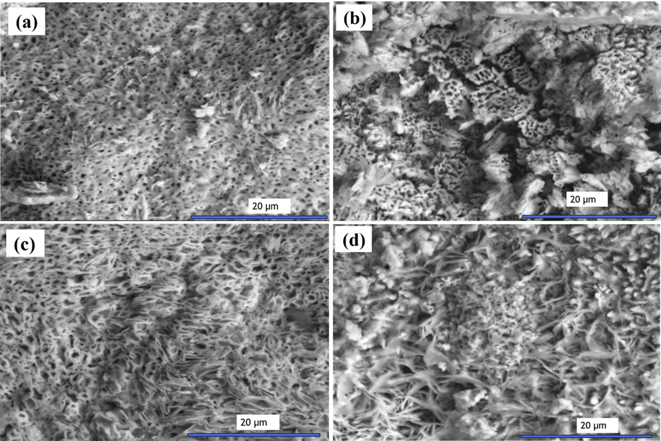 | ||
| Fig. 2 FE-SEM images of doped samples at the original magnification (6000×) (a) Al (Fe–Cr–Si–Ni–Mn–C), (b) Cu (Fe–Cr–Si–Ni–Mn–C), (c) Ti (Fe–Cr–Si–Ni–Mn–C), and (d) W (Fe–Cr–Si–Ni–Mn–C). | ||
3.4. Electrochemical corrosion test
The electrochemical behavior of the synthesized alloys was evaluated using potentio-dynamic polarization and electrochemical impedance spectroscopy (EFM) at both pH 4 and pH 7, with a temperature of 30 °C, as presented in Table 1. The corrosion potential (Ecorr), corrosion current density (Icorr), Tafel slopes (βa and βc), and corrosion rate (k) were determined for each alloy under both acidic and neutral conditions. The analysis of these electrochemical parameters provides valuable insights into the corrosion resistance and stability of these nanocomposite materials in different environments.| pH | Alloy | I corr (μA cm−2) | β a (mV dec−1) | β c (mV dec−1) | CF-2 | CF-3 | k (mpy) |
|---|---|---|---|---|---|---|---|
| (a) | |||||||
| 4 | Al | 1.128 | 117.6 | 122.4 | 1.459 | 3.407 | 0.5156 |
| Cu | 1.248 | 52.1 | 62.98 | 1.239 | 2.762 | 0.5704 | |
| Ti | 43.08 | 228.8 | 256.9 | 2.094 | 1.206 | 19.69 | |
| W | 30.89 | 131.3 | 135.4 | 2.827 | 5.797 | 14.11 | |
| 7 | Al | 1.471 | 86.3 | 87.88 | 1.316 | 9.534 | 0.6723 |
| Cu | 1.612 | 169.9 | 183 | 1.108 | 826.4 | 0.7367 | |
| Ti | 46.15 | 221.8 | 248.2 | 2.065 | 842.1 | 21.09 | |
| W | 33.77 | 148.9 | 155.7 | 1.755 | 9.743 | 15.43 | |
| pH | Alloy | E corr vs. SCE (mV) | I corr (μA cm−2) | β a (mV dec−1) | β c (mV dec−1) | k (mpy) |
|---|---|---|---|---|---|---|
| (b) | ||||||
| 4 | Al | −85.8 | 14.4 | 1679 | 1663 | 6.588 |
| Cu | −104 | 7.88 | 609.2 | 636.6 | 3.602 | |
| Ti | −310 | 39.7 | 588.5 | 471.4 | 18.16 | |
| W | −391 | 154 | 2214 | 865.6 | 70.37 | |
| 7 | Al | −109 | 11.9 | 813.7 | 797.8 | 5.432 |
| Cu | −53.7 | 1.41 | 337.6 | 320.6 | 0.6448 | |
| Ti | −313 | 38.3 | 697.3 | 477.7 | 17.51 | |
| W | −393 | 109 | 1702 | 831.9 | 50.02 | |
| Solutions | Parameters | |||
|---|---|---|---|---|
| β a (mV decade−1) | −βc (mV decade−1) | I corr (μA cm−2) | E corr (V) | |
| (c) | ||||
| Serum | 85 | 50 | 0.35 | −0.480 |
| Urine | 125 | 100 | 0.18 | −0.430 |
| Solutions | Parameters | ||||
|---|---|---|---|---|---|
| E corr,s (V) | E corr,f (V) | E pit (V) | E prot (V) | I corr (μA cm−2) | |
| (d) | |||||
| Serum | −0.460 | −0.312 | −0.180 | −0.150 | 11.4 |
| Urine | −0.430 | −0.115 | 0.2 | −0.040 | 4.1 |
At pH 7, the corrosion potential is less negative for all alloys, with Ecorr values for Cu and Al alloys being significantly more positive compared to Ti and W alloys. This reinforces the conclusion that Cu and Al additions enhance the corrosion resistance of the alloys in neutral conditions. These findings highlight the importance of adjusting alloy composition to optimize performance in specific environmental conditions.
In Table 1c the cyclic voltammograms of Ti and the alloy were analyzed in serum and urine at 37 °C to investigate their corrosion behavior. In serum, the anodic Tafel slope (βa) was 85 mV decade−1, the cathodic Tafel slope (−βc) was 50 mV decade−1, the corrosion current density (Icorr) was 0.35 μA cm−2, and the corrosion potential (Ecorr) was −0.480 V. In urine, βa increased to 125 mV decade−1, −βc doubled to 100 mV decade−1, Icorr decreased significantly to 0.18 μA cm−2, and Ecorr shifted to −0.430 V. These findings suggest a lower corrosion rate in urine compared to serum, highlighting the influence of electrolyte composition on the electrochemical performance of these alloys.
The electrochemical performance of the Ti implant alloy in serum and urine at 37 °C, evaluated using a scan rate of 10 mV s−1 as evaluated in Table 1d. The findings show distinct behaviors. In serum, the corrosion potentials (Ecorr,s and Ecorr,f) were −0.460 V and −0.312 V, with a pitting potential (Epit) of −0.180 V, a protection potential (Eprot) of −0.150 V, and a corrosion current density (Icorr) of 11.4 μA cm−2. In urine, Ecorr,s and Ecorr,f shifted positively to −0.430 V and −0.115 V, respectively, while Epit increased significantly to 0.2 V, Eprot improved to −0.040 V, and Icorr dropped markedly to 4.1 μA cm−2. These results indicate that while the Ti alloy exhibits superior resistance to pitting corrosion in urine, reflected by the higher Epit and Eprot values, the significantly lower Icorr suggests better overall corrosion resistance in urine compared to serum. This behavior highlights the influence of electrolyte composition on the alloy's stability and performance.
 | ||
| Fig. 3 (a) Cyclic voltammograms for the Ti implant, (b) for the Al implant, and (c) for the W implant in two biological solutions at a scan rate of 10 mV s−1: urine and serum at 37 °C. | ||
W alloys displayed higher corrosion current densities in both biological solutions, with values ranging from 0.25 μA cm−2 in serum to 0.34 μA cm−2 in urine. The results suggest that while W may provide adequate corrosion resistance, its performance in biological environments may be surpassed by that of Ti and Al alloys, particularly for applications requiring long-term durability. These electrochemical corrosion tests provide a clear comparison of the corrosion behavior of the various alloys in different pH environments and biological solutions. The data strongly indicate that Cu and Al alloys exhibit superior corrosion resistance, particularly in neutral and acidic conditions, making them promising candidates for applications in harsh environments. The results also emphasize the need to tailor alloy composition and processing conditions to achieve the desired balance of mechanical and corrosion properties for specific applications, such as in biomedical implants or protective coatings.
E corr,f is the final corrosion, and Ecorr,s is the starting corrosion potential after reversing the scan direction. Epit, also known as the pitting potential, is characterized by a continuous increase in the anodic current. Similarly, the protection potential (Eprot) refers to the potential where the reverse scan intersects the forward scan, completing the hysteresis loop. (βa and −βc), and corrosion potentials were obtained from Tafel analyses based on cyclic voltammetry (CV) curves. Log(i)/(A cm−2).
The experimental data of the corrosion potentials are tabulated in Table 2. Table 2(a) shows the cyclic voltammograms recorded for composites doped with Al and the alloy in biological electrolyte solutions (serum and urine) at 37 °C to evaluate their corrosion behavior. In serum, the anodic Tafel slope (βa) was 80 mV decade−1, the cathodic Tafel slope (−βc) was 190 mV decade−1, the corrosion current density (Icorr) was 0.34 μA cm−2, and the corrosion potential (Ecorr) was −0.120 V. In urine, βa increased to 143 mV decade−1, −βc decreased to 129 mV decade−1, Icorr decreased to 0.25 μA cm−2, and Ecorr shifted to −0.194 V. These results indicate that the alloys exhibit distinct electrochemical behaviors in the two electrolytes, reflecting differences in corrosion kinetics and passivation.
| Solutions | Parameters | |||
|---|---|---|---|---|
| β a (mV decade−1) | −βc (mV decade−1) | I corr (μA cm−2) | E corr (V) | |
| (a) | ||||
| Serum | 80 | 190 | 0.34 | −0.120 |
| Urine | 143 | 129 | 0.25 | −0.194 |
| Solutions | Parameters | ||||
|---|---|---|---|---|---|
| E corr,s (V) | E corr,f (V) | E pit (V) | E prot (V) | I corr (μA cm−2) | |
| (b) | |||||
| Serum | −0.812 | −0.815 | −0.80 | −0.250 | 7.4 |
| Urine | −0.294 | −0.298 | −0.285 | −0.155 | 5.12 |
| Solutions | Parameters | |||
|---|---|---|---|---|
| β a (mV decade−1) | −βc (mV decade−1) | I corr (μA cm−2) | E corr (V) | |
| (c) | ||||
| Serum | 92 | 43 | 0.214 | −0.321 |
| Urine | 107 | 92 | 0.318 | −0.374 |
| Solutions | Parameters | ||||
|---|---|---|---|---|---|
| E corr,s (V) | E corr,f (V) | E pit (V) | E prot (V) | I corr (μA cm−2) | |
| (d) | |||||
| Serum | −0.380 | −0.271 | −0.261 | −0.122 | 12.48 |
| Urine | −0.358 | −0.207 | 0.300 | −0.086 | 22.21 |
The results of the electrochemical behavior of a Cu implant alloy were evaluated in serum and urine at 37 °C using a scan rate of 10 mV s−1 presented in Table 2(b). In serum, the start and final corrosion potentials (Ecorr,s and Ecorr,f) were −0.812 V and −0.815 V, respectively, while the pitting potential (Epit) was −0.80 V, the protection potential (Eprot) was −0.250 V, and the corrosion current density (Icorr) was 7.4 μA cm−2. In urine, Ecorr,s and Ecorr,f shifted to −0.294 V and −0.298 V, respectively, with Epit at −0.285 V, Eprot at −0.155 V, and Icorr reduced to 5.12 μA cm−2. These results indicate enhanced corrosion resistance of the Cu alloy in urine compared to serum, as evidenced by the higher Eprot and lower Icorr values.
The electrochemical corrosion behavior of a W implant alloy was investigated in serum and urine at 37 °C using the Tafel diagram analysis depicted in Table 2(c). In serum, the anodic Tafel slope (βa) was 92 mV decade−1, the cathodic Tafel slope (−βc) was 43 mV decade−1, the corrosion current density (Icorr) was 0.214 μA cm−2, and the corrosion potential (Ecorr) was −0.321 V. In urine, βa increased to 107 mV decade−1, −βc more than doubled to 92 mV decade−1, Icorr rose to 0.318 μA cm−2, and Ecorr shifted to −0.374 V. These findings suggest a higher corrosion rate in urine compared to serum, indicated by the increased Icorr and more negative Ecorr values, reflecting the electrolyte's impact on the corrosion kinetics of the W alloy.
The electrochemical behavior of a Ti implant alloy in serum and urine at 37 °C was analyzed using Tafel analyses via cyclic voltammetry (CV) curves, as shown in Table 2(d), with a scan rate of 10 mV s−1. Key parameters, including corrosion and protection potentials as well as current densities, were measured to assess corrosion resistance. In serum, the initial and final corrosion potentials (Ecorr,s and Ecorr,f) were −0.380 V and −0.271 V, respectively, with a pitting potential (Epit) of −0.261 V, a protection potential (Eprot) of −0.122 V, and a corrosion current density (Icorr) of 12.48 μA cm−2. Comparatively, in urine, Ecorr,s and Ecorr,f shifted to −0.358 V and −0.207 V, respectively, while Epit increased significantly to 0.300 V, Eprot rose to −0.086 V, and Icorr was markedly higher at 22.21 μA cm−2. The data indicate that while the Ti alloy exhibited a higher resistance to pitting corrosion in urine, as evidenced by the elevated Epit, the increased Icorr suggests a greater general corrosion rate in urine compared to serum. This highlights the influence of electrolyte composition on the alloy's electrochemical stability and overall corrosion behavior.
The comparison of the data of the electrochemical behavior of Al, Cu, W, and Ti implant alloys in biological solutions at 37 °C reveals distinct corrosion characteristics that can be concluded as: Al alloy exhibits moderate corrosion resistance, with intermediate Icorr and Ecorr values influenced by the electrolyte. But the Cu alloy shows higher corrosion rates in serum (Icorr = 7.4 μA cm−2) compared to urine (Icorr = 5.12 μA cm−2) but demonstrates better pitting resistance in urine. Meanwhile, the W alloy displays a relatively low Icorr in serum (0.214 μA cm−2), indicating good corrosion resistance, though urine increases its corrosion rate. Finally, the Ti alloy, while it has better pitting resistance in urine (Epit = 0.300 V), its higher Icorr in urine (22.21 μA cm−2) suggests increased general corrosion compared to serum. Overall, Ti and Cu alloys show significant sensitivity to electrolyte composition, while W generally offers higher stability.
3.5. Biological and immunogenic effects of the intraperitoneal injection of different alloys
The biological and immunogenic responses of male albino rats following the intraperitoneal (i.p.) injection of various alloy materials at different doses over a subacute duration of 60 days were then explored. The alloys tested include aluminum (Al), copper (Cu), tungsten (W), and titanium (Ti), which were administered at three dose levels: 10 mg kg−1, 20 mg kg−1, and 50 mg kg−1 body weight (BW). Liver function, oxidative stress markers, and immunological responses were monitored throughout the study, with the data presented in Fig. 4. | ||
| Fig. 4 Multivariate analysis of the effects of different alloyed materials on liver function marker enzyme in rats scattered in a dose-dependent manner on serum levels of ALT, serum levels of AST, and oxidative stress markers, SODs, and hepatic level of GSH, in rats scattered in a dose-dependent manner. | ||
The AST levels in Fig. 4(b) support these observations, showing a similar pattern. Significant increases in AST were noted in the aluminum and tungsten groups, especially at higher doses, while copper and titanium exhibited more moderate increases in AST activity. These results align with the observed changes in ALT, suggesting a dose-dependent liver damage associated with the alloys, particularly aluminum and tungsten.
 | ||
| Fig. 5 Multivariate analysis of the effects of different alloy materials on the following: nitrosative stress markers (nitric oxide), serum cortisol level (as a stress marker) in rats scattered in a dose-dependent manner, serum creatinine level (as a renal performance marker), serum IL-1 level (as an immune response marker) and serum IL-10 level in rats scattered in a dose-dependent manner. | ||
4. Conclusions
Nickel-free porous stainless steels composed of F2581 (Fe–17Cr–10Mn–3MX–0.4Si–0.5 N–0.2C in wt%; MX = Al, Cu, Ti, and W) were synthesized via a solid-state reaction method. The XRD pattern revealed the crystal structure of the Fe phase. The crystallite sizes are approximately 73, 27.2, 76, and 98.5 nm for samples with Al, Cu, Ti, and W, respectively. The morphology of the synthesized nanoparticles is as follows: nanowires, square nanotubes, wave-like (growing clover farm) and nanofibers for Al, Cu, Ti, and W ions with nanopores distributed in shapes, respectively. The experimental data revealed that the samples with Cu and Ti seemed to be the most suitable for medical implantation and biological macromolecule applications. Furthermore, the electrochemical kinetic parameters were obtained via two different methods: EFM and polarized techniques. In our practical study of electrochemical corrosion properties of three alloys (Ti, Al, and W) grown in biological solutions by electrochemical techniques, the results revealed that, compared with those of other alloys in serum and urine, the corrosion resistance of Ti alloy in blood serum was the highest. A pilot study evaluating the biological performance and immunogenicity of these alloys revealed that the high dose of each alloy induced biological alterations and introduced immunogenicity at high doses. In contrast, Cu, W, and Ti at low doses are safer and do not present any immune response for subacute doses, and the medium dose exhibited the same response but for Cu only.Author contributions
S. A. Abdelwahab, M. Zewaid and A. I. Ali: fabricated and characterized the samples. Mohamad Warda, Omar A. Ahmed-Farid and Hassan A. M. Hendawy: investigated the results of electrochemical and biological experiments. H. Saleh, J. Y. Son, M. Warda, O. A. Ahmed-Farid, H. A. M. Hendawy, A. I. Ali, A. Negm and E. A. Kamoun: contributed to the analysis of the data and writing and reviewed the final manuscript. All authors approve the final version of the submission.Ethical approval
The experimental procedures adhered strictly to the ethical guidelines established by the Institutional Animal Care and Use Committee (IACUC) at the Faculty of Veterinary Medicine, Cairo University, Egypt (Approval No: Vet CU. 09092023786), as well as the ethical standards outlined by the National Organization for Drug Control and Research. All procedures were performed in accordance with the NC3Rs ARRIVE Guidelines. For the human blood and urine donated samples, the signed consents of the donors (of 5 persons) were first taken before the experiment start according to the research ethical rules in Egyptian Universities.Data availability
The datasets used and/or analyzed during the current study are available from the corresponding author upon reasonable request. In addition, all the data generated or analyzed during this study are included in this article.Conflicts of interest
There are no conflicts of interest.References
- S. Wu, et al., Biomimetic porous scaffolds for bone tissue engineering, Mater. Sci. Eng., R, 2014, 80, 1–36 Search PubMed.
- K.-H. Frosch and K. M. Stürmer, Metallic biomaterials in skeletal repair, Eur. J. Trauma, 2006, 32, 149–159 Search PubMed.
- S. J. Hollister, Porous scaffold design for tissue engineering, Nat. Mater., 2005, 4(7), 518–524 CAS.
- J. Čapek and D. Vojtěch, Effect of sintering conditions on the microstructural and mechanical characteristics of porous magnesium materials prepared by powder metallurgy, Mater. Sci. Eng., C, 2014, 35, 21–28 Search PubMed.
- J. Čapek and D. Vojtěch, Microstructural and mechanical characteristics of porous iron prepared by powder metallurgy, Mater. Sci. Eng., C, 2014, 43, 494–501 CrossRef.
- J. Čapek, D. Vojtěch and A. Oborná, Microstructural and mechanical properties of biodegradable iron foam prepared by powder metallurgy, Mater. Des., 2015, 83, 468–482 CrossRef.
- K. Alvarez and H. Nakajima, Metallic scaffolds for bone regeneration, Materials, 2009, 2(3), 790–832 CrossRef CAS.
- V. Karageorgiou and D. Kaplan, Porosity of 3D biomaterial scaffolds and osteogenesis, Biomaterials, 2005, 26(27), 5474–5491 CrossRef CAS PubMed.
- S. Jayasathyakawin, et al., A Review on Exploration of Magnesium Matrix Composites, Mater. Sci. Forum, 2022, 1068, 63–70 Search PubMed.
- J. Jowsey, Studies of Haversian systems in man and some animals, J. Anat., 1966, 100(Pt 4), 857 CAS.
- V. S. de Viteri and E. Fuentes, Titanium and titanium alloys as biomaterials, Tribol.: Fundam. Adv., 2013, 1(5), 154–181 Search PubMed.
- M. Saini, et al., Implant biomaterials: a comprehensive review, World J. Clin. Cases, 2015, 3(1), 52 CrossRef.
- H. Shi, et al., Functional gradient metallic biomaterials: techniques, current scenery, and future prospects in the biomedical field, Front. Bioeng. Biotechnol., 2021, 8, 616845 CrossRef PubMed.
- X. Zhang, et al., Preparation and mechanical property of a novel 3D porous magnesium scaffold for bone tissue engineering, Mater. Sci. Eng., C, 2014, 42, 362–367 CrossRef CAS PubMed.
- B. Arifvianto and J. Zhou, Fabrication of metallic biomedical scaffolds with the space holder method: a review, Materials, 2014, 7(5), 3588–3622 CrossRef.
- S. Bose, S. Vahabzadeh and A. Bandyopadhyay, Bone tissue engineering using 3D printing, Mater. today, 2013, 16(12), 496–504 CrossRef CAS.
- X. Wang, et al., Porous TiNbZr alloy scaffolds for biomedical applications, Acta Biomater., 2009, 5(9), 3616–3624 CrossRef CAS PubMed.
- E. Babaie and S. B. Bhaduri, Fabrication aspects of porous biomaterials in orthopedic applications: a review, ACS Biomater. Sci. Eng., 2018, 4(1), 1–39 CrossRef CAS PubMed.
- J. Parthasarathy, B. Starly and S. Raman, A design for the additive manufacture of functionally graded porous structures with tailored mechanical properties for biomedical applications, J. Manuf. Process., 2011, 13(2), 160–170 CrossRef.
- V. Geantă, et al., Stainless steels with biocompatible properties for medical devices, Key Eng. Mater., 2014, 583, 9–15 Search PubMed.
- K. Essa, et al., Porosity control in 316 L stainless steel using cold and hot isostatic pressing, Mater. Des., 2018, 138, 21–29 CrossRef CAS.
- M. Resnik, et al., Strategies for improving antimicrobial properties of stainless steel, Materials, 2020, 13(13), 2944 CrossRef CAS PubMed.
- Q. Chen and G. A. Thouas, Metallic implant biomaterials, Mater. Sci. Eng., R, 2015, 87, 1–57 CrossRef.
- E. Davoodi, et al., Additively manufactured metallic biomaterials, Bioact. Mater., 2022, 15, 214–249 CAS.
- T. Murakami, T. Akagi and E. Kasai, Development of porous iron based material by slag foaming and its reduction, Proc. Mater. Sci., 2014, 4, 27–32 CrossRef CAS.
- J. Čapek, et al., Highly porous, low elastic modulus 316 L stainless steel scaffold prepared by selective laser melting, Mater. Sci. Eng., C, 2016, 69, 631–639 CrossRef.
- Z. Li, et al., A sol–gel-derived α-Al2O3 crystal interlayer modified 316 L porous stainless steel to support TiO2, SiO2, and TiO2–SiO2 hybrid membranes, J. Mater. Sci., 2011, 46, 3127–3135 CrossRef CAS.
- S. D. F. F. Mariotto, et al., Porous stainless steel for biomedical applications., Mater. Res., 2011, 14, 146–154 CrossRef CAS.
- F. M. Noor, K. Jamaludin and S. Ahmad, Fabrication of porous stainless steel 316 L for biomedical applications, MATEC Web Conf., 2017, 135, 7 CrossRef.
- M. M. Dewidar, K. A. Khalil and J. Lim, Processing and mechanical properties of porous 316 L stainless steel for biomedical applications, Trans. Nonferrous Met. Soc. China, 2007, 17(3), 468–473 CrossRef.
- E. Salahinejad, et al., Microstructure and wear behavior of a porous nanocrystalline nickel-free austenitic stainless steel developed by powder metallurgy, Mater. Des., 2010, 31(4), 2259–2263 CrossRef CAS.
- A. Dudek and R. Włodarczyk, Effect of sintering atmosphere on properties of porous stainless steel for biomedical applications, Mater. Sci. Eng., C, 2013, 33(1), 434–439 CrossRef CAS PubMed.
- M. Zielecka, et al., Silicone resin-based intumescent paints, Materials, 2020, 13(21), 4785 CrossRef CAS.
- T. K. Tatt, et al., Production of porous stainless steel using the space holder method, Sains Malays., 2021, 50, 507–514 CrossRef CAS.
- W. Zhang, et al., The Effect of Porosity on Mechanical Properties of Porous FeCrN Stainless Steel, J. Phys.: Conf. Ser., 2021, 2044, 012002 CrossRef.
- B. Vandenbroucke and J. P. Kruth, Selective laser melting of biocompatible metals for rapid manufacturing of medical parts, Rapid Prototyping J., 2007, 13(4), 196–203 CrossRef.
- D. K. Pattanayak, et al., Bioactive Ti metal analogous to human cancellous bone: fabrication by selective laser melting and chemical treatments, Acta Biomater., 2011, 7(3), 1398–1406 CrossRef CAS.
- M. Ramirez-del-Solar and E. Blanco, Porous thin films from sol–gel, Submicron porous materials, 2017, 157–188 Search PubMed.
- S. A. Yavari, et al., Fatigue behavior of porous biomaterials manufactured using selective laser melting, Mater. Sci. Eng., C, 2013, 33(8), 4849–4858 CrossRef.
- A. S. Idil and N. Donaldson, The use of tungsten as a chronically implanted material, J. Neural Eng., 2018, 15(2), 021006 CrossRef.
- M. Rahmati and M. Mozafari, Biocompatibility of alumina-based biomaterials–A review, J. Cell. Physiol., 2019, 234(4), 3321–3335 CrossRef CAS PubMed.
- Q. Shen, et al., Advances in copper-based biomaterials with antibacterial and osteogenic properties for bone tissue engineering, Front. Bioeng. Biotechnol., 2022, 9, 795425 CrossRef.
- M. Javanbakht, M. Hadianfard and E. Salahinejad, Microstructure and mechanical properties of a new group of nanocrystalline medical-grade stainless steels prepared by powder metallurgy, J. Alloys Compd., 2015, 624, 17–21 CrossRef CAS.
- G. A. Perchetti, et al., Stability of SARS-CoV-2 in phosphate-buffered saline for molecular detection, J. Clin. Microbiol., 2020, 58(8), 1–3 CrossRef PubMed.
- H. Savaloni, et al., Influence of N+ implantation on structure, morphology, and corrosion behavior of Al in NaCl solution, Chin. Phys. B, 2020, 29(5), 058102 CrossRef CAS.
- D. M. Cocchetto and T. D. Bjornsson, Methods for vascular access and collection of body fluids from the laboratory rat, J. Pharm. Sci., 1983, 72(5), 465–492 CrossRef CAS PubMed.
- S. Reitman and S. Frankel, Calorimetric method for the determination of blood aminotransferase enzymatic activities, Am. J. Clin. Pathol., 1957, 28, 56–63 CrossRef CAS PubMed.
- S. Marklund and G. Marklund, Involvement of the superoxide anion radical in the autoxidation of pyrogallol and a convenient assay for superoxide dismutase, Eur. J. Biochem., 1974, 47(3), 469–474 CrossRef CAS.
- E. Jayatilleke and S. Shaw, A high-performance liquid chromatographic assay for reduced and oxidized glutathione in biological samples, Anal. Biochem., 1993, 214(2), 452–457 CrossRef CAS.
- I. Papadoyannis, V. Samanidou and C. C. Nitsos, Simultaneous determination of nitrite and nitrate in drinking water and human serum by high performance anion-exchange chromatography and UV detection, J. Liq. Chromatogr. Relat. Technol., 1999, 22(13), 2023–2041 CrossRef CAS.
- H. Yang, et al., Facile preparation of super-hydrophobic and super-oleophilic silica film on stainless steel mesh via sol–gel process, Appl. Surf. Sci., 2010, 256(13), 4095–4102 CrossRef CAS.
- E. Salahinejad, R. Amini and M. Hadianfard, Effect of milling time on structure and mechanical properties of porous nickel-free austenitic stainless steels processed by mechanical alloying and sintering, Mater. Sci. Eng., A, 2010, 527(21–22), 5522–5527 CrossRef.
- Y. Zhou, Characterization of the porosity and pore behavior during the sintering process of 420 stainless steel samples produced with gas-and water-atomized powder using powder based 3-d printing, University of Pittsburgh, Swanson School of Engineering, 2014, pp. 1–88.
- E. Salahinejad, et al., Microstructural characterization of medical-grade stainless steel powders prepared by mechanical alloying and subsequent annealing, Adv. Powder Technol., 2013, 24(3), 605–608 CrossRef CAS.
- A. I. Ali and A. Hassen, Synthesis, characterization, ferroelectric, and piezoelectric properties of (1 − x)BaTiO3−x(BaNi0.5Nb0.5O3) perovskite ceramics, J. Mater. Sci.: Mater. Electron., 2021, 32(8), 10769–10777 CrossRef CAS.
- A. I. Ali, et al., Preparation, structural and dielectric properties of nanocomposite Al2O3/BaTiO3 for multilayer ceramic capacitors applications, J. Mater. Res. Technol., 2022, 18, 2083–2092 CrossRef CAS.
- A. M. El Nahrawy, et al., Talented Bi0.5Na0.25K0.25TiO3/oxidized cellulose films for optoelectronic and bioburden of pathogenic microbes, Carbohydr. Polym., 2022, 291, 119656 CrossRef CAS.
- A. M. El-Nahrawy, et al., Influences of Ag-NPs doping chitosan/calcium silicate nanocomposites for optical and antibacterial activity, Int. J. Biol. Macromol., 2016, 93, 267–275 CrossRef CAS.
| This journal is © The Royal Society of Chemistry 2025 |