
 Open Access Article
 This Open Access Article is licensed under a
 Creative Commons Attribution 3.0 Unported Licence
Suckerin based biomaterials for wound healing: a comparative review with natural protein-based biomaterials
Samson Prince
Hiruthyaswamy
 and
Kanagavel
Deepankumar
*
and
Kanagavel
Deepankumar
*
School of Biosciences and Technology, Vellore Institute of Technology, Vellore, India. E-mail: samsonprinceh@gmail.com; deepankumark87@gmail.com; deepankumar.k@vit.ac.in
First published on 22nd January 2025
Abstract
Suckerins, derived from the sucker ring teeth of cephalopods, have emerged as promising biomaterials for wound healing due to their unique structural properties and versatility. Suckerins exhibit a wide range of elasticity, from soft hydrogels to stiff films, controlled by β-sheet content and induced by di-tyrosine crosslinking. Compared to other natural protein-based materials, suckerins offer advantages in terms of mechanical strength, the ability to form robust supramolecular structures, biocompatibility, excellent thermoplastic property and underwater adhesion, making them suitable for wound healing and various biomedical applications. However, the full potential of suckerins in wound healing is yet to be explored. This paper aims to provide a comprehensive understanding of the structural properties, recent findings, advantages, succinylation of biomaterials and applications of natural protein-based biomaterials such as mussel adhesive protein, collagen, gelatin, silk fibroin and chitosan in wound healing, and how they stand in comparison to suckerin protein-based materials. This comprehensive review could pave the way for the development of more effective wound healing therapies. Future research directions are also discussed, emphasizing the need for a deeper understanding of the supramolecular interactions stabilizing suckerins and their potential applications in medicine, tissue engineering, nanotechnology and gaps in other protein-based materials in the context of wound healing.
 Samson Prince Hiruthyaswamy | Samson Prince Hiruthyaswamy is an active researcher at the School of Biosciences and Technology, Vellore Institute of Technology, Vellore, India. His research focuses on biomaterials for regenerative medicine, with an emphasis on protein-based materials for wound healing and drug delivery. He has contributed to the development of suckerin-based biomaterials and their applications in advanced wound care technologies. Samson is actively involved in exploring innovative approaches in nanotechnology, wound care, and biofunctional materials, aiming to address critical challenges in tissue engineering. His dedication to interdisciplinary research is reflected in his collaborative efforts to advance biomedical science. |
 Kanagavel Deepankumar | Dr Kanagavel Deepankumar is an Associate Professor at the School of Bio Sciences and Technology, Vellore Institute of Technology, Vellore, India. With expertise in protein engineering and bioadhesives, he has significantly contributed to the development of biomaterials for advanced biomedical applications. Dr Deepankumar has authored over 15 peer-reviewed publications in high-impact journals, with a current h-index of 16, reflecting his influence in the field. His research focuses on designing biofunctional materials, including suckerin-based biomaterials and nanotechnologies for wound healing, drug delivery, and tissue engineering. |
1. Introduction
Wound healing is a complex and dynamic process involving the restoration of skin integrity after injury. This process includes several overlapping stages: hemostasis, inflammation, proliferation, and remodeling. Efficient wound healing is crucial for preventing infections, minimizing scarring, and restoring normal tissue function.1 Globally, chronic wounds, including diabetic ulcers, pressure ulcers, and venous leg ulcers, pose significant healthcare challenges, affecting millions of people and leading to substantial medical costs. Annually, the incidence of full-thickness wounds is estimated to be between 9.1 and 26.1 million worldwide. Chronic wounds affect approximately 2% of the total population in the United States. The global wound care market is projected to grow at a compound annual growth rate of 4.17% from 2024 to 2030.2 Traditional wound healing treatments, such as wound dressings, antibiotics, and surgical interventions, play a critical role in managing wounds. However, these methods often have limitations, including the risk of infection, delayed healing times, and inadequate functional and aesthetic outcomes. Consequently, there is a growing need for advanced wound care solutions that can overcome these limitations and enhance the healing process.Protein-based biomaterials from natural sources have emerged as promising candidates in the field of regenerative medicine and wound healing. These biomaterials, derived from natural proteins, offer biocompatibility, biodegradability, and the ability to mimic the extracellular matrix (ECM), which is essential for tissue regeneration.3 Recent studies highlight their potential in advancements in dynamic ECM mechanics, applications of 3D aligned collagen fibre platforms for enhancing cell differentiation, and bioprinting methods for screening cell responses to ECM.4–6 Key protein-based biomaterials used in wound healing include collagen, gelatin, silk fibroin, hyaluronic acid, and chitosan. Collagen is the most abundant protein in the ECM, collagen provides structural support and promotes cell adhesion and migration, crucial for tissue repair. Gelatin is derived from collagen, gelatin retains many of its beneficial properties, including biocompatibility and biodegradability, making it a valuable material for wound dressings and scaffolds. Silk fibroin is extracted from silk, which offers excellent mechanical properties, biodegradability, and supports cell proliferation, making it suitable for wound healing applications. Chitosan is derived from chitin, it possesses antimicrobial properties, enhances wound healing, and can form biocompatible and biodegradable films and hydrogels.7,8 Recent advances in chitosan-based hydrogels, such as photoresponsive variants with robust adhesive and antibacterial properties, further highlight their potential in skin wound healing applications.9 Despite their advantages, these protein-based biomaterials have drawbacks like immunogenicity and variable biodegradability and biocompatibility.
Recently, there has been increasing interest in developing advanced biomaterials for biomedical applications. Suckerin-based biomaterials, derived from the sucker rings of squid, represent an emerging class with significant potential in wound healing. Suckerin proteins are characterized by unique structural properties, including tunable mechanical strength, self-assembly capabilities, and robust biocompatibility. These proteins are capable of formulating films and hydrogels that replicate the natural ECM, providing an optimum environment for tissue regeneration and cell proliferation.10,11 Compared to conventional protein-based materials, suckerin-based biomaterials provide several benefits, such as, customised disintegration rates, better mechanical qualities and increased cell-instructive capabilities. These features make them suitable for applications requiring high durability and flexibility, such as tissue engineering and implantable medical devices.
This review aims to investigate, contrast, and leverage suckerin-based biomaterials with other naturally occurring protein-based materials for wound healing applications. Through a comprehensive analysis, this paper elucidates the unique properties, potential benefits, and diverse applications of suckerin proteins, aiming to enhance the understanding of their role in advancing biomaterial science, biomedicine, biotechnology, and materials science.
2. Properties and characteristics of protein-based biomaterials
Protein-based biomaterials have gained significant attention for their diverse applications in biomedical engineering, particularly in wound healing, tissue engineering, and drug delivery. These biomaterials include chitosan, collagen, gelatin, silk fibroin, mussel adhesive protein and suckerin. Each of these materials has unique sources, structural properties, and characteristics that determine their suitability for applications. A comprehensive comparison of these protein-based biomaterials, highlighting their sources, structural properties, and various applications are discussed in Table 1.| Biomaterial | Source | Structural properties | Characteristics | Applications | Ref. |
|---|---|---|---|---|---|
| Chitosan | Derived from chitin | N-Acetyl-D-glucosamine (acetylated unit) and β-(1-4)-linked D-glucosamine (deacetylated unit) are randomly distributed in a linear polysaccharide. | Biocompatible, biodegradable, non-toxic. Able to bind to metal ions, proteins, lipids, and cholesterol. | Agriculture, cosmetology, pharmacy, medicine, food industry, textile and paper industries, and chemistry. | 12 and 13 |
| Collagen | Animal tissues | Triple helix structure composed of three polypeptides of amino acids. | High tensile strength, provides structure to fibrous cartilage, teeth, connective tissue, bones, skin, and tendons. | Wound healing techniques, cosmetic preparations for the skin. | 14 and 15 |
| Gelatin | Derived from collagen | Consists of N-acetyl-D-glucosamine (acetylated unit) and β-(1-4)-linked D-glucosamine (deacetylated unit) dispersed at randomly. | Soluble in glycerol, acetic acid, and hot water—polar solvents. It forms gel when it absorbs 5–10 times its weight in water. | Agriculture, pulp & paper, food industry, medicine, cosmetics, biotechnology, agriculture, textiles, and environmental compounds. | 16 |
| Silk fibroin | Silkworms | Consists of hydrophilic light (L-) chain and the hydrophobic heavy (H-) chain as its two main chains. | Biocompatible, biodegradable, excellent mechanical properties. | 3D bioprinting, tissue engineering, drug delivery, and wound dressings. | 17 and 18 |
| Mussel adhesive protein | Secreted by mussels | Comprises approx. 20% of tyrosine (or DOPA) and lysine, respectively. Enriched with glutamine and glycine. Contains major amounts of polyphenols and repeating amino acid patterns. 3,4-DOPA, a modified amino acid. Modified amino acid 3,4-DOPA. | DOPA-induced interfacial adhesion. Strong adherence to wet surfaces. Both biodegradable and biocompatible. | Utilised in tissue engineering, coatings, bioadhesives, and anti-fouling studies. | 19 and 20 |
| Suckerin | Cephalopods | Block copolymer protein1. The size range is 5–60 kDA, and the isoelectric point is between pI 7 and 10. | High modulus of elasticity and thermoplastic properties. Spider silk-like in structure. | Body armour, artificial ligaments, medical equipment, specialised bandages, stitches, parachutes, sails and aeroplane parts. | 21 and 22 |
2.1 Chitosan
Chitosan is a versatile biomaterial derived from chitin, the second most abundant natural polymer after cellulose. Chitosan is a linear polysaccharide made up of N-acetyl-D-glucosamine (acetylated unit) and randomly distributed β-(1–4)-linked D-glucosamine (deacetylated unit). It is produced by using an alkaline chemical, like sodium hydroxide, to the chitin shells of prawns and other crustaceans. The only distinction between the molecular structures of chitosan and cellulose is the connection between the amino group (–NH2) and the C2-position.23,24 Chitosan is biocompatible, biodegradable, and non-toxic. It forms strong ionic and hydrogen bonds with the paper fibres, demonstrating its affinity for them. Chitosan is a promising natural preservative and agent against bacterial infections because of its strong antibacterial properties, which break microbial membranes and DNA.25 The analgesic properties of chitosan can also be explained by its polycationic composition. In order to understand chitosan's biodegradability, it's highly important to keep in consideration that the material is both a polymer with amino groups and a polysaccharide, meaning it has breakable glycosidic linkages.26,27 Chitosan has a wide range of applications due to its unique properties. It can be used in the chemistry, food, pharmaceutical, medical, cosmetics, textile, and paper sectors. Additionally, the fields of dentistry, ophthalmology, biomedicine and bioimaging, hygiene and personal care, veterinary medicine, packaging, agrochemistry, aquaculture, functional textiles and cosmetotextiles, catalysis, chromatography, beverage industry, photography, wastewater treatment and sludge dewatering, and biotechnology have all gained significant attention to chitosan in recent years.2.2 Collagen
Biomaterials are essential in various applications, including tissue engineering, drug delivery, and regenerative medicine. Among these, collagen stands out due to its unique properties and widespread availability. Collagen is a structural protein that is mainly extracted from the skin of fish and mammals. It is essential for the support of animal organisms.28 Biomaterials based on collagen have attracted a lot of interest due to their remarkable mechanical strength and biocompatibility. These materials offer various advantages over conventional protein-based choices, including increased cell adherence and biodegradability, making them extremely suitable for biomedical applications. The triple helical structure of collagen imparts high tensile strength, contributing to the durability of collagen-based biomaterials. Collagen fibres are formed through the aggregation of tropocollagen molecules, which are left-handed helices composed of three polypeptide chains.14,15,29 Glycine is the predominant amino acid in collagen, occurring at every third residue, while proline and hydroxyproline are also abundant, contributing to the stability of the helical structure.30,31 The strength of collagen fibres is enhanced by cross-links such as lysine–hydroxylysine and hydroxylysine–hydroxyproline, which occur between the fibrils.32 There are different types of collagens, characterized by variations in the polypeptide chains and post-translational modifications, leading to a diversity of functions and locations in the body. As a natural component of the human body, collagen minimizes immune responses, reducing the risk of rejection. This biocompatibility, combined with its biodegradability, makes collagen an excellent choice for temporary scaffolds in tissue engineering, as it supports natural healing processes and degrades as the body heals.33 It undergoes intracellular and extracellular biodegradation. Intracellular biodegradation happens through endocytosis by cells capable of phagocytosis, while extracellular degradation occurs due to matrix metalloproteinases (MMPs). The triple helix structure is destroyed during peptide bond degradation between amino acids, leading to collagen molecule disintegration into two molecules at a ratio of 3![[thin space (1/6-em)]](https://www.rsc.org/images/entities/char_2009.gif) :1.34,35 Additionally, collagen's ability to promote cell adhesion and proliferation, thanks to specific cell adhesion sites within its structure, further enhances its suitability for tissue engineering and regenerative medicine. The ease with which collagen can be modified and functionalized to meet specific biological, clinical, and mechanical requirements underscores its versatility and effectiveness in various biomedical applications.
:1.34,35 Additionally, collagen's ability to promote cell adhesion and proliferation, thanks to specific cell adhesion sites within its structure, further enhances its suitability for tissue engineering and regenerative medicine. The ease with which collagen can be modified and functionalized to meet specific biological, clinical, and mechanical requirements underscores its versatility and effectiveness in various biomedical applications.
2.3 Gelatin
Gelatin is a fascinating biomaterial derived from collagen, a protein found in the bones, skin and connective tissues of animals such as chicken, cattle, fish, and pigs. It is produced when collagen undergoes partial hydrolysis, rupturing some of the protein links that hold it together.18 The primary structure of gelatin is characterized by a repeating sequence of Gly-X-Y triplets, where X and Y are mostly proline and hydroxyproline. One third of the chain is made up of glycine, while another third is proline or hydroxyproline. The amino acid composition and sequence in gelatin are different from one source to another but always consists of large amounts of glycine, proline, and hydroxyproline.36 Gelatin is an amphoteric polymer, meaning it contains both acidic and basic groups. Commercially, there are two types of gelatin: type A, which results from partial acid hydrolysis of collagen, and type B, which derives from alkali-based treatment of collagen.37 Gelatin has a colourless or faintly yellow appearance and is almost completely odourless and tasteless. When dry, it is translucent and brittle; when wet, it takes on a rubbery texture. Gelatin is insoluble in organic solvents like alcohol but soluble in polar solvents like glycerol, hot water, and acetic acid.38,39 When exposed to water, it may absorb five to ten times its own weight and forms gel. Thixotropic gels, which are made of gelatin, have a growing viscosity under stress and can be melted by heating them up. The texture of dishes made with gelatin is significantly influenced by the fact that the top melting point of gelatin is lower than body temperature.40,41 Gelatin's unique properties make it a versatile biomaterial with numerous applications in food industry as widely used as a stabilizer, gelling agent, thickener, and emulsifier in various food products such as, low-fat yogurt, jellies, gelatin dessert, candy, milk powder, sausage, salad, pudding, fruit milk, ham, bean vermicelli, cake, ice cream, drinks.42 In pharmaceuticals, gelatin is used to form gel caps for medications. It's also used in drug delivery systems and nanoparticles.37,43 In cosmetics, due to its water-binding ability, gel formation ability, and film-forming properties, gelatin is used in a variety of cosmetics and hair treatments44 and in biomedicine, gelatin is used in tissue engineering and regenerative medicine due to its biocompatibility, biodegradability, and low immunogenicity.45,462.4 Silk fibroin
Silk-based biomaterials exhibit exceptional structural properties, including high tensile strength, biocompatibility, controlled biodegradability, and the ability to interact with biological systems effectively.47 The major structural components of silk fibroin (SF) are highly repeated sequences made up primarily of serine (12%), alanine (30%), and glycine (43%). It is primarily composed of glycine, alanine, and serine, with a repeating sequence of Gly–Ala–Gly–Ala–Gly–Ser. This unique composition and structure make silk fibroin an ideal material for a wide range of applications, including enzyme immobilization and tissue engineering. It offers a stable matrix for enzyme immobilization, preserving their biological activity.48 These biomaterials are derived from silk proteins, such as silk fibroin, which can serve as templates for the nucleation and growth of inorganic substances, enhancing their properties and expanding their applications in various fields.49,50 The versatility of silk fibroin allows for its manipulation into solutions, gels, membranes, particles, and various other forms, enabling tailored materials with specific performance characteristics for diverse applications. The structural versatility of silk allows for functional modifications to enhance repair performance in cartilage/osteochondral applications.51 Furthermore, conductive materials derived from silk have been produced with excellent mechanical robustness, tissue adhesion ability, biocompatibility, and electrical conductivity, which makes them appropriate for biointerface applications such as tissue scaffolds and artificial epidermal sensors.52 These unique structural properties of silk-based biomaterials pave the way for innovative advancements in personalized healthcare and bioengineering.2.5 Mussel adhesive protein
Aquatic mussels produce amazing natural polymers called mussel adhesive proteins (MAPs), or mussel foot proteins (Mfps), which aid in the mussels’ ability to stick to a variety of moist surfaces. These proteins contain a high concentration of specific amino acids, including 3,4-dihydroxy-L-phenylalanine (DOPA), lysine, and glycine, which are essential for their adhesive capabilities. The presence of DOPA, formed by post-translational modification of tyrosine, is particularly important for their adhesive properties because of its ability to develop strong non-covalent and covalent interactions with both organic and inorganic surfaces.53 MAPs’ molecular structures often feature repeating decapeptide motifs, such as the sequence NH2-Ala-Lys-Pro-Ser-Tyr-DOPA-Lys-Tyr-Ser-Tyr-COOH seen in Mfp-5.19 The high concentration of DOPA residues enhances the proteins’ adhesive and cohesive capabilities, allowing for interactions with surface atoms via hydrogen bonding, π–cation interactions, and metal coordination. MAPs have a strong adhesive bond even in moist and saline conditions, which makes them appropriate for use in maritime and biomedical applications. Naturally biocompatible and biodegradable, they break down into non-toxic components in the body, making them ideal for temporary implants and other biomedical applications. Mussel adhesive proteins (MAPs) possess remarkable versatility, with their adhesive qualities modifiable through alterations in their amino acid sequences or by combining them with other polymers. This adaptability enhances their applicability across diverse fields. Advances in the design of adhesive hydrogels, provide a robust foundation for exploring the potential of MAP-based materials.54 Modern design strategies such as chemical bond-based approaches, including static and dynamic covalent bonds as well as non-covalent interactions like hydrogen bonding and electrostatic forces, to optimize adhesion properties. These hydrogels are engineered with multifunctional properties such as enhanced stretchability, toughness, biocompatibility, and environmental responsiveness, making them ideal for biomedical and industrial applications. Their utility is further demonstrated in fields like tissue repair, biosensors, hemostatic agents, drug delivery systems, and bioelectronics. These advancements illustrate the transformative potential of integrating MAPs with advanced polymer designs, opening new frontiers in innovative technologies and applications.2.5 Suckerin
Suckerin, a block-copolymer peptide derived from squid sucker ring teeth, presents remarkable potential as a biomaterial due to its unique structural and functional properties. Suckerin proteins, which consist of two repeating modules—the glycine-rich M2 and the alanine and histidine-rich M1—have a dynamic structure with domains that fold into anti-parallel β-sheets, indicating a strong affinity for β-strands. The glycine-rich modules contain aromatic residues that participate in π–π stacking interactions, which are crucial for stabilizing the protein's structural core. Identified across various squid species, suckerins are utilized to produce silk-like materials, such as fibres, films, and tissue scaffolds, revealing significant applications in medicine, tissue engineering, and nanotechnology. Suckerin's self-assembly dynamics are particularly noteworthy; M2 is more capable of self-association than M1, which permits microphase separation and the creation of nanoconfined β-sheets in the M1–M2 matrix.21,22 This self-assembly process grants suckerin materials exceptional properties, including excellent thermo-plasticity, pH-responsiveness, biocompatibility, biodegradability, and sustainability. These attributes are further enhanced by manipulating solvent conditions—such as pH, salt type, and protein concentration—allowing control over suckerin self-assembly and mechanical properties. Innovative modifications, such as incorporating non-canonical amino acids (ncAAs), can fine-tune suckerin proteins, improving their mechanical strength and self-assembly behavior.55,56 High biocompatibility, derived from their natural origin and self-assembly characteristics, makes suckerin proteins particularly suitable for various biomedical applications. The biodegradability of suckerin further enhances its appeal for sustainable uses, as it can naturally decompose, reducing environmental impact. One groundbreaking application of suckerin involves the development of a recombinant squid suckerin-spider silk fusion protein. This fusion protein has shown promise in creating biocompatible hydrogels, which are particularly effective for stem cell secretome delivery systems, aiding in chronic wound healing.57 This fusion exemplifies the potential of suckerin in crafting functional biomaterials for biomedical applications. Overall, the insights gained from studying suckerin's structure and function offer substantial guidance for designing novel block-copolymer peptides. These peptides can be tailored for diverse applications in nanotechnology and biomedicine, leveraging their superior mechanical properties and adaptive self-assembly capabilities. By exploring the integration of ncAAs and manipulating environmental factors, researchers can enhance the performance and utility of suckerin-based materials, paving the way for innovative solutions in medical and technological fields. Suckerin's combination of structural sophistication, functional versatility, and environmental sustainability underscores its potential as a foundational biomaterial for future advancements.3. Applications of protein-based biomaterials in wound healing
Protein-based biomaterials such as chitosan, collagen, gelatin, silk fibroin, mussel adhesive protein and suckerin have been extensively studied for their applications in wound healing due to their biocompatibility, biodegradability, and bioactivity. Various fabrication techniques, including 3D printing, electrospinning, and hydrogels, have been employed to utilize these materials effectively. Each technique has its own set of applications, advantages, and disadvantages. The following Table 2 provides a detailed comparison of these aspects.| Biomaterial | Techniques | Applications | Advantages | Disadvantages | Ref. |
|---|---|---|---|---|---|
| Chitosan | Used as hydrogels, films, fibres, membranes, and sponges | Food, agriculture, medicine, pharmacy, cosmetics, paper and textile industries, and chemistry fields | Non-toxic, biocompatible, and biodegradable. Able to attach to proteins, lipids, cholesterol, and metal ions | Only two RCTs reported significant beneficial effects of chitosan on wound healing | 58 and 59 |
| Collagen | Used in the form of sponges, hydrogels, films, and nanoparticles | Wound healing techniques, cosmetic preparations for the skin | High tensile strength. Provides structure to bones, skin, fibrous cartilage, teeth, connective tissue and tendons. Stimulates specific cells that positively affect wound healing | May require further enzymatic breakdown. Clinical trials on its effectiveness are lacking | 14, 15 and 29 |
| Gelatin | Used in the form of hydrogels, sponges, films, and nanoparticles | Food, medicine, biotechnology, agriculture, textiles, pulp & paper, cosmetics, pharmaceuticals, and environmental applications | Promotes wound hemostasis, enhances peri-wound antibacterial and anti-inflammatory properties | Poor mechanical properties and antimicrobial activity | 37 and 43 |
| Silk Fibroin | Used in the form of hydrogels, sponges, films, electrospun nanofibre mats, and hydrocolloid dressings | Wound dressings, drug delivery, tissue engineering, 3D bioprinting | Biocompatible, biodegradable, excellent mechanical properties. Anti-inflammatory, pro-angiogenic properties | Poor tensile strength. Limited use to linear, tension-free, dry wounds without body hair | 17 and 50 |
| Mussel adhesive protein | Used in forms of glue, hydrogels. | Adhesive, wound closure application, tissue engineering | Strong adhesion, versatile, biocompatible | Complex extraction process, limited scalability | 60 and 61 |
| Suckerin | Used in the form of fibres, films, and hydrogels | Adhesives, wound dressings, drug delivery. | High elastic modulus and thermoplastic behavior. Similar structure to spider silk. Promotes skin cell adhesion and proliferation | Limited studies on its use in wound healing | 57, 62 and 63 |
3.1 Chitosan based biomaterials
Chitin, which is made up of 1,4-linked N-acetylglucosamine molecules, is commonly present in insects, molluscs, crustaceans, and fungi. It has been demonstrated that chitosan, an active deacetylated derivative of chitin, accelerates tissue regeneration and aids in hemostasis.64 Numerous studies have demonstrated chitosan's efficacy as an antibacterial agent, preventing wound infections due to its antibacterial and antifungal properties.65 Additionally, chitosan encourages cell multiplication, which is vital for healing, and stimulates the growth of fibroblasts, with the degree of deacetylation positively correlated with fibroblast activation.66 In wound healing applications, hydrogels, foams, and scaffolds containing chitosan and other polymers have demonstrated promising outcomes.67 Being the only cationic polysaccharide identified in nature, chitosan is unique. It demonstrates a number of outstanding characteristics that are advantageous for use in biomedical applications, such as high degrees of hydrophilicity, hemostatic and antibacterial properties, biodegradability, biocompatibility, and bioadhesivity.68,69 However, chitosan has certain limitations, such as chemical properties that restrict its adaptability and therapeutic application. These drawbacks can be mitigated by modifying its chemical composition and creating cross-links with other materials.70 For example, tannic acid (TA) was added to a quaternized chitosan matrix, resulting in good hemostatic action and the capability to reduce reactive oxygen species (ROS). Quaternized chitosan has superior antibacterial and water-soluble qualities compared to regular chitosan.71,72 This modified chitosan hydrogel demonstrated reduced ROS, hemostatic effects, and suppression of S. aureus and E. coli in vitro. In diabetic rats with full-thickness wounds, it promoted coagulation, reduced inflammation, and enhanced collagen deposition.73Additionally, layer-by-layer deposition of TA/QC/TA employing quaternized chitin (QC) and TA was used to construct TA/Ag-modified polylactic acid (PLA)/polyurethane (PU) hybrid nanofibres. When compared to S. aureus and E. coli, the resultant hybrid nanofibres showed decreased ROS and antibacterial activity.74 A composite hydrogel was developed using polyvinyl alcohol (PVA) and chitosan to load EGF-loaded chitosan nanoparticles, perfluorocarbon nanoemulsions, and polyhexamethylene biguanide (PHMB). This composite hydrogel's prolonged release of PHMB and EGF stimulated the growth of human KERTr keratinocytes, which is essential for wound healing, as well as antibacterial activity against S. aureus and S. epidermidis. These hydrogels improved collagen deposition and maturation, promoted re-epithelialization, and reduced inflammatory responses in full-thickness wounds of the diabetic rats.75 Polyvinyl alcohol/sodium alginate/chitosan (PACS) demonstrated good swelling, water vapour permeability, retention, and biocompatibility, but minimal antibacterial activity. A bilayer composite dressing for wound healing that combined the chitosan nanoparticles loaded into an Ag–metal organic framework and PACS demonstrated improved performance. The Ag-MOF/CSNPs layer had strong antibacterial properties, promoting coagulation and cell proliferation. The bilayer dressing demonstrated superior antibacterial activity and significantly accelerated wound healing in vivo compared to PACS, representing a notable advancement in wound care.76 Chitosan, with its potent antibacterial properties, biocompatibility, and ability to enhance hemostasis, collagen deposition, and re-epithelialization, represents a highly effective and promising biomaterial for advanced wound healing applications.
3.2 Collagen based biomaterials
Collagen, the most abundant protein in the human body, plays a crucial role in tissue structure and repair, making it an ideal candidate for biomaterials aimed at wound healing. Recent studies have focused on the development and evaluation of collagen-based biomaterials to enhance the healing process through various techniques and formulations. Collagen's natural biocompatibility and ability to promote cell proliferation and differentiation make it highly effective in wound healing applications. Recent research has demonstrated that collagen-based dressings can significantly accelerate wound closure, reduce inflammation, and improve tissue regeneration compared to conventional treatments. Rats with full-thickness skin defect wounds were treated with an injectable collagen–hyaluronic acid hydrogel featuring antioxidant properties based on the dynamic covalent bond of borate esters. This hydrogel efficiently eliminated excess reactive oxygen species and accelerated wound healing by promoting angiogenesis and enhancing the inflammatory microenvironment of the wound.77 To improve the mechanical properties of collagen-based hydrogels, Zhang et al. utilized graphene oxide and borax as cross-linking agents to incorporate guar gum and poly(N-isopropylacrylamide). This resulted in a hydrogel capable of stretching to 50 times its initial length and self-healing within three minutes without external stimulation, showcasing remarkable hyperductility and self-repair capabilities.78 Additionally, the hydrogel facilitated the repair of full-thickness skin defects in rats and exhibited potential for human sensing applications. Collagen-based hydrogels have seen extensive use in clinical practice, with FDA-approved tissue-engineered skin products like Apligraf being effectively employed for burn wounds, diabetic foot ulcers, and skin replacement. Apligraf is a designed active biomaterial composed of type I collagen hydrogel, allogeneic fibroblasts, and keratinocytes.79Commercial collagen or gelatin sponge dressings are also widely used in wound care due to their excellent hemostatic, biodegradable, and biocompatibility properties. A composite sponge dressing, created by combining collagen, sodium polyacrylate, and biquaternium-conjugated chitosan in an aqueous solution and freeze-drying it, demonstrated antibacterial, hemostatic, anti-inflammatory, and pro-regenerative qualities.80 For full-thickness skin defects in mice, a novel zwitterionic betaine composite collagen sponge dressing with broad antioxidant and anti-inflammatory properties supported wound healing, granulation tissue growth, reepithelialization, collagen deposition, and angiogenesis.81 To mimic the structure of the natural extracellular matrix (ECM), esterified hyaluronic acid was added to electrospun nanofibres to create a collagen-based hydrogel wound dressing. Applied to full-thickness skin defect wounds in mice infected with Staphylococcus aureus, this dressing proved more effective than the hydrogel scaffold alone in promoting angiogenesis, collagen deposition, and wound healing. Its high specific surface area and porosity allowed it to efficiently collect wound exudate and release medication.82 Furthermore, encapsulating the VEGF mimetic peptide Prominin-1 derived peptide in a polylactic acid–glycolic acid copolymer/gelatin-based nanofibre membrane, the dressing was applied to diabetic rats with full-thickness skin defect wounds. The dressing continuously released Prominin-1 derived peptide, encouraged angiogenesis and immune regulation by attracting endogenous VEGF, and ultimately aided in wound healing.83
3.3 Gelatin based biomaterials
Gelatin, a natural polymer derived from collagen, has emerged as a versatile and biocompatible biomaterial for wound healing applications. Its inherent properties, such as biodegradability, non-toxicity, and ability to form hydrogels, make it a promising candidate for developing wound dressings and scaffolds. Recent studies have explored various techniques and formulations of gelatin-based biomaterials, demonstrating their effectiveness and practical implications in enhancing wound healing processes. Gelatin methacrylate (GelMA), a polymer derived from gelatin, has gained widespread use in tissue regeneration due to its biocompatibility and versatility. For instance, a bioprinting-based strategy has been developed to investigate stem cell-extracellular matrix (ECM) interactions using gradient GelMA hydrogels. This approach facilitates the screening of an optimal ECM for the in vivo healing of alveolar bone defects. Studies have demonstrated that GelMA/PEGDA composite hydrogels, engineered through precise bioprinting techniques, support osteogenic differentiation of periodontal ligament stem cells (PDLSCs). The optimized GelMA/PEGDA hydrogels exhibited robust bone formation in rat models of alveolar bone defects.84,85 The use of shape-memory cryogels made of methacrylated gelatin (GelMA) for the controlled release of endothelial progenitor cells (EPCs) and acid fibroblast growth factor (aFGF) is one fostering advancement. Methacrylate groups enable gelatin to be cross-linked into a stable hydrogel under the influence of visible light while maintaining its inherent enzymatic degradability. In a diabetic rat model, these methacrylated gelatin cryogels significantly improved pressure ulcer healing by overexpressing hypoxia-inducible factor 1-alpha (HIF-1α) at the wound site. This overexpression is crucial as HIF-1α plays a vital role in promoting angiogenesis and tissue regeneration under hypoxic conditions typically found in chronic wounds.86 Another innovative approach involves adipose-derived stem cell (ADSC)-loaded gelatin microspheres. These microspheres have been shown to expedite the healing of full-thickness wounds in diabetic rats by promoting M2 macrophage polarization, collagen deposition, angiogenesis, and hair follicle development. The ADSC-loaded gelatin microspheres offer a multifaceted approach to wound healing by addressing various cellular and molecular aspects of tissue repair, thereby enhancing the overall healing process.87Moreover, oxidised dextran and ethylenediamine-modified gelatin have been combined to create hydrogels that can load paeoniflorin (Pf) and zinc oxide nanoparticles (ZnO-NPs). ZnO-NPs possess antimicrobial properties, while Pf has angiogenic properties. These hydrogels are responsive to changes in pH and reactive oxygen species (ROS), exhibiting sequential hemostatic, antibacterial, and angiogenic actions. This multifaceted response is particularly beneficial for treating diabetic wounds, which are often characterized by persistent infections and poor healing. In diabetic rat models, these hydrogels significantly accelerated wound recovery by addressing both microbial infection and promoting tissue regeneration.88 The various formulations and techniques explored in recent studies highlight the effectiveness of gelatin in promoting wound healing through multiple mechanisms, including enhanced angiogenesis, collagen deposition, and antimicrobial action.
3.4 Silk fibroin based biomaterials
Silk fibroin (SF), derived from the Bombyx mori silkworm, is highly valued in the biomedical field for its mechanical properties, biocompatibility, and biodegradability. The efficacy and adaptability of SF-based biomaterials for wound healing have been highlighted in recent studies. Two electrospun SF (ESF) membranes were compared to a commercial wound dressing (3MTM Tegaderm™) in a comparative trial. Poly-dopamine (PESF) was added to one SF membrane, leaving the other unaltered (ESF). All dressings accelerated wound healing in full-thickness skin wound models in Sprague–Dawley rats, but the poly-dopamine-modified SF dressing was found to be much more effective.89 Another study used primary human dermal fibroblasts (PHDFs) in vitro to explore SF scaffolds with fibre sizes ranging from 256 ± 30 to 1214 ± 321 nm. The results showed that SF scaffolds with the smallest fibre diameter (250–300 nm) had the highest PHDF proliferation. This trend persisted throughout the culture period, with larger fibre diameters resulting in decreased proliferation. Ex vivo studies applying a human full-thickness skin model showed that SF scaffolds facilitated re-epithelialization during wound healing, while in vitro investigations revealed a significant increase of collagen I and III in PHDFs grown on the smallest SF fibres.90Sprague–Dawley rats were used in studies to examine full-thickness wound healing using heparinised SF hydrogels containing acidic fibroblast growth factor 1 (FGF1). In vitro, SF hydrogels—both with and without FGF1—boosted the migration and proliferation of L929 cells in comparison with the control group. 3MTM Tegaderm™ was left untreated, heparinised SF hydrogel, heparinised SF hydrogel with FGF1, and a commercial chitosan dressing were the four groups that were given different dressings in vivo. With the FGF1-containing SF hydrogel exhibiting the smallest wound size on day 14, these treatments markedly sped up the healing process.91 Furthermore, a study comparing electrospun SF nanomatrix to Medifoam®, a commercial polyurethane hydrocellular bandage, and medical gauze was conducted on Sprague–Dawley rats with second-degree burn wounds. After seven days, the SF nanomatrix-treated wounds were smaller than the Medifoam®-treated ones. In contrast to control wounds, which shrank by 18% over the course of 28 days, wounds treated with SF nanomatrix and Medifoam® shrank to 4% and 8% of their initial sizes, respectively. According to histological research, the SF nanomatrix promoted skin regrowth and re-epithelialization. It also shown immunomodulatory activity by raising anti-inflammatory marker IL-10 and decreasing pro-inflammatory markers IL-1α and IL-6.92 In another study,93 the effects of three different types of SF hydrogels (SF hydrogel (SFH), SF melanin hydrogel (SFMH), and SF composite hydrogel (SFCH)), with the latter two incorporating anti-inflammatory berberine and antioxidant melanin, were investigated on full-thickness wound healing in diabetic Wistar rats. Compared to untreated wounds, these therapies increased the pace of wound closure, with SFCH showing the quickest rate of healing. The highest levels of re-epithelialization, collagen deposition, and vascular density were observed in wounds treated with SFCH, along with the smallest epithelial gaps. All these studies demonstrate how SF-based dressings can improve wound healing by means of different methodologies, such as growth factor incorporation, structural transformations, and antioxidant and anti-inflammatory effects.
3.5 Mussel adhesive protein-based biomaterials
Significant advancements in wound healing have resulted from attempts to recreate the method of strong underwater adhesion that mussels acquire through the interaction of DOPA and mussel foot proteins. This process involves conjugating catechol groups onto polymers. The ability of DOPA-inspired biomaterials to stick to moist surfaces, including living tissues, is a significant benefit for tissue repair.60 For instance, a biocompatible tissue glue designed for wound closure that was based on a poly(amidoamine) adhesive inspired by mussels showed that its degradability could be altered by changing the amount of zwitterionic sulfobetaine present. Research demonstrated that RAW 264.7 mouse macrophage cells could adhere to and multiply on a substrate covered with tissue adhesive without causing substantial cytotoxicity, and an in vivo investigation demonstrated that the adhesive healed wounds in 28 days with little to no scarring.94 A scar preventing glue made from collagen type I and mussel adhesive proteins (MAPs) was shown to improve early-stage wound healing and prevent scar formation by modulating collagen fibril growth and fibrogenic factor expression.95 In another study, oxidised hyaluronic acid (HA-CHO) and dopa-grafted ε-polylysine (EPL-DOPA) were combined to formulate a bioadhesive hydrogel dressing with antibacterial characteristics that could be tailored, as well as swelling and degradation rates. In comparison to readily available fibrin glue, this hydrogel adhered to wounds rapidly and firmly, accelerated healing, and successfully killed the bacteria at wound site. Histological research also revealed better skin regrowth.96Additionally, by chemically integrating DOPA–methyl ester (DOPA–ME) and DOPA–HA into proteins, namely Candida antarctica fraction B (CAL-B) lipase, researchers have developed a bioadhesive that is inspired by mussels. This study investigated the protein's C-terminal direct DOPA conjugation and N-terminal DOPA–HA polymer conjugation, highlighting the potential uses of these bioadhesives on wet wound surfaces.97 Furthermore, dopamine polymerisation and sodium alginate crosslinking in polyacrylamide networks produced an adhesive hydrogel that demonstrated strong adherence to pig skin (24.5 kPa) and supported cell attachment and proliferation, suggesting that it could be used as a material for skin tissue engineering.98 An enzymatically biodegradable hydrogel inspired by mussels, consisting of a modified PEG coupled to an alanine dipeptide substrate, was created for tissue repair and regenerating purposes. This hydrogel, which was created with elastase-sensitive Ala–Ala linkages to break down in reaction to neutrophil enzyme release, showed sufficient adhesive ability in animal models, achieving a value of 30.4 ± 3.4 kPa and biodegrading over several months.99 Lastly, a pDA-chondroitin sulphate–polyacrylamide hydrogel inspired by mussels was developed for cartilage regeneration in the absence of growth factors. Enriched with catechol groups, this hydrogel demonstrated superior mechanical qualities appropriate for cartilage repair, as well as good cell affinity and promotion of adhesion.100 Bioadhesives inspired by mussels have a great deal of potential in tissue engineering. They act as transporters of biomolecules to improve cell responses in addition to promoting tissue adhesion. The broad range of uses for these biomaterials anticipates assisting future studies in tissue regeneration and repair, enhancing the area.
3.6 Suckerin based biomaterials
Suckerin proteins are a class of structural proteins that resemble block co-polymers and self-assemble into strong supramolecular structures that are used by cephalopods’ arms and tentacles to securely grasp the prey. Because of their aromatic linkage and β-sheet-driven supramolecular pattern, these proteins have great potential as biomimetic protein-based biopolymers. A two-main sequence module designed suckerin protein was studied structurally using multi-dimensional Nuclear Magnetic Resonance (NMR) spectroscopy. According to the study, the protein takes on a dynamic shape, with areas exhibiting β-strand propensity folding into anti-parallel β-sheets in both module 1 (residues A42–A52) and module 2 (residues G30–Y37 and G58–Y65). The glycine (Gly)-rich M2 modules’ aromatic residues stabilise the structural core through π–π stacking interactions.10,21,22,101The elasticity of recombinant suckerin-19 is regulated by the β-sheet composition and induced di-tyrosine crosslinking, which can result in a variety of biomaterials, from extremely soft hydrogels to stiff films. Researchers were able to achieve a wide variety of elastic moduli, from 40 to 500 Pa for hydrogels to several GPa for crosslinked films, by varying the concentration of the initiator ammonium persulfate (APS) during Ru-mediated photo-crosslinking. The mechanical characteristics of the films were validated by nanoindentation testing, and rheological measurements showed that the gels’ storage modulus (G′) improved with APS concentration.56,102 Suckerin assemblies also exhibit excellent thermoplastic properties, retaining their mechanical characteristics through multiple cycles of melting and reshaping. Additionally, suckerin proteins have been utilized in designing and developing protein-based adhesives through genetic engineering. Unaltered recombinant suckerin-12, expressed in semi-crystalline networks formed by self-assembly in E. coli, demonstrated robust underwater adhesion behavior (70 mN m31, Wad ∼15 mJ m2 on mica). This adhesion is attributed to the cross-β sheet domains, as disturbance of these sheets eliminates adherence. Similar characteristics were shown by suckerin-12-DOPA, which was created by directly inserting the DOPA moiety utilising an in vivo residue-specific incorporation technique.62
Microneedles made from suckerin protein, created using a soft lithography technique, precisely replicated their templates and possessed strong mechanical characteristics, enabling high capacity for skin penetration. In vitro skin permeation tests using human cadaver skin samples indicated quicker onset and better skin penetration of medications with suckerin microneedles compared to flat patches. Furthermore, suckerin demonstrated intrinsic antibacterial properties, reducing cell counts from 108 CFU ml−1 to ∼3.02 × 10⁴ CFU ml−1, and showed good cell viability, confirming its biocompatibility and non-toxicity.63 In order to create hydrogels for the encapsulation and release of the stem cell secretome, a recombinant squid suckerin-spider silk fusion protein was created containing cell-adhesion motifs that could thermally gel at physiological temperatures. The secretome-suckerin sample group achieved nearly full wound closure (∼87%) within 24 hours, significantly outperforming the control group (∼59%).57 Overall, suckerin proteins, with their versatile mechanical properties, robust underwater adhesion, and biocompatibility, hold great potential for various biomedical applications as shown in Fig. 1, including biomaterials, adhesives, and drug delivery systems.
 | ||
| Fig. 1 Potential application of suckerin protein in biomedical research. | ||
3.7 Succinylation in biomaterials and their role in wound healing
Succinylated biomaterials are chemically modified materials enriched with succinyl groups (–CO–CH2–COOH), derived from succinic acid. This modification introduces carboxyl (–COOH) and carbonyl (–C![[double bond, length as m-dash]](https://www.rsc.org/images/entities/char_e001.gif) O) groups into biomaterials like proteins and polysaccharides. The resulting changes significantly enhance the material's hydrophilicity and functional versatility, making them ideal for biomedical applications, particularly wound healing as shown in Fig. 2. The carboxyl groups in succinylated materials are highly hydrophilic, forming hydrogen bonds with water molecules, which increases the material's water retention capacity. This hydrophilicity maintains a hydrated wound environment, essential for cell migration, proliferation, and granulation tissue formation, key processes in tissue repair. The carbonyl groups, on the other hand, enhance chemical reactivity, providing active sites for the attachment of bioactive molecules or therapeutic agents, further amplifying the material's functionality. In addition to creating a moist wound environment, hydrophilic surfaces in succinylated biomaterials reduce protein adsorption and bacterial adhesion, thereby lowering the risks of infection and inflammation.103–106 These surfaces also exhibit pH responsiveness, enabling the controlled release of encapsulated drugs like antimicrobial agents or growth factors directly at the wound site. This targeted delivery not only enhances therapeutic efficacy but also accelerates the healing process by mimicking the natural extracellular matrix (ECM), providing a supportive scaffold for cell attachment and tissue regeneration.
O) groups into biomaterials like proteins and polysaccharides. The resulting changes significantly enhance the material's hydrophilicity and functional versatility, making them ideal for biomedical applications, particularly wound healing as shown in Fig. 2. The carboxyl groups in succinylated materials are highly hydrophilic, forming hydrogen bonds with water molecules, which increases the material's water retention capacity. This hydrophilicity maintains a hydrated wound environment, essential for cell migration, proliferation, and granulation tissue formation, key processes in tissue repair. The carbonyl groups, on the other hand, enhance chemical reactivity, providing active sites for the attachment of bioactive molecules or therapeutic agents, further amplifying the material's functionality. In addition to creating a moist wound environment, hydrophilic surfaces in succinylated biomaterials reduce protein adsorption and bacterial adhesion, thereby lowering the risks of infection and inflammation.103–106 These surfaces also exhibit pH responsiveness, enabling the controlled release of encapsulated drugs like antimicrobial agents or growth factors directly at the wound site. This targeted delivery not only enhances therapeutic efficacy but also accelerates the healing process by mimicking the natural extracellular matrix (ECM), providing a supportive scaffold for cell attachment and tissue regeneration.
 | ||
| Fig. 2 Advantages of succinylated biomaterials in wound healing and controlled bioactive release. | ||
Recent advancements highlight the therapeutic potential of succinylated biomaterials. Succinylated chitosan derivatives restored endothelial cell function, increasing HUVEC cell viability from 50% to 80% and improving in vivo wound healing rates from 25% to 65% within 10 days.107 Succinylated chitosan nanoparticle films achieved wound closure rates of up to 90% in 10 days, releasing 85 mg of drugs over 48 hours.108 Similarly, N-succinylated chitosan fibers exhibited a swelling ratio of 5 g g−1 (grams of liquid per gram of fibre), enhancing wound healing rates from 30% to 75% within 7 days.109 Succinylated nanoparticles loaded with basic fibroblast growth factor (bFGF) provided controlled drug release, increasing collagen deposition from 10 mg cm−2 to 25 mg cm−2 and wound healing rates from 35% to 85% in 10 days.110 Other examples include compartmentalized bone graft sponges that reduced inflammatory markers and improved wound closure rates from 30% to 70% within 14 days.111 Pullulan succinate films grafted with chitosan and hyaluronic acid enhanced vascularization, reduced scar formation, and improved healing.112 Succinylated albumin cryogels enabled controlled drug release, enhancing healing rates from 40% to 85% in 12 days.113 Furthermore, biomimetic glycopolypeptide hydrogels with tunable adhesion doubled collagen deposition and increased tissue regeneration from 30% to 75% in 10 days.114 These versatility and efficacy of succinylated biomaterials in promoting wound healing, tissue regeneration, and targeted drug delivery, paving the way for advanced biomedical applications.
4. Challenges and future perspectives
4.1 Challenges and limitations
Suckerin-based biomaterials face challenges in scalability, cost-effectiveness, and functional optimization. The complexity of extraction and production limits their large-scale application, making them less viable for commercial use. Functionalization to enhance biocompatibility and mechanical properties requires advanced techniques that balance flexibility and strength without compromising their structural integrity. Additionally, limited research on suckerin's biological interactions with cells and tissues hinders its clinical translation. Compared to extensively studied biomaterials like collagen or silk fibroin, suckerin's therapeutic potential remains underexplored, leaving gaps in its applicability for wound healing.Hydrophilicity is a key factor in biomaterial performance for wound healing, yet many protein-based materials lack adequate water retention properties. Poor hydrophilicity can lead to desiccation of the wound bed, slowing healing and reducing cell migration. Surface modifications or blending with hydrophilic polymers often compromise mechanical integrity, posing additional challenges. Developing materials that inherently balance hydrophilicity with structural strength remains a major hurdle. This limitation is particularly relevant for materials like suckerin, which require functional enhancements to create a moist wound environment conducive to epithelialization and tissue regeneration while maintaining their other desirable properties. Addressing these issues is crucial for leveraging suckerin's unique properties in regenerative medicine.
4.2 Future perspectives
Looking ahead, several promising avenues exist for the further development of suckerin and other protein-based biomaterials in wound healing as shown in Fig. 3. Decellularized materials, bioactive molecules, and novel approaches like 3D and 4D bioprinting have significantly expanded the scope of wound healing research. These advanced technologies present platforms for enhanced tissue regeneration but still face challenges such as reduced vascularization, poor mechanical integrity, and immune rejection. Promising areas for further exploration include combining suckerin with bioactive molecules (e.g., growth factors, cytokines, and extracellular matrix components) to enhance its functional properties. Such combinations could promote skin regeneration, minimize scar formation, and improve overall healing outcomes.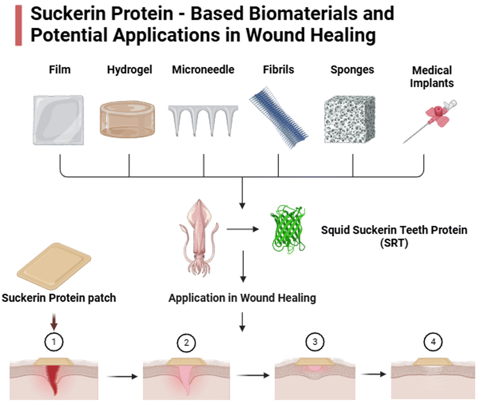 | ||
| Fig. 3 Suckerin protein-based biomaterials and its application in wound healing. | ||
Incorporating succinylation with other chemical modifications, such as phosphorylation or acetylation, may enhance the functional versatility of these materials, while preclinical studies should investigate molecular interactions with immune cells to understand their role in modulating inflammation and promoting tissue regeneration. Succinylation, through the introduction of carboxyl and carbonyl groups, also transforms protein-based biomaterials like suckerin into platforms for controlled bioactive molecule release. Enhanced hydrophilicity and surface charge allow for the encapsulation and sustained release of therapeutic agents in response to environmental cues such as pH changes. Succinylated suckerin, for example, could encapsulate growth factors like VEGF or epidermal growth factor (EGF) to enhance angiogenesis and epithelialization or load anti-inflammatory agents and antibiotics for targeted delivery. These advancements represent a promising future for succinylated biomaterials in advanced wound healing, drug delivery, and tissue engineering, paving the way for precise, efficient, and patient-tailored therapeutic strategies.
5. Conclusion
Suckerin-based biomaterials represent a significant leap forward in the field of wound healing, offering unique structural and functional properties that distinguish them from other natural protein-based biomaterials. Their remarkable mechanical strength, elasticity, biocompatibility, and self-assembly capabilities position them as ideal candidates for developing next – generation wound healing therapies. By mimicking the extracellular matrix and promoting cellular adhesion and proliferation, suckerin-based materials have the potential to enhance tissue regeneration and accelerate healing processes. Furthermore, advancements in the functionalization of suckerin, including the incorporation of bioactive molecules, succinylation, and nanotechnology, promise to unlock new possibilities for targeted drug delivery and infection control. The integration of suckerin with emerging technologies such as 3D and 4D bioprinting, molecular engineering, and adaptive biomaterial systems paves the way for creating highly customizable wound dressings and therapeutic scaffolds tailored to individual needs. Additionally, suckerin's ability to combine with other biomaterials, such as chitosan, collagen, and silk fibroin, offers exciting prospects for hybrid systems with enhanced functionality and versatility.Despite these advances, challenges such as scalability, cost-effectiveness, and understanding of suckerin's long-term interactions with biological systems remain. Addressing these limitations through interdisciplinary collaborations, preclinical studies, and translational research will be critical to achieving clinical adoption. Suckerin-based biomaterials not only expand the frontiers of regenerative medicine but also inspire a new wave of research aimed at creating sustainable, multifunctional biomaterials for advanced wound care. As the field progresses, suckerin holds the potential to revolutionize wound management, offering innovative solutions to persistent clinical challenges.
Author contributions
Kanagavel Deepankumar – developed the initial idea and framework for the review, contributed to original drafting significant portions of the manuscript, oversaw the development of the manuscript and guided the research direction, secured funding for the project and provided the necessary resources for data gathering and writing. Samson Prince Hiruthyaswamy – conducted the literature search and comparative analysis of the biomaterials discussed, wrote the first draft of the manuscript, created and designed figures and tables for data presentation. Organized and analyzed the data included in the review.Disclosure statement
All figures presented in this manuscript have been independently created by the author using Adobe Photoshop. No external sources or third-party images have been used in the preparation of these illustrations. The figures are intended solely for the purpose of enhancing the scientific content and clarity of this paper.Data availability
Suckerin based biomaterials for wound healing: a comparative review with natural protein-based biomaterials, and all data analyzed and discussed are derived from publicly available sources, including peer-reviewed research articles, databases, and reports. No new data were generated for this study. The data supporting the conclusions of this article are fully referenced within the manuscript.Conflicts of interest
The authors report there are no competing interests to declare.Acknowledgements
This review was funded by the Vellore Institute of Technology, Vellore through a seed grant (SG20230131) awarded to Kanagavel Deepankumar. We gratefully acknowledge the support to Vellore institute of technology for financial support, and to our institution for providing resources that made this work possible.References
- H. N. Wilkinson and M. J. Hardman, Wound healing: cellular mechanisms and pathological outcomes, Open Biol., 2020, 10(9), 200223 CrossRef CAS.
- C. K. Sen, Human Wound and Its Burden: Updated 2022 Compendium of Estimates, Adv. Wound Care, 2023, 12(12), 657–670 CrossRef PubMed.
- H. Agnieray, et al., Recent developments in sustainably sourced protein-based biomaterials, Biochem. Soc. Trans., 2021, 49(2), 953–964 CrossRef CAS.
- Y. Ma, T. Han, Q. Yang, J. Wang, B. Feng, Y. Jia and F. Xu, Viscoelastic cell microenvironment: hydrogel-based strategy for recapitulating dynamic ECM mechanics, Adv. Funct. Mater., 2021, 31(24), 2100848 CrossRef CAS.
- Y. Ma, Y. Ji, G. Huang, K. Ling, X. Zhang and F. Xu, Bioprinting 3D cell-laden hydrogel microarray for screening human periodontal ligament stem cell response to extracellular matrix, Biofabrication, 2015, 7(4), 044105 CrossRef.
- N. Shi, Y. Li, L. Chang, G. Zhao, G. Jin, Y. Lyu and F. Xu, A 3D, magnetically actuated, aligned collagen fiber hydrogel platform recapitulates physical microenvironment of myoblasts for enhancing myogenesis, Small Methods, 2021, 5(6), 2100276 CrossRef CAS PubMed.
- D. N. Moholkar, et al., Recent advances in biopolymer-based formulations for wound healing applications, Eur. Polym. J., 2021, 160, 110784 CrossRef CAS.
- W. Peng, et al., Recent progress of collagen, chitosan, alginate and other hydrogels in skin repair and wound dressing applications, Int. J. Biol. Macromol., 2022, 208, 400–408 CrossRef CAS.
- Y. Ma, J. Yao, Q. Liu, T. Han, J. Zhao, X. Ma and F. Xu, Liquid bandage harvests robust adhesive, hemostatic, and antibacterial performances as a first-aid tissue adhesive, Adv. Funct. Mater., 2020, 30(39), 2001820 CrossRef CAS.
- S. H. Hiew, et al., Squid Suckerin Biomimetic Peptides Form Amyloid-like Crystals with Robust Mechanical Properties, Biomacromolecules, 2017, 18(12), 4240–4248 CrossRef CAS PubMed.
- D. Ding, et al., Biomimetic Production of Silk-Like Recombinant Squid Sucker Ring Teeth Proteins, Biomacromolecules, 2014, 15(9), 3278–3289 CrossRef CAS.
- H. Ibrahim and E. El-Zairy, Chitosan as a biomaterial—structure, properties, and electrospun nanofibers, Concepts, compounds and the alternatives of antibacterials, 2015, pp. 81–101 Search PubMed.
- I. Aranaz, et al., Chitosan: An overview of its properties and applications, Polymers, 2021, 13(19), 3256 CrossRef CAS.
- S. Chattopadhyay and R. T. Raines, Collagen-based biomaterials for wound healing, Biopolymers, 2014, 101(8), 821–833 Search PubMed.
- S. Radhakrishnan, et al., Collagen based biomaterials for tissue engineering applications: A review, Processes and phenomena on the boundary between biogenic and Abiogenic nature, 2020, pp. 3–22 Search PubMed.
- Ad. T. Alfaro, et al., Fish gelatin: characteristics, functional properties, applications and future potentials, Food Eng. Rev., 2015, 7, 33–44 CrossRef CAS.
- W. Sun, et al., Silk fibroin as a functional biomaterial for tissue engineering, Int. J. Mol. Sci., 2021, 22(3), 1499 CrossRef CAS PubMed.
- J. A. Rather, et al., A comprehensive review on gelatin: Understanding impact of the sources, extraction methods, and modifications on potential packaging applications. Food Packaging and Shelf, Life, 2022, 34, 100945 Search PubMed.
- M. Kanyalkar, S. Srivastava and E. Coutinho, Conformation of a model peptide of the tandem repeat decapeptide in mussel adhesive protein by NMR and MD simulations, Biomaterials, 2002, 23(2), 389–396 CrossRef CAS.
- R. A. Laursen, et al., Characterization and structure of mussel adhesive proteins, MRS Online Proc. Libr., 1989, 174, 237 CrossRef.
- A. Kumar, et al., Supramolecular propensity of suckerin proteins is driven by β-sheets and aromatic interactions as revealed by solution NMR, Biomater. Sci., 2018, 6(9), 2440–2447 RSC.
- Y. Liu, et al., Molecular Insights into the Self-Assembly of Block Copolymer Suckerin Polypeptides into Nanoconfined β-Sheets, Small, 2022, 18(34), 2202642 CrossRef CAS PubMed.
- W. Wang, C. Xue and X. Mao, Chitosan: Structural modification, biological activity and application, Int. J. Biol. Macromol., 2020, 164, 4532–4546 CrossRef CAS.
- M. Butnariu, Biological and chemical aspects of chitosan, Chitosan Nanocomposites: Bionanomechanical Applications, 2023, Springer, pp. 27–54 Search PubMed.
- P. S. Bakshi, et al., Chitosan as an environment friendly biomaterial–a review on recent modifications and applications, Int. J. Biol. Macromol., 2020, 150, 1072–1083 CrossRef CAS PubMed.
- A. A. Essawy and A. El-Nggar, Biocompatible chitosan in unique applications for tissue engineering, Materials for Biomedical Engineering, 2019, Elsevier, pp. 279–308 Search PubMed.
- F. Croisier and C. Jérôme, Chitosan-based biomaterials for tissue engineering, Eur. Polym. J., 2013, 49(4), 780–792 Search PubMed.
- M. R. Alam, et al., Sources, extractions and applications of bio-maker collagen–A review, Biomed. Eng. Adv., 2022, 4, 100064 CrossRef.
- K. Lin, et al., Advanced collagen-based biomaterials for regenerative biomedicine, Adv. Funct. Mater., 2019, 29(3), 1804943 CrossRef.
- J. Engel and H. P. Bächinger, Structure, stability and folding of the collagen triple helix, Collagen: primer in structure, processing and assembly, 2005, pp. 7–33 Search PubMed.
- B. Brodsky, et al., Triple-helical peptides: An approach to collagen conformation, stability, and self-association. Biopolymers: Original Research on, Biomolecules, 2008, 89(5), 345–353 CAS.
- D. R. Eyre and J.-J. Wu, Collagen cross-links, Collagen: primer in structure, processing and assembly, 2005, pp. 207–229 Search PubMed.
- A. Fertala, Three decades of research on recombinant collagens: reinventing the wheel or developing new biomedical products?, Bioengineering, 2020, 7(4), 155 CrossRef CAS.
- C. Tang, et al., Mechanism behind the deterioration in gel properties of collagen gel induced by high-temperature treatments: A molecular perspective, Food Res. Int., 2023, 171, 112985 CrossRef CAS.
- A. Owczarzy, R. Kurasiński, K. Kulig, W. Rogóż, A. Szkudlarek and M. Maciążek-Jurczyk, Collagen-structure, properties and application, Eng. Biomater., 2020, (156), 17–23 Search PubMed.
- Kt Nijenhuis, Thermoreversible networks: viscoelastic properties and structure of gels, Adv. Polym. Sci., 1997, 130(1), 160–193 Search PubMed.
- F. Milano, et al., Current trends in gelatin-based drug delivery systems, Pharmaceutics, 2023, 15(5), 1499 CrossRef CAS PubMed.
- D. R. Bagal-Kestwal, M. Pan and B. H. Chiang, Properties and applications of gelatin, pectin, and carrageenan gels, Bio monomers for green polymeric composite materials, 2019, pp. 117–140 Search PubMed.
- F. Mushtaq, et al., Preparation, properties, and applications of gelatin-based hydrogels (GHs) in the environmental, technological, and biomedical sectors, Int. J. Biol. Macromol., 2022, 218, 601–633 CrossRef CAS.
- P. Binsi, et al., Rheological and functional properties of gelatin from the skin of Bigeye snapper (Priacanthus hamrur) fish: Influence of gelatin on the gel-forming ability of fish mince, Food Hydrocolloids, 2009, 23(1), 132–145 CrossRef CAS.
- J. Ahmed, Rheology of gelatin and advances in rheological measurements, in Advances in food rheology and its applications, 2023, Elsevier, pp. 591–636 Search PubMed.
- J. Alipal, et al., A review of gelatin: Properties, sources, process, applications, and commercialisation, Mater. Today: Proc., 2021, 42, 240–250 CAS.
- M. Foox and M. Zilberman, Drug delivery from gelatin-based systems, Expert Opin. Drug Delivery, 2015, 12(9), 1547–1563 CrossRef CAS.
- S. Al-Nimry, et al., Cosmetic, biomedical and pharmaceutical applications of fish gelatin/hydrolysates, Mar. Drugs, 2021, 19(3), 145 CrossRef CAS PubMed.
- I. Lukin, et al., Progress in gelatin as biomaterial for tissue engineering, Pharmaceutics, 2022, 14(6), 1177 CrossRef CAS.
- K. Su and C. Wang, Recent advances in the use of gelatin in biomedical research, Biotechnol. Lett., 2015, 37, 2139–2145 CrossRef CAS.
- G. H. Altman, et al., Silk-based biomaterials, Biomaterials, 2003, 24(3), 401–416 CrossRef CAS PubMed.
- C. Belda Marín, et al., Silk polymers and nanoparticles: a powerful combination for the design of versatile biomaterials, Front. Chem., 2020, 8, 604398 CrossRef.
- Y. Qi, et al., A review of structure construction of silk fibroin biomaterials from single structures to multi-level structures, Int. J. Mol. Sci., 2017, 18(3), 237 CrossRef PubMed.
- N. Kasoju and U. Bora, Silk fibroin in tissue engineering, Adv. Healthcare Mater., 2012, 1(4), 393–412 CrossRef CAS.
- Z. Zhou, et al., Silk fibroin-based biomaterials for cartilage/osteochondral repair, Theranostics, 2022, 12(11), 5103 CrossRef CAS.
- F. Fu, D. Liu and Y. Wu, Silk-based conductive materials for smart biointerfaces, Smart Med., 2023, 2(2), e20230004 Search PubMed.
- A. C. Vale, P. R. Pereira and N. M. Alves, Polymeric biomaterials inspired by marine mussel adhesive proteins, React. Funct. Polym., 2021, 159, 104802 Search PubMed.
- S. Tang, K. Feng, R. Yang, Y. Cheng, M. Chen, H. Zhang, N. Shi, Z. Wei, H. Ren and Y. Ma, Multifunctional Adhesive Hydrogels: From Design to Biomedical Applications, Adv. Healthcare Mater., 2025, 14, 2403734 CrossRef CAS.
- Y. Sun and F. Ding, Thermo- and pH-responsive fibrillization of squid suckerin A1H1 peptide, Nanoscale, 2020, 12(11), 6307–6317 RSC.
- C. C. Buck, et al., Anion-Mediated Effects on the Size and Mechanical Properties of Enzymatically Crosslinked Suckerin Hydrogels, Macromol. Biosci., 2019, 19(3), 1800238 CrossRef.
- K. Koh, et al., Squid suckerin-spider silk fusion protein hydrogel for delivery of mesenchymal stem cell secretome to chronic wounds, Adv. Healthcare Mater., 2023, 12(1), 2201900 CrossRef CAS PubMed.
- X. Che, et al., Application of Chitosan-Based Hydrogel in Promoting Wound Healing: A Review, Polymers, 2024, 16(3), 344 CrossRef CAS.
- P. Feng, et al., Chitosan-based functional materials for skin wound repair: Mechanisms and applications, Front. Bioeng. Biotechnol., 2021, 9, 650598 CrossRef PubMed.
- N. R. Barros, et al., Recent developments in mussel-inspired materials for biomedical applications, Biomater. Sci., 2021, 9(20), 6653–6672 RSC.
- N. Pandey, et al., Mussel-inspired bioadhesives in healthcare: design parameters, current trends, and future perspectives, Biomater. Sci., 2020, 8(5), 1240–1255 RSC.
- K. Deepankumar, et al., Supramolecular β-sheet suckerin–based underwater adhesives, Adv. Funct. Mater., 2020, 30(16), 1907534 CrossRef CAS.
- D. Ding, et al., Squid suckerin microneedle arrays for tunable drug release, J. Mater. Chem. B, 2017, 5(43), 8467–8478 RSC.
- F. I. Khan, et al., Implications of molecular diversity of chitin and its derivatives, Appl. Microbiol. Biotechnol., 2017, 101, 3513–3536 CrossRef CAS.
- V. P. Santos, et al., Seafood waste as attractive source of chitin and chitosan production and their applications, Int. J. Mol. Sci., 2020, 21(12), 4290 CrossRef CAS PubMed.
- Z. Deng, et al., Applications of chitosan-based biomaterials: a focus on dependent antimicrobial properties, Mar. Life Sci. & Technol., 2020, 2(4), 398–413 Search PubMed.
- M. A. Matica, et al., Chitosan as a wound dressing starting material: Antimicrobial properties and mode of action, Int. J. Mol. Sci., 2019, 20(23), 5889 CrossRef CAS.
- D. R. Baidamshina, et al., Anti-biofilm and wound-healing activity of chitosan-immobilized Ficin, Int. J. Biol. Macromol., 2020, 164, 4205–4217 CrossRef CAS PubMed.
- H. Rodríguez-Acosta, et al., Chronic wound healing by controlled release of chitosan hydrogels loaded with silver nanoparticles and calendula extract, J. Tissue Viability, 2022, 31(1), 173–179 CrossRef.
- B. Blanco-Fernandez, et al., Nanotechnology approaches in chronic wound healing, Adv. Wound Care, 2021, 10(5), 234–256 CrossRef PubMed.
- C. Abueva, et al., Quaternary ammonium N,N,N-trimethyl chitosan derivative and povidone-iodine complex as a potent antiseptic with enhanced wound healing property, Int. J. Biol. Macromol., 2021, 182, 1713–1723 CrossRef CAS PubMed.
- J.-Y. Wu, et al., Antibacterial efficacy of quaternized chitosan/poly(vinyl alcohol) nanofiber membrane crosslinked with blocked diisocyanate, Carbohydr. Polym., 2021, 262, 117910 CrossRef CAS PubMed.
- W. Pan, et al., Facile formation of injectable quaternized chitosan/tannic acid hydrogels with antibacterial and ROS scavenging capabilities for diabetic wound healing, Int. J. Biol. Macromol., 2022, 195, 190–197 CrossRef CAS PubMed.
- A. Zhou, et al., Quaternized chitin/tannic acid bilayers layer-by-layer deposited poly(lactic acid)/polyurethane nanofibrous mats decorated with photoresponsive complex and silver nanoparticles for antibacterial activity, Int. J. Biol. Macromol., 2022, 201, 448–457 CrossRef CAS PubMed.
- Y.-H. Lee and S.-J. Lin, Chitosan/PVA hetero-composite hydrogel containing antimicrobials, perfluorocarbon nanoemulsions, and growth factor-loaded nanoparticles as a multifunctional dressing for diabetic wound healing: synthesis, characterization, and in vitro/in vivo evaluation, Pharmaceutics, 2022, 14(3), 537 CrossRef CAS PubMed.
- Y. Li, et al., A tough chitosan-alginate porous hydrogel prepared by simple foaming method, J. Solid State Chem., 2021, 294, 121797 CrossRef CAS.
- C. Yang, et al., An injectable, self-healing, and antioxidant collagen-and hyaluronic acid-based hydrogel mediated with gallic acid and dopamine for wound repair, Carbohydr. Polym., 2023, 320, 121231 CrossRef CAS.
- M. Zhang, et al., Super-ductile, injectable, fast self-healing collagen-based hydrogels with multi-responsive and accelerated wound-repair properties, Chem. Eng. J., 2021, 405, 126756 CrossRef CAS.
- T. Baltazar, et al., Three dimensional bioprinting of a vascularized and perfusable skin graft using human keratinocytes, fibroblasts, pericytes, and endothelial cells, Tissue Eng., Part A, 2020, 26(5–6), 227–238 Search PubMed.
- W. Liu, et al., Polymer composite sponges with inherent antibacterial, hemostatic, inflammation-modulating and proregenerative performances for methicillin-resistant Staphylococcus aureus-infected wound healing, Adv. Healthcare Mater., 2021, 10(22), 2101247 CrossRef CAS PubMed.
- A. Chen, et al., Highly water-preserving zwitterionic betaine-incorporated collagen sponges with anti-oxidation and anti-inflammation for wound regeneration, Front. Cell Dev. Biol., 2020, 8, 491 Search PubMed.
- Y. Zou, et al., Hydrophobic Tetracycline Immobilized in Fibrous Hyaluronan Regulates Adhesive Collagen-Based Hydrogel Stability for Infected Wound Healing, Small, 2023, 19(45), 2303414 CrossRef CAS.
- Y. Chen, et al., Vascular endothelial growth factor-recruiting nanofiber bandages promote multifunctional skin regeneration via improved angiogenesis and immunomodulation, Adv. Fiber Mater., 2023, 5(1), 327–348 CrossRef CAS.
- Y. Ma, Y. Ji, T. Zhong, W. Wan, Q. Yang, A. Li and M. Lin, Bioprinting-based PDLSC-ECM screening for in vivo repair of alveolar bone defect using cell-laden, injectable and photocrosslinkable hydrogels, ACS Biomater. Sci. Eng., 2017, 3(12), 3534–3545 CrossRef CAS PubMed.
- N. Shi, J. Wang, S. Tang, H. Zhang, Z. Wei, A. Li and F. Xu, Matrix nonlinear viscoelasticity regulates skeletal myogenesis through mrtf nuclear localization and nuclear mechanotransduction, Small, 2024, 20(9), 2305218 CrossRef CAS.
- H. Zhu, et al., Methacrylated gelatin shape-memorable cryogel subcutaneously delivers EPCs and aFGF for improved pressure ulcer repair in diabetic rat model, Int. J. Biol. Macromol., 2022, 199, 69–76 CrossRef CAS.
- M. Shi, et al., Adaptive Gelatin Microspheres Enhanced Stem Cell Delivery and Integration With Diabetic Wounds to Activate Skin Tissue Regeneration, Front. Bioeng. Biotechnol., 2022, 10, 813805 CrossRef PubMed.
- C. Guo, et al., Development of a Microenvironment-Responsive Hydrogel Promoting Chronically Infected Diabetic Wound Healing through Sequential Hemostatic, Antibacterial, and Angiogenic Activities, ACS Appl. Mater. Interfaces, 2022, 14(27), 30480–30492 CrossRef CAS PubMed.
- Y. Zhang, et al., Polydopamine modification of silk fibroin membranes significantly promotes their wound healing effect, Biomater. Sci., 2019, 7(12), 5232–5237 RSC.
- T. Hodgkinson, X.-F. Yuan and A. Bayat, Electrospun silk fibroin fiber diameter influences in vitro dermal fibroblast behavior and promotes healing of ex vivo wound models, J. Tissue Eng., 2014, 5, 2041731414551661 CrossRef PubMed.
- S. He, et al., Heparinized silk fibroin hydrogels loading FGF1 promote the wound healing in rats with full-thickness skin excision, Biomed. Eng. Online, 2019, 18(1), 97 CrossRef PubMed.
- H. W. Ju, et al., Wound healing effect of electrospun silk fibroin nanomatrix in burn-model, Int. J. Biol. Macromol., 2016, 85, 29–39 CrossRef CAS PubMed.
- B. Maity, et al., Antioxidant Silk Fibroin Composite Hydrogel for Rapid Healing of Diabetic Wound, Macromol. Biosci., 2022, 22(9), 2200097 CrossRef CAS PubMed.
- B. Peng, et al., Scarless wound closure by a mussel-inspired poly(amidoamine) tissue adhesive with tunable degradability, ACS Omega, 2017, 2(9), 6053–6062 CrossRef CAS PubMed.
- E. Y. Jeon, et al., Natural healing-inspired collagen-targeting surgical protein glue for accelerated scarless skin regeneration, Biomaterials, 2017, 134, 154–165 CrossRef CAS PubMed.
- S. Liu, et al., Mussel-inspired dual-cross-linking hyaluronic acid/ε-polylysine hydrogel with self-healing and antibacterial properties for wound healing, ACS Appl. Mater. Interfaces, 2020, 12(25), 27876–27888 CrossRef CAS PubMed.
- J. Jaramillo, et al., Specific chemical incorporation of l-DOPA and functionalized l-DOPA-hyaluronic acid in Candida antarctica lipase: creating potential mussel-inspired bioadhesives, SN Appl. Sci., 2020, 2, 1–12 Search PubMed.
- M. Suneetha, K. M. Rao and S. S. Han, Mussel-inspired cell/tissue-adhesive, hemostatic hydrogels for tissue engineering applications, ACS Omega, 2019, 4(7), 12647–12656 CrossRef CAS.
- C. E. Brubaker and P. B. Messersmith, Enzymatically degradable mussel-inspired adhesive hydrogel, Biomacromolecules, 2011, 12(12), 4326–4334 CrossRef CAS.
- L. Han, et al., Mussel-inspired tissue-adhesive hydrogel based on the polydopamine–chondroitin sulfate complex for growth-factor-free cartilage regeneration, ACS Appl. Mater. Interfaces, 2018, 10(33), 28015–28026 CrossRef CAS.
- J. M. Hershewe, et al., Characterizing and controlling nanoscale self-assembly of suckerin-12, ACS Synth. Biol., 2020, 9(12), 3388–3399 CrossRef CAS.
- D. Ding, et al., From soft self-healing gels to stiff films in suckerin-based materials through modulation of crosslink density and β-sheet content, Adv. Mater., 2015, 27(26), 3953–3961 Search PubMed.
- Z. Xie, J. Dai, L. Dai, M. Tan, Z. Cheng, Y. Wu and Y. Zhao, Lysine succinylation and lysine malonylation in histones, Mol. Cell. Proteomics, 2012, 11(5), 100–107 CrossRef CAS.
- W. Wang, F. Shu, L. Pan, S. Huang, X. Tu, P. Li and H. Wang, Impact of grafting density on the self-assembly and hydrophilicity of succinylated collagen, Macromol. Res., 2020, 28(6), 636–643 CrossRef CAS.
- Z. Chen, M. Yuan, H. Li, L. Li, B. Luo, L. Lu and S. Ding, Succinylated chitosan derivative restore HUVEC cells function damaged by TNF-α and high glucose in vitro and enhanced wound healing, Int. J. Biol. Macromol., 2024, 265, 130825 CrossRef CAS.
- M. Li, W. Xia, Y. M. Khoong, L. Huang, X. Huang, H. Liang and T. Zan, Smart and versatile biomaterials for cutaneous wound healing, Biomater. Res., 2023, 27(1), 87 CrossRef PubMed.
- N. T. Thao, H. M. S. M. Wijerathna, R. S. Kumar, D. Choi, S. H. S. Dananjaya and A. P. Attanayake, Preparation and characterization of succinyl chitosan and succinyl chitosan nanoparticle film: In vitro and in vivo evaluation of wound healing activity, Int. J. Biol. Macromol., 2021, 193, 1823–1834 CrossRef CAS PubMed.
- G. Sun, C. Feng, M. Kong, X. Cheng, J. Bing, G. Xia and X. Chen, Development of part-dissolvable chitosan fibers with surface N-succinylation for wound care dressing, Front. Mater. Sci., 2015, 9, 272–281 Search PubMed.
- A. Butko, G. Bonat Celli, A. Paulson and A. Ghanem, Entrapment of basic fibroblast growth factor (bFGF) in a succinylated chitosan nanoparticle delivery system and release profile, J. Biomater. Sci., Polym. Ed., 2016, 27(10), 1045–1057 Search PubMed.
- J. S. Lee, H. S. Kim, H. Nah, S. J. Lee, H. J. Moon, J. B. Bang and D. N. Heo, The effectiveness of compartmentalized bone graft sponges made using complementary bone graft materials and succinylated chitosan hydrogels, Biomedicines, 2021, 9(12), 1765 CrossRef CAS PubMed.
- Z. Wang, K. Li, Q. Xu, G. Fu, H. Li and W. Yang, Preparation and evaluation of chitosan-and hyaluronic acid-grafted pullulan succinate films for skin wound healing, Int. J. Biol. Macromol., 2022, 223, 1432–1442 Search PubMed.
- V. I. Lozinsky, A. O. Shchekoltsova, E. S. Sinitskaya, O. I. Vernaya, A. V. Nuzhdina, I. V. Bakeeva and M. Y. Melnikov, Influence of succinylation of a wide-pore albumin cryogels on their properties, structure, biodegradability, and release dynamics of dioxidine loaded in such spongy carriers, Int. J. Biol. Macromol., 2020, 160, 583–592 CrossRef CAS PubMed.
- L. Teng, Z. Shao, Q. Bai, X. Zhang, Y. S. He, J. Lu and C. M. Dong, Biomimetic glycopolypeptide hydrogels with tunable adhesion and microporous structure for fast hemostasis and highly efficient wound healing, Adv. Funct. Mater., 2021, 31(43), 2105628 Search PubMed.
- S. Bashir, Y. Y. Teo, S. Ramesh, K. Ramesh and A. A. Khan, N-succinyl chitosan preparation, characterization, properties and biomedical applications: a state of the art review, Rev. Chem. Eng., 2015, 31(6), 563–597 Search PubMed.
| This journal is © The Royal Society of Chemistry 2025 |