
 Open Access Article
 This Open Access Article is licensed under a
 Creative Commons Attribution 3.0 Unported Licence
Correction: Melatonin as an inducer of arecoline and their coordinated roles in anti-oxidative activity and immune responses
Xiaojian
Yin
a,
Yunxie
Wei
b,
Wei
Song
a,
He
Zhang
b,
Guoyin
Liu
b,
Yan
Chen
a,
Lan-Zhu
Li
a,
Raphael N.
Alolga
a,
Gaoxiang
Ma
a,
Russel J.
Reiter
c,
Jia
Li
*a and
Haitao
Shi
*b
aState Key Laboratory of Natural Medicines, Department of Pharmacognosy, China Pharmaceutical University, No. 639 Longmian Road, Nanjing, Jiangsu Province 211198, China. E-mail: lijia0803@126.com
bHainan Key Laboratory for Sustainable Utilization of Tropical Bioresources, College of Tropical Crops, Hainan University, Haikou, Hainan Province 570228, China. E-mail: haitaoshi@hainanu.edu.cn
cDepartment of Cellular and Structural Biology, UT Health San Antonio, San Antonio, TX, USA
First published on 13th December 2024
Abstract
Correction for ‘Melatonin as an inducer of arecoline and their coordinated roles in anti-oxidative activity and immune responses’ by Xiaojian Yin et al., Food Funct., 2020, 11, 8788–8799, https://doi.org/10.1039/D0FO01841D.
The authors regret that there was an error in Fig. 6. The incorrect panels were displayed in images of 4 d in Fig. 6A (400 μM Melatonin, 100 μM Arecoline and 500 μM Arecoline). The authors confirm that the original raw images were correct, and found that the error was caused by deleting the correct result layer during image organization.
The correct Fig. 6A image is shown below.
 | ||
| Fig. 1 (A) The effects of arecoline or melatonin on PPD. Visual examination of areca fruits at different time points. Bar = 1 cm. | ||
When assessing this correction request, the authors were unable to provide the raw western blot images containing a protein marker ladder for Fig. 7B. Therefore, to verify the reliability of the published results, the authors repeated the western blot assay and obtained results that showed a similar trend to the previous Fig. 7B. The following images with protein marker ladders were provided as supplementary information for the verification of the previous Fig. 7B, and the full blots of the repeated experiment were added as Fig. S6 in the updated ESI.
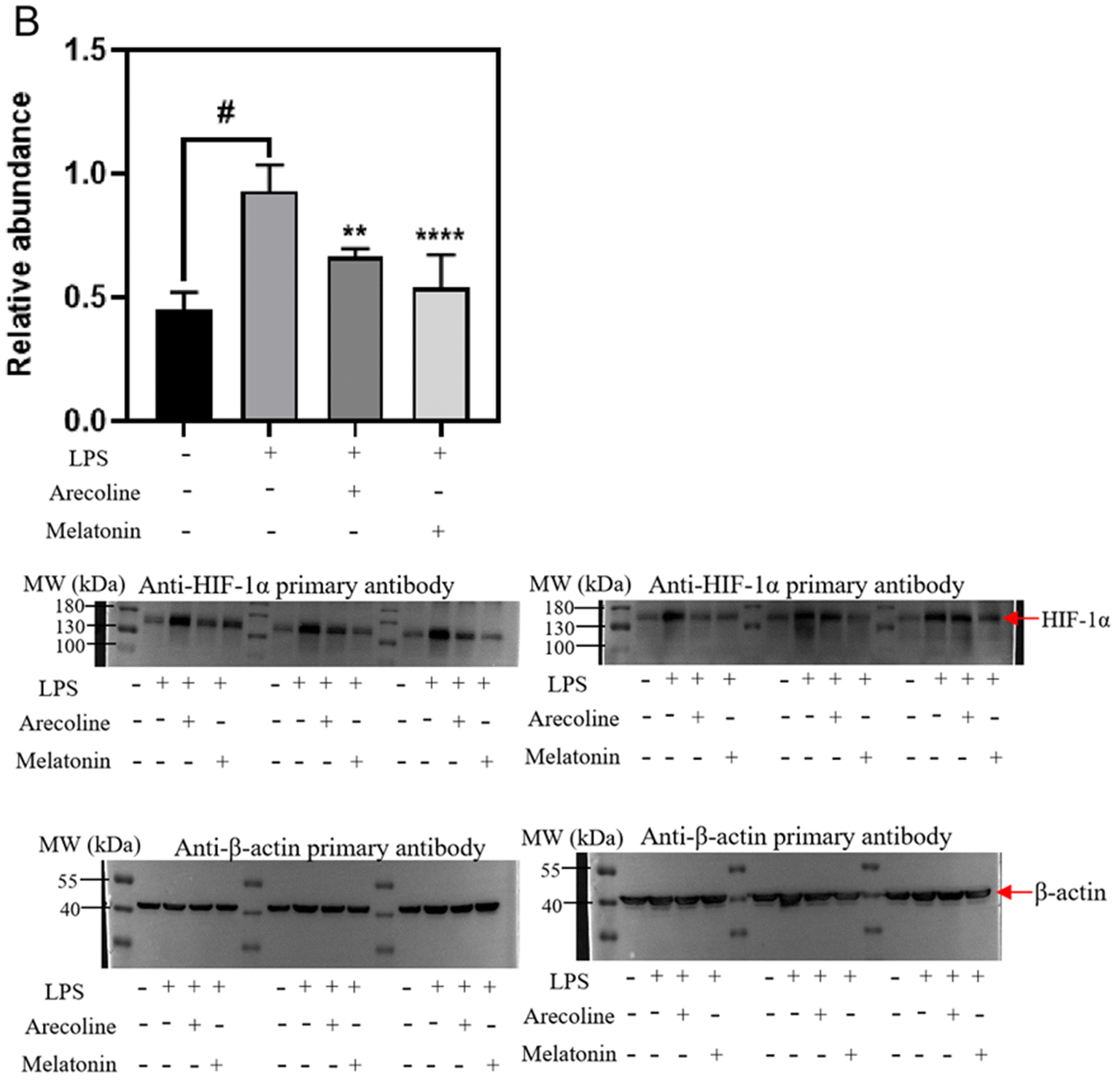 | ||
| Fig. 2 The anti-inflammatory effects of arecoline or melatonin on macrophage. RAW 264.7 macrophages were cultured in DMEM (containing 10% heat-inactivated FBS and 1% (v/v) penicillin/streptomycin) at 37 °C with 95% humidity and 5% CO2. RAW 264.7 macrophages were treated with LPS (1 μg mL−1) for 24 h to set up the inflammation model (control). To check the anti-inflammatory effects of melatonin and arecoline, RAW 264.7 cells were treated with melatonin (40 μM) or arecoline (40 μM) along with LPS stimulation. The protein expression of HIF-1α (B) was determined. The statistical analysis was performed using one-way ANOVA with Bonferroni correction. **P < 0.01 or ****P < 0.0001: arecoline or melatonin treatment vs. the control group; #P < 0.0001: the blank group vs. the control group. | ||
The Royal Society of Chemistry apologises for these errors and any consequent inconvenience to authors and readers.
| This journal is © The Royal Society of Chemistry 2025 |